
All iLive content is medically reviewed or fact checked to ensure as much factual accuracy as possible.
We have strict sourcing guidelines and only link to reputable media sites, academic research institutions and, whenever possible, medically peer reviewed studies. Note that the numbers in parentheses ([1], [2], etc.) are clickable links to these studies.
If you feel that any of our content is inaccurate, out-of-date, or otherwise questionable, please select it and press Ctrl + Enter.
The causative agent of pheohyphomycosis
Medical expert of the article

Last reviewed: 06.07.2025
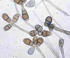
Phaeohyphomycosis is a mycosis (pheomycotic cyst) caused by many demacium (brown-pigmented) fungi that form hyphae (mycelium) in tissues. Demacium fungi are brown-pigmented fungi, unlike non-demacium fungi - hyalohyphomycetes (hyaline - non-pigmented hyphomycetes) that form mycelium and cause hyalohyphomycosis. Demacium fungi that cause phaeohyphomycosis are representatives of the genera Exophiala, Pbialaphora, Wangielta, Bipoiaris, Exscrohilum, Ctadophiahphora, Phaeoatmelhtnyccs, Altemaria, Aureobasidium, Cladosporium, Curvularia, Phoma.
Phaeohyphomycosis develops after demacium fungi from the soil enter microdamages in the skin of the extremities. A painless encapsulated mass is formed, which necrotizes, and a subcutaneous abscess develops. Brown yeast-like cells, pseudohyphae, and hyphae are found in the tissues. These fungi can cause opportunistic infections, including sinusitis (for example, Bipolaris, Exserohilum, Curvularia, Altemaria species in patients with chronic allergic rhinitis or immunosuppression), and brain abscess in immunodeficiencies after inhalation of conidia. Most often, brain damage is caused by the neurotropic fungus Cladophiatophora bantiana. Special care must be taken when working with these fungi.
 [
[