
All iLive content is medically reviewed or fact checked to ensure as much factual accuracy as possible.
We have strict sourcing guidelines and only link to reputable media sites, academic research institutions and, whenever possible, medically peer reviewed studies. Note that the numbers in parentheses ([1], [2], etc.) are clickable links to these studies.
If you feel that any of our content is inaccurate, out-of-date, or otherwise questionable, please select it and press Ctrl + Enter.
Mycetoma causative agents
Medical expert of the article

Last reviewed: 06.07.2025
Mycetoma (maduromycosis, malursky foot) is a chronic purulent-inflammatory process of the subcutaneous tissue and adjacent tissues. Mycetoma is caused by demacium fungi (eumycotic mycetoma) or actinomycetes (actinomycetes and cetomies) of the genera Actinomyces, Nocardia, Sireptomyces, Actimomadura. Among the fungi there are Pseudallescheria boydii, Aeremomum (Cephalosporium) Jidciforme, Madurella grisea, Phialophora cryanescem, Exophiala jrcmselmei, Scedosporium apiospermum, Leptosphaeria senegcdensis.
 [
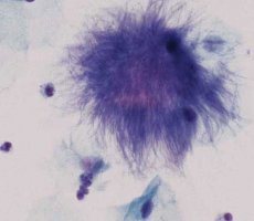
[ Microbiological diagnostics of mycetoma
In pus and biopsy treated with KOH solution, characteristic multi-colored grains (0.5-2 μm in diameter), septate hyphae and chlamydospores of fungi are revealed. Pseudallescheria boydii hyphae are difficult to distinguish from Aspergillus. In the presence of actinomycetes, druses and branching thin bacterial threads are visible. The sexual stage of P. boydii is accompanied by the formation of cleistothecia (100-200 μm), which rupture and release pale brown elliptical ascospores.