
All iLive content is medically reviewed or fact checked to ensure as much factual accuracy as possible.
We have strict sourcing guidelines and only link to reputable media sites, academic research institutions and, whenever possible, medically peer reviewed studies. Note that the numbers in parentheses ([1], [2], etc.) are clickable links to these studies.
If you feel that any of our content is inaccurate, out-of-date, or otherwise questionable, please select it and press Ctrl + Enter.
Open fracture
Medical expert of the article

Last reviewed: 12.07.2025
If an open wound has formed over the site of a bone fracture, that is, a violation of its anatomical integrity, then this is an open fracture, which, like a closed fracture, is classified as an injury: according to ICD-10, class XIX, code - S00-S99.
The size of the wound varies from a small puncture in the skin to an extensive rupture of all layers of the skin and gaping of damaged soft tissues, often with their separation and exposure of bone fragments that come out into the cavity of the open wound.
 [
[ Causes open fracture
The causes of an open fracture are a strong external traumatic (deforming) impact of residual energy of destruction during an impact, fall, collision, accelerated compression, etc. Most specific situations in which all these impacts are manifested are known to everyone and do not need to be listed: this is a fairly large list, including all accidents.
Regardless of how this case occurred, the pathogenesis of a skeletal bone fracture is associated with the fact that the force of external action (specific surface energy) on a particular skeletal structure at the moment of the fracture greatly exceeds the limit of the bone's biomechanical strength - its ability to withstand the impact energy (which bone tissue absorbs just like any other material). The biomechanical resistance of bone is viscoelastic in nature and, in addition, changes depending on the rate of application of forces: at high rates of mechanical action, bone tissue retains more energy, which leads to the destruction of their layered-crystalline structure.
The causes of any bone fractures are also seen in the fact that the bone structure is heterogeneous in the transverse and longitudinal directions, due to which the bone has different mechanical properties when loaded along different axes. And most fractures are the result of simultaneous impact on the bone in several directions.
Thus, stretching leads to a transverse fracture, with longitudinal action of dynamic compression - an oblique (diagonal) fracture. For example, an open fracture of the femur, as a rule, occurs with deformation in bending, when the forces of compression and tension acting towards each other are combined. But, since the bones are asymmetrical, the compressive and tensile stresses cannot be balanced, and the bone breaks.
Symptoms open fracture
The first signs of an open fracture of any location are sharp, severe pain (up to the development of pain shock), deformation of the broken limb, and bleeding due to damage to blood vessels.
Rapidly appearing symptoms of an open fracture of any location include the formation of edema (the nearby joint also swells) and hematomas at the fracture site.
When an open fracture of the lower extremities (thigh, shin, ankle) or an open fracture of the pelvis occurs, the person is immobilized and feels some numbness of the limb (due to nerve damage) and general weakness. The skin turns pale, chills begin. According to traumatologists, a characteristic sign of an open fracture of the tubular bones of the extremities is the mobility of bone fragments and a crunching sound when palpating the fracture site.
An open skull fracture is accompanied by a leak of cerebrospinal fluid from the subarachnoid space (through the ears and nose), loss of consciousness, and bleeding from the venous collectors of the dura mater adjacent to the bones. If the temporal bone is broken, bleeding occurs from the ear, and it stops hearing. Bleeding from the ears and nose, as well as liquorrhea (discharge of cerebrospinal fluid from the nose) occurs with open fractures of the occipital, ethmoid, and sphenoid bones of the skull.
Distinctive symptoms of an open fracture of the lower jaw: inability to close the mouth, blood or hematoma in the oral cavity, bloody saliva, the lower teeth may be broken. See also - Fracture of the lower jaw
If an open fracture of the nose occurs, then against the background of intense pain syndrome, nosebleeds are observed (possibly the release of mucous exudate from the nasal passages), hematomas in the area of the bridge of the nose and sphenoid sinuses, swelling of the mucous membrane of the nasal passages with the loss of the ability to breathe through the nose.
Forms
The Kaplan-Markova classification of open fractures determines the degree of tissue damage in cases of disruption of the integrity of tubular bones, identifying categories (A, B and C) with subcategories (I, II, III, IV):
Category A – minor local injury: IA (wound size less than 1.5 cm), IIA (wound size from 2 to 9 cm), IIIA (wound size over 10 cm);
Category B – contused lacerated wounds of soft tissues of moderate severity: IB (wound size up to 1.5 cm), IIB (wound 2-9 cm), IIIB (more than 10 cm);
Category B – severe crushed and crushed soft tissue injuries: IB (with a wound up to 1.5 cm), IIB (2-9 cm), IIIB (over 10 cm).
Categories AIV, BIV and BIV are open fractures with bone fragmentation, destruction of large areas of soft tissue and damage to large blood vessels.
The Gustilo-Anderson classification of open fractures also determines the severity of the limb fracture based on the size of the wound, the level of its contamination, and the degree of soft tissue damage and the level of contamination:
- Type I – simple transverse or oblique short fracture, wound <1 cm in diameter, practically clean, damage to soft tissues is minimal (no crushing);
- Type II – wound size from 1 cm to 10 cm in length, moderately contaminated lacerated wound without significant crushing of soft tissues;
- Type III – open segmental fractures with an extensive lacerated wound >10 cm, damage to soft tissues and the degree of wound contamination are significant, blood vessels are also damaged;
- type IIIA – fractures with a contaminated wound, extensive crushing of soft tissues and moderate exposure of the periosteum;
- Type IIIB – fractures with a heavily contaminated wound, extensive crushing of soft tissues and significant exposure of the periosteum; vascular restoration is required to preserve the limb.
Regardless of the size of the wound, this classification of open fractures automatically includes an open segmental fracture with displacement, an open fracture from a gunshot wound, fractures of the extremities in transport accidents, and injuries contaminated with soil in agricultural work as type III. This also includes traumatic amputations and open fractures that occurred 8 hours before seeking medical care.
It should be borne in mind that the occurrence of a defect in the skin and soft tissues as a result of the same traumatic impact that led to a bone fracture is defined in clinical traumatology as a primary open fracture. And when the skin and soft tissues are damaged by bone fragments, the open fracture is usually called secondary, and in such cases the wound from the perforation is small (although this does not exclude its infection).
Localization of open fracture
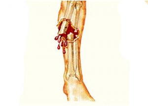
Trauma of the musculoskeletal system - open fracture of the limbs - can affect the fibula, tibia and femur tubular bones of the lower limbs; the humerus, ulna or radius of the upper limbs.
Open fracture of the femur - the proximal end of the femur, the body (diaphysis) of the femur; supracondylar and supracondylar fractures; open fracture of the ankle (bones of the ankle joint).
An open fracture of the humerus is a fracture of the shaft of the humerus or a supracondylar fracture of the humerus.
An open forearm fracture is an injury to the ulna or olecranon (the end of the bone); there may also be an open fracture of the radius. However, in severe injuries, both the ulna and radius are broken at the same time.
An open fracture of the clavicle is very rare, and in most cases – due to a fall on the side or on outstretched arms – the clavicle bone breaks in the middle third of the diaphysis.
An open pelvic fracture is diagnosed in cases of fractures of the bones of the pelvic ring - the pubic, iliac, sciatic, sacrum - if the fragments perforate the skin.
An open skull fracture is a fracture of the bones of the cranial vault (neurocranium); most often, open fractures (with indentation of a bone fragment) are suffered by the thinner temporal and parietal bones, the ethmoid bone, and the area of the occipital foramen near the base of the skull. And an open fracture of the jaw, a fracture of the orbital vault, and an open fracture of the nose are the most common open fractures of the facial bones of the skull.
Complications and consequences
What is the danger of an open fracture? The main danger is that such an injury is accompanied by bleeding and internal hemorrhages, leading to significant blood loss. Also, with such complex fractures, traumatic pain shock occurs, and infection penetrates the wound. Infection, in turn, is fraught with necrosis of soft tissues and the development of gas gangrene and sepsis.
With any localization of an open fracture, there may be certain consequences and complications.
First of all, complications include displacement of bone fragments, which is defined in the diagnosis as an open fracture with displacement. Displacement relative to the physiological position of the bone can be longitudinal, lateral, at an angle, with rotation of bone fragments, and also combined. As a result of displacement of fragments, there is an infringement or rupture of muscles, tendons, blood vessels and nerve fibers. In addition, there may be avulsion - a detachment of a fragment of a broken bone (splinter) from the main mass of bone tissue.
Among the general negative consequences of open fractures, experts note the closure of the lumen of the vessels of the lungs or brain by particles of fat from the bone marrow of tubular bones that have entered the blood (fat embolism), which leads to death.
Intracerebral hematoma and cerebral hemorrhage accompany open fractures of the skull bones.
As a result of damage to peripheral nerves, neurological complications of varying severity develop. For example, in cases of fracture in the foramen magnum area, conduction of the vagus, hypoglossal and glossopharyngeal nerves is disrupted, which causes speech, swallowing and breathing disorders.
Due to the rupture of the femoral nerve in an open fracture of the femur, leg extension is blocked, and after an open fracture of the tibia, it is often impossible to straighten the foot and lean on the heel when walking. And an open fracture of the radius can cause dysfunction of the radial nerve, and then problems with extension of the hand and fingers arise.
Complications of an open ankle fracture may include post-traumatic deforming osteoarthrosis of the ankle, the formation of a false joint in the bone fusion zone, and the development of habitual dislocation of the foot. An open forearm fracture may result in the fusion of the radius with the ulna.
An open pelvic fracture may result in hematomas in the retroperitoneal space and may also cause complications such as rupture of the bladder, urethra, or rectum; a fracture of the ischial tuberosity may result in shortening of the leg on the side of the fracture and significant limitation of its mobility in the hip joint.
Such consequences and complications as a deviated nose or nasal septum, impaired nasal breathing, inflammation of the trigeminal nerve are typical for an open fracture of the nose. An open fracture with displacement of the lower jaw bone can disrupt the closure of the dental arches and deform the bite.
In addition, for all open fractures, especially those with displacement and avulsion, there is a risk of developing inflammation and necrosis of bone tissue – post-traumatic osteomyelitis.
Diagnostics open fracture
For traumatologists and surgeons, injuries of this nature are obvious. And the diagnosis of an open fracture, which begins with an examination when the victim is admitted to the emergency room or trauma department, does not cause any difficulties.
However, only instrumental diagnostics can accurately determine the extent of damage to bones and soft tissues, as well as identify the presence of displacements and fragments - examination of the patient by X-ray (the image must be taken in two projections), computed tomography, and in the case of skull bone fractures - MRI.
Treatment open fracture
First aid provided at the site of injury for an open fracture consists of the following:
- it is necessary to stop the bleeding: in case of severe arterial bleeding - by applying a tourniquet above the fracture site and the wound (indicating the time of its application), in case of minor bleeding - a pressure bandage on the wound area;
- the wound above the fracture should be covered with a sterile bandage, but nothing should be touched in the wound itself;
- Give the victim any pain reliever.
First aid for an open fracture is provided until the ambulance team arrives. During this time, it is not recommended to move or transfer a victim with an open fracture of the hip, pelvis or skull to another location, so that bone fragments do not damage larger areas of soft tissue.
To prevent displacement of bone fragments, proper transport immobilization is necessary for open fractures. For example, when the radius is broken, a splint is applied that holds not only the forearm bones in a motionless position, but also the joints - the elbow and wrist. And in the case of a shin fracture, the knee and ankle joints should be immobilized using a splint made from improvised materials.
If the victim has an open fracture of the pelvic bones, he should be placed so that the part of the body above the waist is slightly raised, and under the knees (so that they are half-bent) a small elevation is needed, which can be made from a rolled-up piece of clothing.
In case of an open fracture of the lower jaw, transport immobilization is provided by tying the jaw over the head, and the victim is transported lying down.
Given the complexity of the injury, treatment of open fractures is carried out in a comprehensive manner.
Wound treatment is necessary – see more details – Treatment of open wounds, pain relief, anti-inflammatory therapy, reposition – anatomically precise unification (matching) of bone fragments – and their fixation in the most appropriate way for each case.
This can be a plaster or plastic splint - when there is an open fracture of the limbs without displacement. But in the presence of displacement and bone fragments (in particular, with an open fracture of the femur or tibia), they resort to skeletal traction under load (traction), which ensures their stable position and thereby promotes normal healing of the fracture.
In most cases, surgical treatment is necessary to properly treat the wound and to perform the most precise alignment of the broken bones. After anatomical reposition, fixation is required, for which trauma surgeons have special pins, pins, clamps, and plates in their arsenal. The most famous device used for external fixation of bone fragments is the Ilizarov device. Although the pioneer of KDO – compression-distraction osteosynthesis (i.e. surgical reposition of fragments with fixing structures) – is the Belgian surgeon Albin Lambotte, who worked in the Netherlands, and who, back in the early 20th century, used the first metal compression-distraction device he developed – a simple one-sided external fixator for a broken bone.
After the bone has healed, the fixing structures are removed and the soft tissues are sutured. Surgical treatment of open fractures also includes the elimination of damage to the peripheral nerves, which can be carried out at a later date - within three months after the injury (after identifying certain dysfunctions). Such operations are performed by neurosurgeons.
Drug treatment of open fractures
Drug treatment of open fractures is carried out using antibacterial, analgesic, decongestant, immunostimulating, and neuroprotective agents.
By using antibiotics – Amoxiclav, Cefazolin, Ceftriaxone, Metronidazole (Flagyl), etc. – doctors prevent or significantly reduce inflammatory complications. Amoxiclav is administered intravenously at 1.2 g (for children under 12 years old at 0.03 g per kilogram of weight) at intervals of no more than 8 hours. A single dose of Cefazolin is 0.5-1 g (for adults), administered in the same way. Side effects of all antibiotics of the named drugs include nausea, diarrhea and enterocolitis; urticaria; blood changes (anemia and leukopenia); increased levels of liver enzymes and nitrogen in the urine.
To relieve pain, non-steroidal anti-inflammatory drugs (NSAIDs) are used by injection or orally: Indomethacin, Ketoprofen, Ibuprofen, etc. Thus, Indomethacin can be administered intramuscularly for two weeks - once or twice a day (60 mg), and then you can switch to taking tablets - 25 mg twice a day, always after meals. Among the side effects of NSAIDs are headache, gastrointestinal manifestations with pain in the stomach. Therefore, these drugs are contraindicated in the presence of ulcerative diseases of the gastrointestinal tract, as well as in a history of bronchial asthma.
Capillary stabilizing drugs are used against edema, such as Methyl ethyl pyridinol or L-lysine excinate. L-lysine is administered intravenously once a day at 5-10 ml (twice a day for open TBI) for 3-7 days; the dose for children is calculated based on body weight. This drug is not used in renal failure and simultaneously with cephalosporin antibiotics; in rare cases, there may be side effects in the form of an allergic reaction.
In addition, in case of open fractures - to stimulate tissue metabolism and regeneration of damaged tissues - it is considered advisable to use the immunomodulatory agent Timalin. Intramuscular injections of this drug (single dose from 5 to 20 mg) are made once a day; the course of treatment lasts up to five days.
Calcium gluconate and calcium hydroxyapatite (Osteogenon) help restore bone tissue and fix calcium in it. After fractures, Osteogenon is recommended to be taken twice a day (1-2 tablets) for 2.5-3 months. This drug is contraindicated for kidney problems and for patients under 18 years of age.
The drug Gliatilin (Cereton) is a neuroprotector and is used to regenerate damaged peripheral nerves, especially in open fractures and other craniocerebral injuries: one capsule per day; in severe cases, the drug is used parenterally (in IV drips).
Rehabilitation after an open fracture
The duration of the rehabilitation period, which begins after the removal of the splint or compression-distraction apparatus, as well as the prognosis for the further condition, depends on the location of the open fracture and the degree of its complexity.
The modern complex of rehabilitation measures that help restore physiological functions to the affected skeletal structures includes various physiotherapeutic procedures, special therapeutic exercises, massage, as well as mechanotherapy or prolonged passive development of joints – Continuous passive motion, CPM therapy.
This method, the concept of which was created by Canadian orthopedic surgeon Robert B. Salter in the 1970s, is aimed at developing joints after injuries using special devices. CPM devices force joints to bend at a predetermined degree without the participation of the patient's muscle strength. In this case, the degree of joint flexion increases as rehabilitation after an open fracture proceeds, and the range of motion gradually expands.
Rehabilitation doctors advise eating right during the recovery period after an open fracture, consuming enough protein, vitamins A, C, D and group B, as well as calcium-rich dairy products and products containing phosphorus (vegetable oils, legumes, oats, almonds, nuts).