
All iLive content is medically reviewed or fact checked to ensure as much factual accuracy as possible.
We have strict sourcing guidelines and only link to reputable media sites, academic research institutions and, whenever possible, medically peer reviewed studies. Note that the numbers in parentheses ([1], [2], etc.) are clickable links to these studies.
If you feel that any of our content is inaccurate, out-of-date, or otherwise questionable, please select it and press Ctrl + Enter.
Hip MRI: what does it show and how is it done?
Medical expert of the article

Last reviewed: 03.07.2025
Among the visualization methods of hardware diagnostics, MRI of the hip joint plays a leading role in identifying damage and pathological changes in the largest joint of the human musculoskeletal system.
Magnetic resonance imaging provides the clearest and most detailed images, that is, the maximum information for making the correct diagnosis, and also facilitates the differential diagnosis of joint syndromes.
 [
[ Indications for the procedure
In traumatology, orthopedics and rheumatology, indications for examination of the hip joint using an MRI scanner include detection of:
- injuries (fractures, cracks, dislocations and ligament ruptures) and anomalies (hip dysplasia or congenital dislocation);
- coxarthrosis (deforming hip osteoarthritis);
- osteomyelitis of the femur and/or ilium;
- rheumatic joint lesions (arthritis), including systemic autoimmune diseases;
- osteoporosis, degenerative and necrotic changes in joint structures;
- foci of inflammation of periarticular tissues in tendonitis, bursitis of the hip joint, etc.;
- bone metastases of cancer.
MRI of the pelvis and hip joints is prescribed if there is a suspicion of the development of ankylosing spondylitis of the sacroiliac joints (Bechterew's disease).
MRI can be used to evaluate the results of corrective orthopedic procedures. This examination is mandatory before the upcoming installation of a hip replacement.
Preparation
No special preparation is required before magnetic resonance imaging of this joint: you just need to remove any metal products and change clothes (usually disposable medical clothing is provided or you bring it with you).
This examination is completely painless, the patient is in a lying position, does not move, so there is no need to administer pain relief before the MRI procedure of the hip joint. However, if the patient feels intense pain after an injury or a recent operation, then shortly before the procedure, analgesics are taken, and in case of severe anxiety - light sedatives.
When an MRI with contrast is prescribed, the doctor warns the patient about the need to stop eating and drinking liquids five to six hours before the examination.
Technique MRI of the hip joint
Magnetic resonance imaging creates images using a combination of a strong electromagnetic field around the body with induced resonant pulses of radio waves that are sensed by a scanner connected to a computer system that records the response signals and processes them – a visual transformation.
The patient is positioned on a surface that slides into the large, circular bore of the MRI scanner. Straps and cushions may be used to prevent the patient from moving during the procedure (since any movement can distort the image).
The technician who controls the movements of the scanner required by the MRI scanning technique is in the next room, but he or she is monitoring the patient and is connected to the patient for communication.
The examination lasts 15-20 minutes; for MRI with contrast, 25-30 minutes.
Contraindications to the procedure
Due to the use of a strong magnet, MRI of the pelvis and hip joints is contraindicated in patients with surgical staples, plates, pins, screws, clips or implanted devices made of metal and metal alloys, including
Pacemaker or cochlear implant. MRI is not performed for hip replacement.
Contraindications to this diagnostic procedure concern people with mental illnesses and severe somatic pathologies.
MRI of the hip joint during pregnancy is never performed in the first half of the term, and MRI with contrast is strictly prohibited for pregnant women.
In cases of renal failure and hemolytic anemia, as well as in patients on renal dialysis, MRI with a contrast agent is contraindicated, which helps determine the condition of periarticular tissues and vessels.
For patients suffering from claustrophobia (fear of enclosed spaces), as well as when it is necessary to perform MRI of the hip joint in children (especially younger children who find it difficult to remain still), an open-type MRI of the hip joint is an alternative. This examination is performed on a different modification of the MRI scanner - with an open design of the scanning part of the device (without placing the patient in a tunnel chamber). For example, the mother can be next to the child, who will restrain his attempts to change the position of the body or a separate limb.
Normal performance
There are atlases of normal MRI and CT anatomy (for all systems and organs), human anatomy in sections and images on CT and MRI, as well as sectional anatomy using CT and MRI sections as an example. Their images are compared with the MRI anatomy of the hip joint of specific patients, and this allows specialists to accurately determine pathological deviations that appear as a result of various diseases or traumatic injuries.
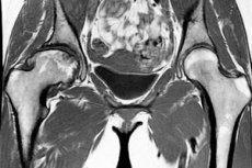
MRI shows all structures of the hip joint: the articular head of the femur with the topography of the bone and cartilage tissue; the acetabulum (where the femur and pelvic bones articulate); the femoral neck; the joint capsule with the internal synovial membrane (as well as the presence or absence of inflammatory exudate in it); the bone marrow canal of the femur; the entire ligamentous apparatus of the joint; adjacent soft tissues and blood vessels.
Also shown are the ilium, pubis, ischium and their ligaments, which are part of the hip apparatus.
Complications after the procedure
MRI does not use ionizing radiation, so if the scanning protocol is followed accurately, there are no negative consequences after the procedure.
There is also no special care after the procedure, and patients do not receive any restrictive recommendations from doctors. Simply – to avoid dizziness – do not make any sudden movements when getting up from the scanner table.
Possible complications after the procedure concern only MRI with a contrast agent, which at best can cause an allergic reaction, attacks of difficulty breathing and a decrease in blood pressure, and in the case of kidney problems - nephrogenic fibrosis and sickle cell anemia.
Patient reviews after MRI scanning of the pelvis and hip joints indicate the absence of discomfort or deterioration in well-being.
Which is better: X-ray, CT or MRI of the hip joint?
Experts in the field of hardware diagnostics believe that when choosing CT or MRI of the hip joint, most orthopedists prescribe MRI: due to the absence of radiation in MRI and the high quality of the volumetric layered image.
X-ray images are incomparable with the visualization of all structures and tissues that MRI scanners provide. So, when choosing an X-ray or MRI of the hip joint for examination, doctors take into account the degree of complexity of each specific case and assess the likelihood of an erroneous diagnosis in the absence of a detailed tomogram of the joint.