
All iLive content is medically reviewed or fact checked to ensure as much factual accuracy as possible.
We have strict sourcing guidelines and only link to reputable media sites, academic research institutions and, whenever possible, medically peer reviewed studies. Note that the numbers in parentheses ([1], [2], etc.) are clickable links to these studies.
If you feel that any of our content is inaccurate, out-of-date, or otherwise questionable, please select it and press Ctrl + Enter.
Metastatic melanoma
Medical expert of the article

Last reviewed: 04.07.2025
The last (fourth) stage of one of the most aggressive forms of cancer, when the deep layers of the skin are already affected and secondary neoplasms have spread not only to the nearest lymph nodes, but also to the distal ones, is diagnosed as metastatic melanoma. If vital internal organs are affected, only a miracle can save the patient.
What is this?
The surface layer of the skin contains cells containing melanin, a pigment substance that gives us a beautiful tan, unique hair and eye color, and unique moles and freckles on our skin.
Uncontrolled progressive proliferation of melanocytes, occurring in a certain place of the body, not only on open skin, but also on mucous membranes, under the mutagenic action of ultraviolet rays (the dose of which is individual for each) - this is melanoma. At the beginning of the process, when it is best to treat it, it often looks like a new, ordinary flat mole of irregular shape and does not manifest itself in any special way. Therefore, melanoma is often detected at later stages, which leads to disappointing results.
Does melanoma metastasize? Yes, and quite quickly. It is the ability to metastasize that is the defining characteristic of the aggressiveness of malignant neoplasms. Compared to other forms of skin cancer, which are curable even in relatively advanced stages, with melanoma "delay is like death."
Epidemiology
Among all malignant tumors, melanoma accounts for one to four cases out of a hundred. Residents of southern countries of the Caucasian race, who are constantly exposed to increased natural insolation, are more likely to get sick. Other types of skin cancer are ten times more common, but melanoma surpasses them in aggressiveness several times over. Every year, about 50 thousand people die from melanoma worldwide (according to the World Health Organization).
The highest incidence rates are registered among white Australians and New Zealanders (23-29.8 cases per 100,000 residents). Among Europeans, this rate is 2-3 times lower - about 10 primary visits per 100,000 residents annually. Ethnic Africans and Asians suffer from melanoma 8-10 times less often than representatives of the white race, regardless of their place of residence. Statistics show that the number of cases of malignant skin neoplasms is growing, including the number of patients on the planet diagnosed with melanoma, which doubles every decade.
Melanoma is diagnosed in children very rarely. Most sources say that the most likely age for melanoma manifestation is 30-50 years, medical statistics of the Russian Federation notes that most of their patients first sought help for a neoplasm after they had already passed the half-century mark (in 2008, the average age of those who first sought help was 58.7 years).
The risk of developing “black skin cancer,” as melanoma is also called, on apparently healthy and clear skin is approximately equal to the probability of malignancy of existing nevi.
Melanocyte degeneration can occur anywhere on the skin, but the most common site of neoplasm is the skin of the back in male patients, the skin of the shin in female patients, and the face in elderly patients. Female patients with melanoma of the skin are twice as common as male patients.
Melanoma always metastasizes to the lymph nodes, statistics say, not counting the initial stages, when there are simply no metastases yet. This is the main target organ. Then, in about 60% of cases, metastases are found in the skin.
The frequency of occurrence of metastatic lesions of internal organs is as follows: lungs (about 36%), liver (approximately a third of cases, sometimes called the first target organ), brain - a fifth of cases of secondary melanoma; bone tissue - up to 17%; digestive tract - no more than 9%.
Causes metastatic melanoma
Ultraviolet rays stimulate the production of melatonin. Excessive exposure to radiation is blamed for the occurrence of mutations in melanocytes, triggering the process of their uncontrolled growth and reproduction.
The origin of ultraviolet radiation can also be important. Natural sunlight (usually burns) can trigger the development of melanoma. In this case, the quantitative factor is dangerous. Artificial ultraviolet rays, especially those obtained in any modern and positioned as safe solariums, regardless of the time of exposure, increase the risk of developing melanoma by 74%. This conclusion was made by American oncologists from Minnesota based on the results of a three-year study. They found that melanoma develops 2.5-3 times more often in solarium lovers than in people who have never visited them.
The risk group includes fair-skinned people - blondes, albinos, redheads. Those who have a family history of melanoma or many moles on the body should take care. The increased risk of developing this neoplasm is associated with a hereditary disorder in the activity of the gene that suppresses tumor changes in cells.
Pigment nevi already present on the skin are dangerous in terms of malignant transformation: giant, complex, borderline, blue. Also, nevus of Ota, Dubreuil's melanosis, pigment xeroderma pose a melanogenic danger.
Risk factors for the development of malignant proliferation of melanocytes include living in areas with high levels of radioactivity or insolation, working in hazardous industries, periodic or even single sunburns to the point of blisters, trauma to birthmarks, and metabolic disorders.
Any of the above mentioned causes, often in combination, can trigger the pathogenesis of the appearance of atypical melanocytes and their hyperproliferation. Most patients with melanoma, especially at the metastatic stage, have a violation of the normal sequence of the signaling cascade of the BRAF gene, but not all of them. This is not the only molecular target in the pathogenesis of melanoma. Others have not yet been identified, but significant efforts are being made to this end.
The mechanism of malignancy of existing nevi includes both hereditary and external factors – excessive insolation, trauma, etc.
In the pathogenesis of melanoma, two main phases are distinguished - superficial or horizontal, when the spread occurs along the same plane as the skin surface, in the epithelium, and vertical, when the tumor begins to grow inward, into the deep layers of the skin and subcutaneous fat layer. Metastases appear when the process moves to the phase of vertical spread and reaches the lymphatic and blood vessels. Cancer cells are carried by the lymph flow to nearby, and later - to distant lymph nodes, and with the blood flow they reach even remote vital organs. Melanoma with multiple metastases not only to the distal lymph nodes, but also to the internal organs has the most unfavorable prognosis. The main reason for the diagnosis of "metastatic melanoma" is late diagnosis. It reflects a deeply neglected process.
Metastases after melanoma removal are most often detected in the first year. However, it happens that metastases appear much later. The process of metastasis has not yet been fully studied, but it is known that, even having penetrated from the vascular bed into the target organ, degenerated cells and their conglomerates can remain in a clinically undetectable state for a long time and manifest their presence unexpectedly, many years later.
The more time has passed since the radical treatment, the lower the estimated risk of metastasis. After seven years, it reaches a minimum. However, there are cases of late metastasis (after a ten-year relapse-free interval). A unique case of a secondary tumor appearing 24 years after the primary tumor was removed is known.
At what stage does melanoma metastasize?
Clinicians distinguish five main stages of melanoma (0-IV), in addition, they distinguish intermediate stages that take into account the thickness, the rate of cell division in the lesion, the presence of ulcers and different types of metastases.
At the third stage of melanoma, secondary formations are already detected in the lymph nodes, vessels and/or skin areas (satellites) closest to it. At stages IIIA and IIIB, the presence of altered cells can only be determined by microscopy of the smear-print and punctured lymph, at stages IIIC and IIID, an increase in regional lymph nodes is determined by palpation, and skin lesions are determined by visual examination.
Stage IV corresponds to the appearance of palpable secondary tumors at least in the lymph nodes located at a distance from the primary focus. At this stage, any distant areas of the skin and muscle tissue, as well as internal organs, can be affected. The most typical places are the lungs, liver, brain, bones. The diagnosis of metastatic melanoma is made when metastases are detected.
In the initial (in situ), first and second stages of melanoma, its spread to the nearest skin and lymph nodes cannot be detected even with microscopy. However, the modern oncological concept suggests that with the appearance of a malignant tumor, there is almost immediately a probability of metastasis. Modified cells constantly break away from the primary formation and are sent to new places by the lymphogenous (hematogenous) route, stop and grow, forming metastases. This process is quite complex, cells in the vascular bed interact with each other, other factors, and most of them die without turning into a metastasis. At first, metastasis occurs slowly and imperceptibly, but with melanoma that has spread to a depth of more than 1 mm, and this corresponds to only the second stage, there is already a risk of detecting secondary tumors some time after its removal.
This neoplasm is most often classified using the TNM classification developed by the American Cancer Society, which reflects three categories:
- T (tumor translation: tumor) – reflects the depth of the spread of the process, the presence (absence) of surface damage, the rate of division of the nuclei of modified cells (metastatic melanoma is coded T3-T4 with letter additions);
- N (Node Lymph – lymph node) – reflects the presence of lesions in the lymph nodes, the digital index indicates their number, the letter index, in particular b, indicates that lymphadenopathy is palpable or even visible visually;
- M (metastasis) – distant metastasis (M1 metastases present, M0 – none detected).
Melanoma primarily affects the lymph nodes located close to each other, the so-called sentinel nodes. At the stage of early metastasis, they are removed; this stage of the disease is prognostically relatively favorable.
A metastasis to the skin located at a distance of no more than 2 cm from the mother tumor is called a satellite. There are usually several of them, they are clusters of cancer cells (determined under a microscope) or look like small or large nodules. Secondary neoplasms on the skin located outside the two-centimeter zone are called transit metastases. Metastasis to the skin, especially transit, is considered an unfavorable sign, as well as to internal organs.
 [ 9 ]
[ 9 ]
Symptoms metastatic melanoma
To avoid the diagnosis of "metastatic melanoma", you need to periodically examine the moles on your body and, if any of them raises doubts about its benignity, you should consult a dermato-oncologist.
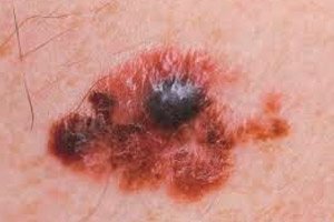
The first signs that should alert you are a noticeable increase in the size of the mole in the plane of the skin (more than 5 mm) and/or vertically above it; asymmetrical shape, uneven scalloped borders; noticeable changes in shape and color - asymmetrical depigmented areas, dots and areas of different colors. There is usually more than one alarming symptom; rapid growth means that the mole adds about a millimeter per month in any direction.
Later symptoms include itching in the area, inflammation of the skin around the questionable mole, depigmentation, loss of hair that previously grew on it, peeling of the surface of the mole, and the appearance of nodules on it.
A wet, ulcerated or bleeding surface, just like that, without trauma, are unfavorable symptoms. A varnished surface without a skin pattern is also unfavorable, as is a palpatory sensation of a change in the density of the formation.
The appearance of satellites on the surface of the skin surrounding a questionable mole – pigmented (flesh-pinkish) nodules or spots, that is, metastases to nearby skin, indicates that the melanoma stage is at least IIIC.
Melanoma can develop in several forms. The following are distinguished:
- the most common (more than 2/3 of cases) - superficially spreading, looking like a brown, almost flat spot of irregular shape and uneven color (darker, flesh-colored pinkish-gray areas), localized more often on the trunk and limbs; over time, the surface darkens, becomes glossy, is easily damaged, bleeds, ulcerates; the horizontal phase can last from several months to seven to eight years (it has a more favorable prognosis); after the onset of the vertical phase, the tumor begins to grow upward and inward, rapid metastasis occurs;
- nodular (nodular) melanoma immediately grows vertically (there is no horizontal growth phase) - it rises above the skin in a dome-shaped manner, has different, often uneven, pigmentation (sometimes depigmented), clear boundaries and the shape of a circle or oval, a smooth, shiny, easily damaged surface; sometimes it looks like a polyp on a stalk; it develops quickly - from six months to one and a half years;
- lentigo melanoma (malignant melanosis) - spots without a specific shape and clear boundaries, resembling large freckles, horizontal growth is very slow from ten to twenty years, more often found in older people on exposed parts of the body and face, the vertical phase is manifested by the fact that the borders become zigzag or wavy, the spot begins to rise above the skin, nodules, ulcers, crusts, cracks appear on its surface - this phase is fraught with the appearance of metastases;
- spotted (acral-lentiginous) melanoma is a rare type, mainly affects dark skin, develops on the fingers, palms, feet, under the nail (a dark stripe is formed).
There is a high probability of metastasis in melanomas developing on mucous membranes. They are usually detected accidentally during examinations by a dentist, otolaryngologist, proctologist and gynecologist. The pigmentation of such formations is usually noticeable and uneven.
Amelanoma without pigment is extremely rare. It is often diagnosed at late stages. It can be of any type – superficial, nodular, lentiginous.
General signs of metastatic melanoma, like all cancers in the late stages, are manifested by constant malaise, anemia, thinness, pallor, decreased immunity and, as a consequence, endless sluggish acute respiratory viral infections and exacerbations of existing chronic pathologies.
What do melanoma metastases look like?
Secondary neoplasms on the skin are visually visible. Satellites look like small multiple dark spots or nodules located near the parent tumor or the place of its removal. This form is typical for localizations of the primary formation on the skin of the trunk or limbs. Satellite metastases in melanoma spread through the lymphatic vessels, appear in about 36% of cases. They can be combined with nodal metastases, which occur in more than half of patients with metastatic melanoma.
Nodular (subcutaneous metastases of melanoma), spread with the lymph flow, usually look like sub- or intradermal tumors, often with an ulcerated, bleeding surface. Usually regional. Secondary nodular foci, appearing as a result of hematogenous spread, look like multiple round or oval nodes scattered over any part of the body, but their favorite places are the chest, back and abdomen. The skin above them is intact, flesh-colored or bluish, when accumulated melanin shines through under its thin layer. The size most often fluctuates from 50 mm to 4 cm, with larger sizes, tumors can merge, the skin becomes thinner, becomes glossy, the integrity of the integument is damaged (cracks, ulcers). At first glance, secondary skin tumors can resemble lipomas, epidermoid cysts, scars, dermatoses. Melanoma metastases to the subcutaneous fat tissue may not be noticeable during external examination, but they can be determined by palpation.
Erysipelas-like skin metastases in melanoma are rare, less than 1.5% of cases. In this case, the skin surface is seeded with defective melanocytes via the lymphatic route. They are typical for the location of the parent tumor on the temporal areas of the scalp, wrists, legs and chest. They resemble erysipelas in appearance - the skin around the primary lesion hurts, has a bluish tint and is swollen. They can be combined with satellites.
Rarely, but somewhat more often than erysipelas-like (up to 4% of cases, mainly with melanoma localized on the shin), thrombophlebitic skin metastases occur. Painful seals are hyperemic, with dilated superficial veins. The location is regional, the route of spread of cancer cells is lymphogenous.
When melanoma cells break off and enter the lymph flow, they first attack the sentinel lymph nodes. They are the first barrier to the spread of cancer cells and are the first to suffer. At first, melanoma metastases to the lymph nodes are detected by microscopy of their contents obtained by puncturing. At later stages, the nodes closest to the parent tumor are already enlarged and easily palpated, and later even visible. However, while 2-3 sentinel lymph nodes are affected and there is no further spread, they can still be removed. If metastases are detected in distant nodes of the lymphatic system, the patient's situation is interpreted as much worse, although much depends on their number and location.
The most severe degree of damage corresponds to the situation when wandering cancer cells have settled in the internal organs. They are carried hematogenously throughout the body and affect vital organs, which, or even part of them, are generally impossible to remove. With regard to metastases to internal organs, the expression "look" is not correct. They manifest themselves symptomatically and are visualized using various instrumental methods - ultrasound, MRI, radiography, and are also detected through laboratory tests.
Melanoma metastases to the brain are clusters of continuously dividing melanocytes in different parts of the brain, so different symptoms will manifest. Metastatic brain tumors are characterized by general malaise, loss of appetite and body weight, fever. General cerebral manifestations can be expressed by headaches, nausea, vomiting, sleep disorders, gait, coordination of movements, memory, speech, personality changes. Melanoma metastases to the brain can cause intracranial hemorrhage, seizures, paresis and paralysis, other neurological disorders depending on the lesion. For example, melanoma metastases to the pituitary gland are manifested by headache, ophthalmoplegia (oculomotor nerve paralysis) and other visual impairments, severe thirst and polyuria (neurogenic diabetes insipidus). For diagnostic purposes, magnetic resonance imaging of the brain is prescribed, but it cannot always provide an accurate answer about the origin and quality of the neoplasm.
Melanoma metastases to the liver, in addition to general symptoms of malaise, are manifested by constant nausea and vomiting, especially after eating non-dietary products, discomfort in the liver area, jaundice. Palpation also reveals an increase and compaction of the organ, in addition, splenomegaly is observed. Ultrasound examination shows that the surface of the liver is covered with dense tubercles.
The biochemical composition of the blood is disrupted. Uncontrollable vomiting lasting more than a day, especially with blood, black stool, visual enlargement of the abdomen are symptoms that require urgent help.
Melanoma often metastasizes to the lungs, in some sources this organ is called the main target, in others - the liver or brain. This localization of the secondary tumor manifests itself, in addition to general symptoms, with shortness of breath, wheezing, uneven breathing, a constant dry cough with poorly discharged sputum, sometimes with an admixture of blood, chest pain, and there may be a high temperature.
The neoplasm is usually visualized by radiation methods. Metastases can be focal, round in shape. With a small spread, they are most favorable. They have a hematogenous origin. More often, with melanoma, infiltrative metastases of lymphogenous origin are observed, which look like local darkening or a mesh entangling the lungs on the image. In practice, mixed forms are mainly observed.
Melanoma metastases to bones are manifested by local, intractable pain and frequent fractures. The appearance of malignant cells in bones and tumor growth disrupts the equilibrium state of metabolic processes between osteoblasts, synthesizing young cells of the bone matrix, and osteoclasts, destroying bone tissue. In most cases, osteoclasts and bone resorption processes are activated under the influence of cancer cells, however, osteoblastic activity sometimes predominates, which contributes to abnormal bone compaction, although mixed forms are most common.
Melanoma metastasizes to bones less frequently than to the liver, lungs, and brain. First of all, melanoma metastases appear in the spine, then in the ribs, skull, hip bones, and sternum. After that, cancer cells seed the pelvic bones (typical for the localization of the parent tumor in the groin) and, last of all, the scapular bones. Secondary tumors are localized in the medullary parts, which serve to accumulate calcium, and they prefer spongy bones, which are well supplied with blood. Tubular bones are extremely rarely involved in the pathological process, when all the “favorite” places are already occupied.
Osteolytic processes lead to hypercalcemia, which negatively affects the course of various processes in the body - the kidneys, central nervous and cardiovascular systems, and the gastrointestinal tract suffer.
Metastases of melanoma to the heart appear at an advanced stage of the disease. In melanoma, such localization is more common than in other primary foci. Cancer cells often migrate to the heart from the lung, getting there both through the lymphatic system and through the bloodstream. Most often, metastases are found in the pericardium, then in any cardiac chamber. The valves and endocardium are rarely affected. Metastatic tumors in the heart manifest themselves as a violation of cardiac activity, they are detected late, they do not affect the mechanism of death and survival.
If metastasis spreads to the gastrointestinal tract, dyspeptic symptoms appear. Against the background of general manifestations of cancer intoxication - exhaustion, weakness, abdominal pain, flatulence, nausea, vomiting are felt. With localization in the esophagus, first of all, a violation of the ability to swallow is observed. Pain is localized behind the sternum and in the upper abdomen, there may be perforation of the walls and bleeding. A tumor in the stomach is characterized by pain in the epigastrium, nausea, vomiting, black tarry feces. Secondary pancreatic tumor manifests itself with symptoms of chronic pancreatitis. Melanoma metastases to the intestine are extremely rare, however, they are the most malignant. They manifest themselves with symptoms of intestinal dysfunction, can lead to through perforations of its walls or intestinal obstruction.
It is extremely rare for melanoma as a primary neoplasm to develop on the mucous membrane of the digestive tract; secondary formations occur there much more often.
Achromatic, i.e. uncolored melanoma is often detected at late stages, when metastases have already appeared. It is characterized by the same clinical symptoms, only there is no specific dark color, which, first of all, attracts attention. Achromatic (non-pigmented) melanoma appears on a clean area of the skin, its forms correspond to the usual, the color is flesh-colored with a reddish, pinkish, grayish tint. It, like pigmented, grows quickly and changes shape, is asymmetrical, with uneven edges, or nodular, can bleed, itch, become covered with scabs and ulcers.
Metastases of non-pigmented melanoma spread in the same ways and to the same organs. Many consider this form of melanoma to be more malignant; it is believed that metastases appear and spread throughout the body much earlier than with ordinary "black" cancer. Perhaps this opinion is created because patients with an achromatic tumor often come to the attention of doctors with already pronounced metastases, without having any idea that they have melanoma.
Pain often occurs with melanoma with metastases, sometimes they require constant pain relief. The most painful are metastases to the brain and bone tissue.
Complications and consequences
Melanoma metastases are almost always multiple, which makes it very difficult to fight them. In addition, the metastasis stage occurs when the body no longer has the strength to resist. Secondary tumors disrupt the functioning of all vital organs and lead to the death of patients.
After removal of melanoma, even in a favorable initial stage in the absence of detectable metastases, there is no guarantee that the tumor will not relapse. Up to 90% of such events occur in the first two years after treatment, but it is recommended to undergo periodic examinations by a dermato-oncologist, since there are cases when the disease made itself known after a long-term relapse-free period.
Metastatic melanoma of the skin is already a complicated form in itself. In addition, common postoperative complications are possible - suppuration, infection, intractable pain localized in the incision sites.
Of great importance in the prognosis is such an indicator as the mitotic index, reflecting the ability of cells to divide. A high mitotic index indicates intensive cell division, and given that we are talking about cancer cells, lentigo melanoma with a high metastatic index (apparently mitotic) has a high probability of having metastases.
Diagnostics metastatic melanoma
The earliest diagnostic measure is an external examination of the patient, palpation of the lymph nodes and dermatoscopy, especially in a special immersion medium, which allows for a good view of the stratum corneum of the epidermis and a fairly accurate determination of whether a questionable mole is dangerous. To do this, its parameters (shape, size, borders, uneven color, presence of white-blue structures) are analyzed using the ABCDE rule. There is also a computer program that allows comparing photographs of a questionable mole with those in the database, but such diagnostics have not yet become widespread. In the presence of a suspicious nevus, in addition to a thorough examination of the skin and visible mucous membranes, the patient undergoes chest X-ray in two projections (direct and lateral), as well as an ultrasound examination of the lymph nodes, abdominal organs and pelvis.
Invasive methods of examination (biopsy) of the primary formation itself in melanoma are not allowed. Cytological analysis of a smear-print from the surface of the formation can be performed.
The final conclusion about the stage and morphology of the formation is made after a histological examination of the removed mole, the exact depth of its germination and the mitotic index are determined.
However, to detect micrometastases in sentinel lymph nodes that have not yet enlarged, the method of aspiration fine-needle biopsy under ultrasound control is increasingly used, making it possible to abandon traumatic prophylactic lymph node dissections.
Biopsy is used for certain locations of metastases, for example, in the lungs.
Before the operation, the patient undergoes standard clinical tests to assess his health.
If clinical signs indicate the presence of metastases in the liver, liver tests are performed and the level of lactate dehydrogenase (LDH) is assessed.
Melanoma metastases are usually multiple. Modern instrumental diagnostics are used to find them – radiation (radiography, computed tomography), magnetic resonance imaging, ultrasound, fibrogastroscopy, scintigraphy.
Differential diagnosis
Differential diagnostics are performed with melanoma-hazardous and benign nevi, according to the stages of the disease, the presence of single or multiple metastases. This is of great importance for choosing the tactics of providing the most effective assistance.
In the initial stages, as well as in solitary and single metastatic tumors, surgical treatment is fundamental, even in the presence of micrometastases - in combination with drug therapy.
Disseminated melanoma of the skin is distinguished, for which surgical treatment is no longer relevant, but palliative drug therapy is carried out.
Secondary tumors are differentiated from other neoplasms, often benign, such as lipoma or melanoma metastasis to the subcutaneous fat, melanotic schwannoma of the Gasserian ganglion of the brain, or metastatic melanoma of the base of the middle cranial fossa. Metastases to the heart are distinguished from the clinical consequences of chemotherapy and radiation exposure.
[ 24 ], [ 25 ], [ 26 ], [ 27 ], [ 28 ], [ 29 ], [ 30 ], [ 31 ], [ 32 ]
Who to contact?
More information of the treatment
Prevention
Any disease is easier to prevent than to cure. Melanoma is generally incurable in its later stages, so early and correct diagnostics is the main prevention of disease progression and the appearance of metastases, when the survival prognosis is less optimistic.
Experts recommend removing any mole that bothers you, even the most harmless one, not in beauty salons, but in specialized medical institutions, using removal methods that make it possible to conduct a subsequent histological examination of the removed tissue.
Prevention of melanoma formation is proper behavior in the sun - avoiding sunburn, burning to redness. Sunbathing should be done early in the morning or after 4 p.m., when the sun's rays are not so aggressive. Time spent in the sun should also be limited.
If you have moles on your body, you should try to protect them from the sun's rays, wear wide-brimmed hats on sunny days, light, natural, but closed clothing, use high-quality sunglasses and creams with a light filter of at least SPF15.
In light of new research, it is better to avoid visiting solariums altogether, since artificial ultraviolet light, even obtained from the most modern and safe sources and when the recommended time intervals are observed, is not absolutely harmless to the skin.
People at risk should be doubly careful.
Nutrition should be complete, many products have antitumor properties - fresh carrots, parsley, tomatoes, pumpkin. Coffee lovers are less likely to get skin cancer, concluded Boston researchers. It is useful to eat foods containing selenium (meat and offal, mushrooms, onions, garlic, black bread, Brazil nuts) and vitamin E (vegetable oils, sunflower seeds and most nuts, peas, beans, cabbage, eggs).
People after early stage melanoma removal are recommended to undergo a course of treatment with herbal preparations that have cytostatic activity and prevent the spread of metastatic formations. These are birch mushroom chaga, veselka, herbs - golden root, celandine, common thistle, white mistletoe, Siberian liana (priest) and others. Homeopathic treatment after surgery can also bring tangible benefits and prevent relapses.
Forecast
If we talk about metastatic melanoma with distant metastases, then patients with such a diagnosis have a high risk of death in the first five years after diagnosis. It is estimated at more than 80%. However, still not 100%!
How long do people with stage IV melanoma live? The data is disappointing: despite all the efforts of doctors, even patients from research groups on average do not survive even a year. Although different cases are known, even a complete cure is possible, so you should not give up.
Five-year survival is slightly higher in the group of patients with a milder stage of melanoma. At stage III of the disease with metastatic tumors in regional lymph nodes, as well as in patients with vertical spread of melanoma to a depth of more than 4 mm (stage II b and c) after radical treatment, the probability of relapse is estimated at 50-80%.