
All iLive content is medically reviewed or fact checked to ensure as much factual accuracy as possible.
We have strict sourcing guidelines and only link to reputable media sites, academic research institutions and, whenever possible, medically peer reviewed studies. Note that the numbers in parentheses ([1], [2], etc.) are clickable links to these studies.
If you feel that any of our content is inaccurate, out-of-date, or otherwise questionable, please select it and press Ctrl + Enter.
Lumbar puncture
Medical expert of the article

Last reviewed: 04.07.2025

Lumbar puncture (lumbar puncture, puncture of the subarachnoid space of the spinal cord, spinal puncture, lumbar puncture) is the insertion of a needle into the subarachnoid space of the spinal cord for diagnostic or therapeutic purposes.
Lumbar puncture is one of the widely used methods of examination in neurology. In some cases (infectious diseases of the central nervous system, subarachnoid hemorrhage) the diagnosis is entirely based on the results of lumbar puncture. Its data complement the clinical picture and confirm the diagnosis in polyneuropathies, multiple sclerosis and neuroleukemia. It should be noted that the widespread introduction of neuroimaging techniques has sharply reduced the number of diagnostic lumbar punctures. Puncture can sometimes be used for therapeutic purposes for intrathecal administration of antibiotics and chemotherapeutic drugs, as well as to reduce intracranial pressure in benign intracranial hypertension and normotensive hydrocephalus.
The total volume of cerebrospinal fluid in adults is about 120 ml. When speaking about extracting small volumes of it (from 10 to 20 ml) for diagnostic purposes, it should be borne in mind that the daily secretion volume is 500 ml. Thus, a complete renewal of cerebrospinal fluid occurs 5 times a day.
Indications for the procedure
Lumbar puncture is performed for diagnostic or therapeutic purposes.
- For diagnostic purposes, a puncture is performed to examine the cerebrospinal fluid. When analyzing the cerebrospinal fluid, the color, transparency, and cellular composition are determined. It is possible to study the biochemical composition of the cerebrospinal fluid, conduct microbiological tests, including its sowing on special media. During a lumbar puncture, the cerebrospinal fluid pressure is measured, and the patency of the subarachnoid space of the spinal cord is examined using compression tests.
- For therapeutic purposes, lumbar puncture is performed to remove cerebrospinal fluid and normalize cerebrospinal fluid circulation, control conditions associated with communicating hydrocephalus, as well as to sanitize cerebrospinal fluid in meningitis of various etiologies and administer medications (antibiotics, antiseptics, cytostatics).
There are absolute and relative indications for lumbar puncture.
- Absolute indications: suspected CNS infection ( meningitis, encephalitis, ventriculitis), oncological lesions of the membranes of the brain and spinal cord, normotensive hydrocephalus; diagnosis of cerebrospinal fluid leakage and detection of cerebrospinal fluid fistulas by introducing dyes, fluorescent and radiopaque substances into the subarachnoid space; diagnosis of subarachnoid hemorrhage when CT is not possible.
- Relative indications: fever of unknown genesis in children under 2 years of age, septic vascular embolism, demyelinating processes, inflammatory polyneuropathies, paraneoplastic syndromes, systemic lupus erythematosus, etc.
Technique lumbar puncture
Lumbar puncture can be performed with the patient lying down or sitting. The latter position is rarely used today. Usually, the puncture is performed with the patient lying on his side with the head tilted forward and the legs bent at the hip and knee joints. The spinal cord cone in a healthy adult is usually located between the middle sections of the L 1 and L 2 vertebrae. The dural sac usually ends at the S 2 level. The line connecting the iliac crests intersects the spinous process of L 4 or the space between the spinous processes of L 4 and L 5 (Jacobi's line).
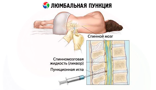
In adults, lumbar puncture is usually performed in the L3-L4 space ; in children, the procedure should be performed through the L4-L5 space . The skin in the puncture area is treated with an antiseptic solution, followed by local anesthesia by administering an anesthetic intradermally, subcutaneously, and along the puncture. A special needle with a mandrel is used to puncture the subarachnoid space in the sagittal plane parallel to the spinous processes (at a slight angle). The bevel of the needle should be oriented parallel to the long axis of the body. A bone obstruction usually occurs when deviating from the midline. Often, when the needle passes through the yellow ligaments and the dura mater, a feeling of failure is noted. In the absence of such a landmark, the position of the needle can be checked by the appearance of cerebrospinal fluid in the needle pavilion; for this, the mandrel must be periodically removed. If typical radicular pain occurs during needle insertion, the procedure should be stopped immediately, the needle should be removed to a sufficient distance, and the puncture should be performed with the needle slightly tilted toward the contralateral leg. If the needle rests against the vertebral body, it should be pulled up by 0.5-1 cm. Sometimes the lumen of the needle can cover the spinal cord root, in which case slight rotation of the needle around its axis and its pulling up by 2-3 mm can help. Sometimes, even if the needle enters the dural sac, it is not possible to obtain cerebrospinal fluid due to severe cerebrospinal fluid hypotension. In this case, raising the head end helps, the patient can be asked to cough, and compression tests can be used. With multiple punctures (especially after chemotherapy ), a rough adhesive process develops at the puncture site. If, despite following all the rules, it is not possible to achieve the appearance of cerebrospinal fluid, an attempt to perform a puncture at another level is advisable. Rare reasons for the impossibility of performing a lumbar puncture include a tumor of the spinal canal and an advanced purulent process.
Measurement of cerebrospinal fluid pressure and compression tests
Immediately after the cerebrospinal fluid appears in the needle pavilion, it is possible to measure the pressure in the subarachnoid space by connecting a plastic tube or a special system to the needle. The patient should be as relaxed as possible during the pressure measurement. Normal fluid pressure in a sitting position is 300 mm H2O, lying down - 100-200 mm H2O. Indirectly, the pressure level can be estimated by the rate of cerebrospinal fluid outflow (60 drops per minute conventionally corresponds to normal pressure). The pressure increases with inflammatory processes of the meninges and vascular plexuses, impaired fluid outflow due to increased pressure in the venous system (venous congestion). Liquorodynamic tests are used to determine the patency of the subarachnoid spaces.
- Queckenstedt's test. After determining the initial cerebrospinal fluid pressure, the jugular veins are compressed for no longer than 10 seconds. In this case, the pressure normally increases by an average of 10-20 cm H2O and returns to normal 10 seconds after the compression is stopped.
- During the Stukey test, the abdomen is pressed with a fist in the navel area for 10 seconds, creating congestion in the inferior vena cava system, where blood flows from the thoracic and lumbosacral sections of the spinal cord, and the epidural veins. Normally, the pressure also increases, but more slowly and not as significantly as during the Queckenstedt test.
Blood in the cerebrospinal fluid
Blood in the cerebrospinal fluid is most typical for subarachnoid hemorrhage. In some cases, a vessel may be damaged during lumbar puncture, and an admixture of "traveling blood" appears in the cerebrospinal fluid. In case of intense bleeding and if it is impossible to obtain cerebrospinal fluid, it is necessary to change the direction or puncture another level. When obtaining cerebrospinal fluid with blood, differential diagnostics should be performed between subarachnoid hemorrhage and an admixture of "traveling blood". For this purpose, the cerebrospinal fluid is collected in three test tubes. In case of subarachnoid hemorrhage, the cerebrospinal fluid in all three test tubes is colored almost the same. In case of traumatic puncture, the cerebrospinal fluid from the first to the third test tube will gradually clear. Another method is to evaluate the color of the supernatant: yellow cerebrospinal fluid (xanthochromic) is a reliable sign of hemorrhage. Xanthochromia appears within 2-4 hours after subarachnoid hemorrhage (result of hemoglobin degradation from broken red blood cells). A small subarachnoid hemorrhage can be difficult to visually distinguish from inflammatory changes, in which case one should wait for laboratory test results. Rarely, xanthochromia can be a consequence of hyperbilirubinemia.
Contraindications to the procedure
In the presence of a volumetric formation of the brain, occlusive hydrocephalus, signs of severe cerebral edema and intracranial hypertension, there is a risk of axial wedging during lumbar puncture, its probability increases when using thick needles and removing a large amount of cerebrospinal fluid. In these conditions, lumbar puncture is performed only in cases of extreme necessity, and the amount of cerebrospinal fluid removed should be minimal. If symptoms of wedging appear during the puncture (currently an extremely rare situation), urgent endolumbar administration of the required amount of fluid is recommended. Other contraindications to lumbar puncture are not considered so absolute. These include infectious processes in the lumbosacral region, blood clotting disorders, taking anticoagulants and antiplatelet agents (risk of epidural or subdural hemorrhage with secondary compression of the spinal cord). Caution when performing a lumbar puncture (removing a minimal amount of cerebrospinal fluid) is necessary if there is a suspicion of hemorrhage from a ruptured aneurysm of the cerebral vessels (risk of repeated rupture) and blockade of the subarachnoid space of the spinal cord (risk of the appearance or worsening of neurological deficit).
 [ 9 ]
[ 9 ]
Normal performance
For a standard study, cerebrospinal fluid is taken into three test tubes: for general, biochemical and microbiological analysis.
Standard clinical analysis of cerebrospinal fluid includes assessment of density, pH, color and transparency of cerebrospinal fluid before and after centrifugation, assessment of total cytosis (normally no more than 5 cells per 1 μl), determination of protein content. Depending on the need and capabilities of the laboratory, the number of lymphocytes, eosinophils, neutrophils, macrophages, altered cells, polyblasts, plasma cells, arachnoendothelial cells, epidermal cells, granular balls, tumor cells are also examined.
The relative density of the cerebrospinal fluid is normally 1.005-1.008, it is increased in inflammatory processes, decreased in excess fluid formation. Normally, pH is 7.35-7.8, it decreases in meningitis, encephalitis, paralysis, increases in paralysis (before treatment), syphilis of the brain, epilepsy, chronic alcoholism.
Yellow color of cerebrospinal fluid is possible with high protein content, in case of previous subarachnoid hemorrhage and hyperbilirubinemia. In case of melanoma metastases and jaundice, cerebrospinal fluid may be dark. Significant neutrophilic cytosis is characteristic of bacterial infection, lymphocytic - of viral and chronic diseases. Eosinophils are characteristic of parasitic diseases. With 200-300 leukocytes in 1 μl, cerebrospinal fluid becomes cloudy. To differentiate leukocytosis caused by subarachnoid hemorrhage, it is necessary to count leukocytes, taking into account that in the blood there is approximately 1 leukocyte for 700 erythrocytes. The protein content normally does not exceed 0.45 g/l and increases in meningitis, encephalitis, spinal cord and brain tumors, various forms of hydrocephalus, subarachnoid space block of the spinal cord, carcinomatosis, neurosyphilis, GBS, inflammatory diseases. Colloidal reactions also play a significant role - the Lange reaction ("golden reaction"), colloidal mastic reaction, Takata-Ara reaction, etc.
During biochemical analysis of cerebrospinal fluid, the glucose content (normally within 2.2-3.9 mmol/l) and lactate (normally within 1.1-2.4 mmol/l) are assessed. The assessment should be carried out taking into account that the glucose content of cerebrospinal fluid depends on the concentration of blood glucose (40-60% of this value). A decrease in glucose content is a common symptom of meningitis of various etiologies (usually bacterial in origin, including tuberculosis), an increase in the concentration of cerebrospinal fluid glucose is possible with ischemic and hemorrhagic stroke.
A decreased chloride content in the cerebrospinal fluid is characteristic of meningitis, especially tuberculosis, neurosyphilis, brucellosis, and an increase is characteristic of brain tumors, brain abscesses, and echinococcosis.
In a microbiology laboratory, a smear or sediment of cerebrospinal fluid can be stained depending on the suspected etiology of the pathogen: according to Gram - if a bacterial infection is suspected, for acid-fast microorganisms - if tuberculosis is suspected, with India ink - if a fungal infection is suspected. Cerebrospinal fluid cultures are carried out on special media, including media that absorb antibiotics (in the case of massive antibiotic therapy).
There are a large number of tests for identifying specific diseases, such as the Wasserman reaction, RIF and RIBT to exclude neurosyphilis, tests for various antigens for typing tumor antigens, determining antibodies to various viruses, etc. During bacteriological examination, it is possible to identify meningococci, pneumococci, Haemophilus influenzae, streptococci, staphylococci, listeria, and mycobacterium tuberculosis. Bacteriological studies of cerebrospinal fluid are aimed at identifying pathogens of various infections: coccal group (meningo-, pneumo-, staphylo- and streptococci) in meningitis and brain abscesses, pale treponema - in neurosyphilis, mycobacterium tuberculosis - in tuberculous meningitis, toxoplasma - in toxoplasmosis, cysticercus vesicles - in cysticercosis. Virological studies of cerebrospinal fluid are aimed at establishing the viral etiology of the disease (some forms of encephalitis).
Complications after the procedure
The total risk of complications is estimated at 0.1-0.5%. Possible complications include the following.
- Axial wedging:
- acute wedging during puncture in conditions of intracranial hypertension;
- chronic wedging as a consequence of repeated lumbar punctures;
- Meningismus.
- Infectious complications.
- Headaches usually go away when lying down.
- Hemorrhagic complications, usually associated with blood clotting disorders.
- Epidermoid cysts as a result of using low-quality needles or needles without a mandrin.
- Damage to the roots (possible development of persistent pain syndrome).
- Damage to the intervertebral disc with the formation of a disc herniation.
The introduction of contrast agents, anesthetics, chemotherapeutic agents, and antibacterial agents into the subarachnoid space may cause a meningeal reaction. It is characterized by an increase in cytosis to 1000 cells during the first day, an increase in protein content with normal glucose content and sterile seeding. This reaction usually regresses quickly, but in rare cases it may lead to arachnoiditis, radiculitis, or myelitis.
[ 15 ]