
All iLive content is medically reviewed or fact checked to ensure as much factual accuracy as possible.
We have strict sourcing guidelines and only link to reputable media sites, academic research institutions and, whenever possible, medically peer reviewed studies. Note that the numbers in parentheses ([1], [2], etc.) are clickable links to these studies.
If you feel that any of our content is inaccurate, out-of-date, or otherwise questionable, please select it and press Ctrl + Enter.
Leukoderma: causes, symptoms, treatment
Medical expert of the article

Last reviewed: 12.07.2025
Leukoderma - like leukocytes, leukemia and adhesive plaster - is a term of Greek etiology, and leukos means "white". Although, you must admit, if you do not know what leukoderma is, then the name of this skin disease (similar to blood cancer - leukemia) looks ominous.
Perhaps this is why dermatologists often use names such as hypopigmentation, hypochromia or hypomelanosis in cases of leukoderma.
Four pigments are involved in skin coloring – pigmentation – but the main role is played by the well-known melanin. Its synthesis and accumulation occurs in special cells – melanocytes. The initial “material” of melanogenesis is the essential amino acid tyrosine. Tyrosine enters the body from the outside, but under the influence of pituitary hormones and the enzyme phenylalanine-4-hydroxylase, it can be formed from the amino acid L-phenylalanine found in muscle tissue proteins. When any failure occurs in this complex biochemical process, keratinocytes (the main cells of the epidermis) stop receiving melanin, and dyschromia – a disorder of skin pigmentation – occurs. One of such disorders is a decrease in the amount of melanin or its complete absence in the skin – leukoderma.
 [ 1 ]
[ 1 ]
Causes of Leukoderma
Despite the fact that the biochemical mechanism of skin pigmentation disorders - amino acid metabolism disorders - is known to science, the causes of leukoderma in many cases remain unclear.
According to some experts, hypomelanosis is a secondary dyschromia. Others distinguish between primary, secondary, acquired and congenital hypochromia. And today, most of them consider various dermatological inflammations, as well as disorders of the nervous or endocrine systems of the body, to be the causes of this disease. Some dermatologists divide all causes of leukoderma into two groups. The first group includes all infections, and the second - unknown causes...
The primary form of hypomelanosis is chemical hypochromia and drug leukoderma. Chemical leukoderma, which is also called professional, is a diagnosis for those who are forced to constantly deal with chemicals that have a negative effect on the skin during their work. For example, hypopigmentation can be caused by hydroquinone and its derivatives, which are used in the production of rubber, plastics and dyes. And the cause of drug hypochromia is the effect of some medical drugs.
Primary leukoderma is such a common dermatological pathology as vitiligo. Specialists are still working on finding out the exact causes of vitiligo, and so far two versions of the etiology of this form of hypochromia have been accepted: congenital (i.e. genetic) and autoimmune.
Among the congenital forms of leukoderma, which manifests itself in childhood and disappears without a trace in adulthood, it is worth noting achromatic incontinence of pigment or Ito's hypomelanosis. This pathology reveals itself as colorless spots of various shapes, which are scattered throughout the body and form all sorts of "patterns" with clear boundaries. Rare autosomal dominant forms of primary hypomelanosis also include incomplete albinism (piebaldism) and complete albinism, which people have to put up with their entire lives.
Secondary leukoderma is not an independent disease, but only one of the symptoms or consequences of another pathology. For example, syphilitic leukoderma, which usually manifests itself six months after infection with this venereal disease, refers specifically to secondary hypochromia. And the loss of melanin pigment by skin rashes when the body is affected by the causative agent of syphilis, pale treponema, is a key sign of secondary syphilis.
The situation is similar with leprous leukoderma. The symptom of leprosy is pink-red spots with a "rim" that fade as the infectious disease progresses, then lose their color and atrophy. And pigmented leprida (spots on the skin) in tuberculoid leprosy are much lighter than the rest of the skin from the very beginning of the disease.
Fortunately, the cause of secondary hypochromia is more prosaic in most cases. Discolored spots on the skin appear where there were rashes of various nature in people suffering from such dermatological diseases as keratomycosis (scaly lichen, versicolor, pink), seborrheic eczema, trichophytosis, psoriasis, parapsoriasis, focal neurodermatitis, etc. That is, the loss of melanin in certain areas of the skin is the result of their primary lesions.
Typical symptoms of the so-called solar leukoderma, etiologically also associated with other skin diseases (most often with lichen), are manifested by depigmented spots that replace various rashes under the influence of sunlight. By the way, many dermatologists are convinced that ultraviolet rays contribute to the regression of skin rashes, although discolored spots remain on the skin for a very long time, but they no longer bother patients with peeling and itching.
Symptoms of Leukoderma
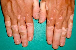
The main symptom of leukoderma is the appearance of discolored spots of various shapes, sizes, shades and locations on the skin. In some cases, the edges of melanin-deprived areas of the skin are framed by a more intensely colored "border".
Symptoms of syphilitic leukoderma include such varieties as lacy (net), marbled and spotted. In the first case, small depigmented spots merge into a mesh, which is located on the neck and is called the "necklace of Venus". With marbled syphilitic hypomelanosis, whitish spots do not have clear boundaries and seem to "blur". And spotted syphilitic leukoderma appears as a large number of practically identical in size light spots of round or oval shape on a background of darker skin. These spots can be both in the neck area and on the skin of other parts of the body.
Localization of symptoms of leprosy leukoderma - hips, lower back, buttocks, arms. This hypochromia behaves differently: it can remain for years without any changes, can capture new areas of the body, or can disappear on its own with the possibility of distant relapses.
The symptom of leukoderma in chronic systemic lupus erythematosus is inherent in the discoid form of this autoimmune disease. In the third stage of lupus dermatosis, white spots with characteristic cicatricial atrophy appear in the center of the rash.
Leucoderma scleroderma (lichen sclerosus atrophicus) is a secondary dyschromia and appears as small light spots, localized mainly on the neck, shoulders and upper chest. White spots may appear at the site of rashes and scratches in neurodermatitis (atopic dermatitis). And this is perhaps one of the few cases when, after successful treatment of this neurogenic-allergic skin disease, its normal color is restored - gradually and without any medication.
But the restoration of normal pigmentation of discolored areas of skin in vitiligo is a rare case. In this hypomelanosis, which does not cause any other symptoms, the colorless areas of the skin have clearly defined boundaries, and typical places of their localization are the upper chest, face, hands from the back side, feet, elbows and knees. As the disease progresses, the area of hypopigmentation increases, involving the hair growing on the affected areas of the skin in the pathological process.
Among the symptoms of such a rare type of leukoderma as piebaldism, that is, incomplete albinism, are the presence of a strand of completely white hair on the crown of the head, whitish spots on the forehead, chest, in the area of the knee and elbow joints, as well as dark spots on areas of discolored skin of the abdomen, shoulders and forearms.
Probably, everyone knows the external symptoms of albinism, which is closer to anomalies than to diseases. But in addition to obvious signs, albinos have nystagmus (involuntary rhythmic movements of the eyeballs), photophobia, and functional weakening of vision in one or both eyes (amblyopia) due to congenital underdevelopment of the optic nerve. According to scientists, the incidence of albinism in the world is approximately one person per 17 thousand. And most people with this congenital form of leukoderma are born in Africa - south of the Sahara Desert.
Diagnosis of leukoderma
In determining dermatological pathology in syphilis or lupus, the main thing is the diagnosis of these diseases. The diagnosis of leukoderma is based on a comprehensive examination of patients, which includes a thorough examination of the skin, a detailed biochemical blood test, dermatoscopy, differentiation of the clinical picture of the disease, collection of anamnesis, including the closest relatives. The doctor also necessarily finds out what medications the person took, and the connection of his work with chemicals.
Examination of the skin in primary or secondary leukoderma allows the dermatologist to determine the nature of hypomelanosis and identify its etiology.
An auxiliary method in diagnosing leukoderma is luminescent diagnostics using a Wood's lamp, which makes it possible to detect invisible lesions. However, according to the doctors themselves, luminescent diagnostics is applicable only when there is a suspicion of lichen, and it cannot guarantee a correct diagnosis in case of hypochromia.
Who to contact?
Treatment of leukoderma
In cases of solar leukoderma or drug-induced hypochromia, no treatment is required, as skin depigmentation in the affected areas resolves over time.
There is no treatment for chemical leukoderma as such, and the main thing here is to remove the provoking factor, that is, to stop contact with the chemicals that caused the pigmentation disorder.
Treatment of syphilitic hypochromia or leukoderma in lupus is associated with the general treatment of the underlying disease with the help of appropriate medications.
Therapy for secondary leukoderma is determined by a specific dermatological disease that has caused hypochromia and is prescribed by a doctor exclusively individually - using various medications for internal and external use: glucocorticosteroid and furocoumarin drugs, synthetic substitutes for natural amino acids tyrosine and phenylalanine, etc. Vitamins of group B, A, C and PP are prescribed. In the treatment of vitiligo, special PUVA therapy is widely practiced: application of photoactive medicinal substances - psoralens to the skin with irradiation with soft long-wave ultraviolet rays. However, this method of treatment does not help all patients get rid of leukoderma.
Prevention of leukoderma
Since tyrosine is necessary for the synthesis of melanin, it is recommended to eat foods that contain this amino acid to prevent leukoderma. Namely:
- cereals (especially millet, oatmeal, buckwheat);
- meat, liver, eggs;
- milk and dairy products (butter, cheese);
- sea fish and seafood;
- vegetable oils;
- pumpkin, carrots, beets, tomatoes, radishes, cauliflower, spinach;
- legumes (beans, soybeans, lentils, chickpeas);
- raisins, dates, bananas, avocados, blueberries;
- walnuts, hazelnuts, peanuts, pistachios, almonds, sesame and flax seeds, pumpkin and sunflower seeds