
All iLive content is medically reviewed or fact checked to ensure as much factual accuracy as possible.
We have strict sourcing guidelines and only link to reputable media sites, academic research institutions and, whenever possible, medically peer reviewed studies. Note that the numbers in parentheses ([1], [2], etc.) are clickable links to these studies.
If you feel that any of our content is inaccurate, out-of-date, or otherwise questionable, please select it and press Ctrl + Enter.
Esophageal varices
Medical expert of the article

Last reviewed: 12.07.2025
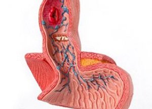
Esophageal varices are located in the distal esophagus or proximal stomach and are caused by increased pressure in the portal venous system, which is characteristic of liver cirrhosis. Varices may be complicated by massive bleeding without prior symptoms. Diagnosis is made by endoscopy, and treatment primarily involves endoscopic suturing and intravenous octreatide. Sometimes transjugular intrahepatic portosystemic (portocaval) shunting is necessary.
Causes of esophageal varices
The main symptom of any vascular disease of the esophagus is almost always the symptom of esophageal bleeding. These bleedings can occur when the esophagus and a large vessel lying nearby are injured, for example, when a large foreign body with sharp and cutting edges is fixed in the esophagus; when an esophageal tumor grows into a large vessel of the mediastinum and breaks through, for example, into the descending aorta. Most often, bleedings are observed from the vessels of the esophagus itself when its wall is damaged by a rigid esophagoscope, a sharp foreign body, erosion of the vessel by an ulcerative process or a disintegrating tumor; with congenital or acquired varicose veins of the esophagus. Acquired varicose veins of the esophagus are much more common than congenital ones, and sometimes reach significant sizes. Bleeding from these saccular venous formations can occur both spontaneously and even during very careful fibroesophagoscopy.
The cause of esophageal varices in the lower esophagus is congestion in the hepatic portal vein system, which occurs with liver cirrhosis and thrombosis v. portae. In the upper section, esophageal varices occur with malignant goiter. Among other causes, it is necessary to note angioma of the esophagus and vascular changes in Rendu-Osler disease.
Bleeding can occur spontaneously, when straining, lifting heavy objects, with increased blood pressure, common gastrointestinal diseases, and feverish conditions. It can be repeated, occur without any symptoms in the midst of "perfect health" and, if it becomes profuse, lead to death. A harbinger of such bleeding may be the appearance of a slight tickling in the throat, a peculiar salty-sour taste in the mouth, and then sudden vomiting of scarlet, and sometimes blood resembling coffee grounds. With significant blood loss, anxiety, weakness, darkening of the eyes with photopsies, dizziness, and other signs of increasing blood loss appear.
Esophageal varices are quite common compared to other causes of esophageal bleeding, especially in people with liver cirrhosis.
Liver cirrhosis is a chronic disease characterized by disruption of the liver structure due to the proliferation of connective tissue and pathological regeneration of the parenchyma, manifested by pronounced signs of failure of multiple liver functions and portal hypertension. The most common causes of liver cirrhosis in adults are chronic alcoholism and viral hepatitis, mainly hepatitis B. The development of liver cirrhosis can be caused by taking certain medications (methotrexate, isoniazid, etc.), exposure to a number of hepatotoxic agents, less often they are observed in some hereditary diseases - galactosemia, beta1-antitrypsin deficiency, hepatocerebral dystrophy, hemochromatosis, etc. Liver cirrhosis caused by venous congestion in the liver (congestive liver cirrhosis) is observed in long-term heart failure, disease of the hepatic veins and inferior vena cava. Liver cirrhosis in children can be observed already in the neonatal period due to liver damage in the antenatal period (fetal hepatitis). The cause may be viral infections suffered by the mother (hepatitis, cytomegalovirus, rubella, herpes infection), in which the virus is transmitted to the fetus through the placenta.
The cause and pathogenesis of varicose veins of the esophagus are determined by the anatomical connection of the veins of the esophagus with the venous system of the portal vein and the veins of the spleen, as well as other organs of the abdominal cavity, diseases of which lead to blockage of their venous networks and the development of venous collaterals, aneurysms and varicose veins of the esophagus. The development of these pathological formations in the area of the esophageal veins can be caused by compression of the portal vein also in diseases such as tumors, peritonitis, adenopathy, portal vein thrombosis, its angiomas, splenomegaly, etc. Circulatory disorders in the venous system of the spleen can be caused by diseases such as Banti syndrome (secondary splenogenic splenohepatomegalic symptom complex - anemia, thrombocytopenia, leukopenia, congestive splenomegaly, portal cirrhosis of the liver with symptoms of portal hypertension; more often observed in women under 35 years of age; the disease, according to modern concepts, is polyetiological in nature; this syndrome can develop as a result of intoxication and various infections, especially malaria, syphilis, brucellosis, leishmaniasis, etc.), Laennec's atrophic cirrhosis, chronic lymphocytic leukemia, etc. Other causes that can cause esophageal varices include certain diseases of the stomach and pancreas, as well as hemodynamic disorders in the superior vena cava. Age is not important for the development of esophageal varices. The entire process is determined by the emerging condition that prevents normal blood flow in the portal vein system.
Symptoms of esophageal varices
The symptoms of esophageal varices and the clinical course are determined by the cause of this gastrointestinal disease. Most often, the evolution of the disease is characterized by progressive development. Most often, the initial period of the disease is asymptomatic until bleeding from the esophagus develops. Bleeding can be from minor to profuse with a fatal outcome. Chronic blood loss of even small amounts of blood leads to hypochromic anemia, general weakening of the body, adynamia, shortness of breath, pallor, and emaciation. Melena is often observed.
The evolution of the disease can proceed very slowly or develop very quickly. With a slow development of varicose veins of the esophagus, patients remain unaware for a long time about the development of a terrible disease, in other cases, with a rapid development of the varicose process in the esophagus, a few days before bleeding, patients experience a feeling of compression in the chest. Sometimes a feeling of heaviness and compression in the chest can be harbingers of fatal bleeding. The data of some foreign researchers indicate a high legality from bleeding with varicose veins of the esophagus, on average 4 fatal cases per 5 patients. Hence the importance of early diagnosis of this disease.
What's bothering you?
Diagnosis of esophageal varices
Esophageal varices are diagnosed using fibroesophagoscopy, which establishes the causes of bleeding, the presence or absence of extraesophageal factors, determines the degree of vein dilation and the condition of their walls, and predicts the rupture of another aneurysm. In the case of ongoing bleeding, it is often difficult to establish its cause due to the impossibility of effectively performing esophagoscopy. Many other causes may be involved in the hyoid bone, information about which is given in the following sections on esophageal diseases. Certain information about the nature of esophageal varices can be obtained from X-ray examination of the esophagus with contrast.
Because varices are commonly associated with severe liver disease, evaluation for possible coagulopathy is important. Laboratory tests include complete blood count withplatelet count, prothrombin time, activated partial thromboplastin time, and liver function tests. Patients with bleeding should have blood type, Rh factor, and cross-matching of 6 units of packed red blood cells.
Who to contact?
Treatment of esophageal varices
Treatment of esophageal varices is aimed at compensating for hypovolemia and hemorrhagic shock. Patients with coagulation disorders (eg, elevated INR) require intravenous transfusion of 1-2 units of fresh frozen plasma and 2.5-10 mg of vitamin K intramuscularly (or intravenously in severe bleeding).
Because esophageal varices are initially diagnosed by endoscopy, the primary treatment involves endoscopic hemostasis. Endoscopic suturing of the veins is preferable to injection sclerotherapy. At the same time, intravenous octreotide (a synthetic analogue of somatostatin) should be administered. Octreotide increases visceral vascular resistance by inhibiting the release of visceral vasodilator hormones (eg, glucagon and vasoactive intestinal peptide). The usual dose is 50 mcg intravenously, followed by an infusion of 50 mcg/hour. Octreotide is preferable to other drugs such as vasopressin and terlipressin, since this drug has fewer side effects.
If, despite the treatment, bleeding continues or recurs, emergency methods of shunting (dumping) blood from the portal system into the inferior vena cava can reduce portal pressure and decrease bleeding. Transjugular intrahepatic portosystemic shunting (TIPS) is the emergency intervention of choice: the method is an invasive endovascular procedure under radiological control, in which a metal guidewire from the vena cava penetrates through the liver parenchyma into the portal bloodstream. The resulting anastomosis is widened with a balloon catheter and a metal stent is installed, creating a shunt between the portal bloodstream and the hepatic veins. The size of the stent is of fundamental importance: if it is too wide, hepatic encephalopathy develops due to too much portal blood being dumped from the liver into the systemic bloodstream. On the other hand, small stents tend to become occluded. Surgical portocaval shunting, such as distal splenorenal anastomosis, has a similar mechanism but is riskier and carries a higher mortality rate.
In case of severe bleeding, rubber inflatable probes are used to stop bleeding by pressing the bleeding vessel, for example, the Sengstaken-Blakemore probe. Currently, there are corrugated obturator probes for this purpose, which are used to stop bleeding from varicose veins of the esophagus and with a bleeding gastric ulcer.
Through a probe inserted below the bifurcation, the esophagus can be washed with hot water (40-45°C), which sometimes stops bleeding. Repeated bleeding requires the use of all the same measures for any prolonged bleeding (intravenous administration of 10-20 ml of 10% calcium chloride solution, intramuscular - vikasol). Do not administer agents that increase blood pressure until bleeding has completely stopped due to the risk of increasing the latter.
In case of massive blood loss, intravenous administration of blood, plasma, blood-substituting fluids, pituitrin, platelet mass, etc. is performed.
In case of repeated bleeding, intervention on the vessels of the hepatic portal vein system may be required. If very large vessels are damaged, patients die quickly.
Forecast
In approximately 80% of patients, variceal bleeding stops spontaneously. However, esophageal varices have a high mortality rate, often greater than 50%. Mortality depends primarily on the severity of the underlying liver disease rather than the severity of bleeding; bleeding is often fatal in patients with severe hepatocellular insufficiency (eg, advanced cirrhosis), whereas patients with good liver function usually recover.
In patients who survive at high risk of variceal bleeding, rebleeding typically occurs in 50-75% of cases within the next 1-2 years. Chronic endoscopic and medical treatment of esophageal varices significantly reduces this risk, but overall the effect on long-term survival remains very low, mainly due to the underlying liver disease.
 [ 6 ]
[ 6 ]