
All iLive content is medically reviewed or fact checked to ensure as much factual accuracy as possible.
We have strict sourcing guidelines and only link to reputable media sites, academic research institutions and, whenever possible, medically peer reviewed studies. Note that the numbers in parentheses ([1], [2], etc.) are clickable links to these studies.
If you feel that any of our content is inaccurate, out-of-date, or otherwise questionable, please select it and press Ctrl + Enter.
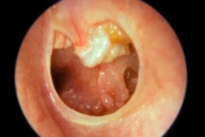
Cholesteatoma of the middle ear
Medical expert of the article

Last reviewed: 12.07.2025
Most often, cholesteatoma is defined as a type of epidermoid cyst that is localized in the middle ear and mastoid process of the temporal bone of the skull. So histologically, it is not a tumor. According to ICD-10, this pathological formation of the middle ear has the code H71.
Epidemiology
According to experts from the European Academy of Otology and Neuro-otology (EAONO), out of more than 20 million people worldwide with chronic inflammatory diseases of the ears, up to 25% of patients have cholesteatoma. [ 1 ]
The prevalence of acquired cholesteatoma is estimated at 95-98%; congenital accounts for 2-5% of cases.
The annual incidence of this middle ear mass is three per 100,000 children and nine per 100,000 adults. In the United States, one study reported six cholesteatomas per 100,000 people. The average age of children diagnosed with acquired cholesteatoma was 9.7 years. Acquired cholesteatomas are approximately 1.4 times more common in males than in females. One English study found an increased incidence of cholesteatoma in socioeconomically disadvantaged areas, suggesting that the incidence of acquired cholesteatoma is higher in low-income patients, although more research is needed in this area. [ 2 ]
Causes cholesteatomas
By origin, cholesteatomas are divided into primary (congenital), secondary (acquired, occurring at any age) and idiopathic (when it is impossible to accurately determine the etiology). [ 3 ]
Experts name such key causes of the most common secondary form of this pathology as perforation of the eardrum of inflammatory, traumatic or iatrogenic etiology; exudative otitis media and purulent otitis, or more precisely, chronic purulent otitis media.
Among the etiological factors, purulent inflammation of the middle ear, which develops in the supratympanic (epitympanic-antral) region, is also distinguished - epitympanitis with cholesteatoma.
Often, cholesteatoma is the result of problems with the auditory (Eustachian) tube: a disruption of its function caused by inflammation - tubootitis or infections of the middle ear and paranasal sinuses.
Congenital cholesteatoma is a rare diagnosis. Primary cystic formation of the intact eardrum (membrana tympani) usually forms in its weakly stretched part (pars flaccida), but can occur in the middle ear (near the cochlear process of the tympanic cavity or near the Eustachian tube), as well as in the adjacent bones of the skull. [ 4 ]
Congenital cholesteatoma in a child is a heteroplastic epidermoid formation formed during intrauterine development. In more than half of cases in children and adolescents, such a formation is detected when visiting an otolaryngologist about hearing loss.
Risk factors
In otology, risk factors for the development of cholesteatoma are usually associated with frequent acute infectious and chronic diseases of the middle ear; perforation and other damage to the eardrum; obstruction of the auditory tubes (often seen in patients with a history of chronic nasopharyngitis, allergic rhinitis, or enlarged adenoids); and some otological procedures (e.g., drainage of the middle ear with tympanostomy tubes). [ 5 ]
There is an increased risk of developing this pathology in children with ear developmental anomalies, which are observed in congenital syndromes of Treacher Collins, Crouzon, Goldenhar, and also occur in children with Down syndrome, Jessner-Cole syndrome and cleft palate.
Pathogenesis
In appearance, cholesteatoma is a whitish-pearlescent elastic formation of oval shape - a thin-walled cyst containing layered waxy or caseous keratin fragments (called keratin debris by doctors). And inside the congenital cholesteatoma, keratinized cells of stratified squamous epithelium of exodermal origin are found histologically. [ 6 ]
Studying the clinical picture, etiology and pathogenesis of this formation, otologists and otoneurologists put forward different theories of the formation of cholesteatoma.
According to the most convincing version, the mechanism of formation of congenital cholesteatoma is caused by abnormal movement of mesenchyme cells of the dorsal part of the neural crest during the formation of the pharyngeal arches and the middle ear rudiment during embryogenesis or during the formation of the auditory canal and eardrum of the fetus in the early stages of gestation. Another hypothesis suggests penetration of cells of the extraembryonic ecto and mesoderm of the amnion into the middle ear space. [ 7 ]
One theory explaining the occurrence of acquired cholesteatomas links increased keratinization of the epithelium of the mucous membrane of the middle ear with an inflammatory reaction leading to the release of cyclooxygenase-2, interleukins, vascular endothelial growth factors and epidermal growth factors, which stimulate the proliferation of epithelial keratinocytes. In addition, researchers have found that osteoclastic resorption of the auditory ossicles of the middle ear or mastoid bone during the formation of cholesteatoma occurs due to the action of prostaglandins, collagenolytic and lysosomal enzymes, which are synthesized by connective (granulation) tissue formed around bone structures,
Another theory is that in cases of Eustachian tube dysfunction, negative pressure in the middle ear pulls the eardrum inward (towards the auditory ossicles) to form a fold (called a retraction pocket) that fills with exfoliated squamous epithelial cells and turns into a cyst.
Another theory suggests that when the eardrum is perforated, the squamous epithelium lining the external auditory canal spreads (migrations) into the middle ear cavity, that is, it grows along the edges of the membrane defect.
Symptoms cholesteatomas
As clinical practice shows, cholesteatomas, especially congenital ones, can remain latent for a long time, and the symptoms that appear usually affect only one ear.
In the case of acquired cholesteatoma, the first signs are constant or periodic otorrhea – watery discharge from the ear, which, if there is an infection, can be purulent (with an unpleasant odor), and sometimes bloody. In advanced inflammation of the middle ear, there may be ear pain. [ 8 ]
As the cystic formation grows, the list of patient complaints expands and includes:
- a feeling of discomfort and pressure in one ear;
- tinnitus (constant noise or ringing in the ear);
- headache;
- dizziness;
- pain in the ear or behind the ear;
- unilateral hypoacusis (hearing loss);
- muscle weakness on one side of the face (in rare cases).
The severity of symptoms varies, and some patients may experience only minor discomfort in the ear.
In addition to all the listed symptoms, when the cerebellopontine angle cholesteatoma reaches a significant size, involuntary contraction of the facial muscles and progressive paralysis of the facial nerve are observed.
Forms
There are also different types of cholesteatoma based on the location of their formation. Cholesteatoma of the outer ear is rarely diagnosed, but can spread to the eardrum, middle ear or mastoid process, and damage to the facial nerve canal located in the temporal bone (os temporale) is also possible.
Cholesteatoma of the external auditory canal is a cystic mass in the area of damaged bone cortex in the wall of the bony part of the external auditory canal (meatus acusticus externus).
Cholesteatoma of the middle ear or cholesteatoma of the tympanic cavity (which is located in the center of the middle ear - between the eardrum and the inner ear) is in most cases a complication of chronic otitis.
Congenital cholesteatoma of the temporal bone occurs in its mastoid process (processus mastoideus) or in the thin tympanic part (pars tympanica) fused with it, which limits the external auditory canal and the auditory opening. If a cystic formation is formed in the process of the temporal bone of the skull located behind the auricle and having air cavities, then cholesteatoma of the mastoid process is diagnosed.
The middle ear cavity with the tympanic membrane is located in the petrous (petrous) part of the temporal bone, which is called a pyramid due to its triangular shape. Part of its anterior surface is the upper wall (roof) of the tympanic cavity. And this is the place where cholesteatoma of the temporal bone pyramid, that is, its petrous part (pars petrosa), can form. And cholesteatoma of the apex of the temporal bone pyramid means its localization in the upward facing anterior surface of the pyramid, where the semi-canal of the Eustachian tube is located.
The upper wall of the tympanic cavity of the middle ear separates it from the cranial cavity, and if a cholesteatoma formed in the middle ear or the pyramid of the temporal bone spreads into the brain - through the elytra of the middle cranial fossa - a cerebral cholesteatoma may be observed, which experts classify as an otogenic intracranial complication.
And cholesteatoma of the cerebellopontine angle is a congenital formation that slowly grows in the cerebrospinal fluid-filled space between the brainstem, cerebellum and the posterior surface of the temporal bone.
Specialists determine the stages of cholesteatoma of the middle ear: cholesteatoma of the pars flaccida (weakly stretched part of the tympanic membrane), cholesteatoma of the stretched part (pars tensa); congenital and secondary cholesteatoma (with perforation of the tympanic membrane).
At stage I, cholesteatoma is localized in one place; at stage II, two or more structures may be affected; at stage III, there are extracranial complications; stage IV is determined by intracranial spread of the formation. [ 9 ]
Complications and consequences
Aggressive growth of cholesteatoma – including congenital – can cause dangerous consequences and complications:
- destruction of the ossicular chain with hearing impairment (conductive or mixed hearing loss);
- destruction of the wall of the bony part of the external auditory canal and erosion of the walls of the tympanic cavity;
- development of the inflammatory process and its spread to surrounding areas, including the inner ear (labyrinth). Due to the penetration of cholesteatoma into the labyrinth, its inflammation (labyrinthitis) may occur, as well as a fistula (fistula) of the inner ear.
- Spread of the formation beyond the ear can lead to:
- obstruction of the antrum (cave) of the mastoid process of the temporal bone, which is fraught with its inflammation - mastoiditis;
- thrombosis of the cavernous sinus of the dura mater of the brain;
- development of purulent meningitis;
- intracranial (epidural or subdural) abscess;
- brain abscess.
Diagnostics cholesteatomas
Clinical diagnosis of cholesteatoma is made during a thorough examination of the ear.
For this purpose, instrumental diagnostics are used:
- otoscopy and otomicroscopy;
- X-ray of the ear and temporal bone;
- tympanometry.
A hearing test is performed (using audiometry or impedancemetry).
Detection or visual confirmation of cholesteatoma requires computed tomography or magnetic resonance imaging. If cholesteatoma is suspected, all patients should undergo diffusion-weighted MRI. Cholesteatoma appears as a hyperintense (bright) area on MRI (on T2-weighted images in the frontal and axial planes).
A cholesteatoma of the middle ear is visualized on CT as a sharply defined accumulation of homogeneous soft tissues (low density) in the middle ear cavity, but due to the low specificity of computed tomography, it is almost impossible to distinguish it from the surrounding bone structures of granulation tissue. However, CT shows all bone changes, including defects of the auditory ossicles and erosion of the temporal bone, so this examination is necessary for planning an operation to remove this formation.
It is difficult to distinguish congenital cholesteatoma from acquired cholesteatoma, so the diagnosis is primarily based on the anamnesis and clinical signs.
Differential diagnosis
Of great importance is the differential diagnosis of cholesteatoma with keratosis and erosive tumor of the external auditory canal, atheroma and adenoma of the middle ear, eosinophilic granuloma, oto- and tympanosclerosis, glomangioma of the tympanic cavity, ectopic meningioma, squamous cell carcinoma.
Who to contact?
Treatment cholesteatomas
To suppress inflammation in cases of secondary cholesteatoma, treatment is carried out, which involves cleaning the ear, taking antibiotics and using ear drops. All the details are in the publications:
- Antibiotics for otitis
- Drops for otitis
- Treatment of chronic purulent otitis in hospital and at home
- Physiotherapy for otitis media
There is no medicine that can remove this formation, so the only way is surgical treatment, the tactics of which are determined by the stage of the disease at the time of surgery.
The usual method of removing cholesteatoma is mastoidectomy (opening the air cells of the mastoid process of the temporal bone). The standardized microsurgical procedure is a down-canal mastoidectomy (contraindicated in children) - a modified radical mastoidectomy with removal of the bony wall of the external auditory canal (also requiring reconstruction of the eardrum). Another technique is an up-canal mastoidectomy, which removes all pneumatized areas of the mastoid process while preserving the posterior wall of the auditory canal. [ 10 ]
At the same time, surgeons can perform tympanoplasty – restoration of the eardrum (with cartilage or muscle tissue from another part of the ear).
Examination for the operation to remove cholesteatoma includes X-ray and CT of the ear and temporal bone, ECG. It is also necessary to take blood tests (general, biochemical, coagulation).
How long does cholesteatoma removal surgery take? The average duration of such surgery, performed under general anesthesia, is two to three hours.
In the postoperative period (for several weeks), patients should not remove the bandage (until the doctor’s permission); it is recommended to sleep with the head elevated (this will reduce swelling and improve the outflow of exudate from the ear cavity); water should be avoided in the operated ear, physical activity and air travel. [ 11 ]
Quite often, even a successful operation is not able to prevent recurrence of cholesteatoma, which is observed in 15-18% of cases in adults and in 27-35% of cases in children.
Taking this into account, 6-12 months after surgery, a revision is performed after removal of the cholesteatoma - either surgically or using MRI. According to some data, in almost 5% of cases there is a need for a repeat operation. [ 12 ]
Prevention
It is impossible to prevent the formation of congenital cholesteatoma, and the prevention of secondary epidermoid formation of the middle ear is the timely detection and treatment of its inflammatory diseases.
Forecast
In general, the prognosis of cholesteatoma depends on its location, ethology, stage of development and the age of the patient.
Almost always, this formation can be removed, but its uncontrolled growth can cause serious problems, primarily with hearing.
When asked whether disability is granted for cholesteatoma, specialists answer as follows. This diagnosis is not on the list that gives the right to disability, but there is a hearing disability, including third-degree hypoacusis, provided that its compensation with a hearing aid is insufficient for full-fledged professional activity.