
All iLive content is medically reviewed or fact checked to ensure as much factual accuracy as possible.
We have strict sourcing guidelines and only link to reputable media sites, academic research institutions and, whenever possible, medically peer reviewed studies. Note that the numbers in parentheses ([1], [2], etc.) are clickable links to these studies.
If you feel that any of our content is inaccurate, out-of-date, or otherwise questionable, please select it and press Ctrl + Enter.
Cerebellar damage
Medical expert of the article

Last reviewed: 04.07.2025
 [
[ Causes cerebellar lesions
Of all tumor-like formations of the brain, both benign and malignant processes, damage to the cerebellum is observed most often. Strokes and traumatic hemorrhages also most often damage the basal part of the brain (in trauma, the mechanism of a direct blow to the back of the head is typical). Inflammatory pathology is characterized by the transition of the otogenic process, especially in mastoiditis, to the posterior cranial fossa.
Structure of the cerebellum
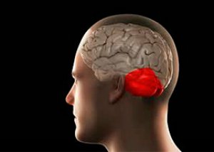
The cerebellum is located in the posterior cranial fossa above the medulla oblongata and the pons. It is separated from the occipital lobes of the cerebral hemispheres by the cerebellar tentorium. The surface of the cerebellar cortex is significantly increased by deep parallel arcuate grooves dividing the cerebellum into sheets. Physiologically, the cerebellum is divided into the ancient part (flock and node), the old part (worm), and the new part (hemispheres).
In the white matter of the cerebellar hemisphere and vermis there are several nuclei. Paramedian is the paired nucleus of the tent (nucl. fastigii), lateral to it are small islands of gray matter - the spherical nucleus (nucl. globusus), even more laterally, protruding into the white matter of the hemisphere, is the cork-shaped nucleus (nucl. emboliformis). In the white matter of the hemisphere are located the dentate nuclei (nucl. dentatus).
The cerebellum has three pairs of peduncles. In the inferior peduncles of the cerebellum pass afferent (posterior spinocerebellar tract, from the superior nucleus of the vestibular nerve - vestibulocerebellar tract, from the nuclei of the thin and cuneate fasciculi - bulbocerebellar tract, from the reticular formation - reticulocerebellar tract, from the inferior olive - olivocerebellar tract) and efferent tracts (cerebellar-reticulospinal, cerebellar-vestibulospinal - through the lateral nucleus of the vestibular nerve, cerebellar-olivospinal), mainly associated with the structures of the cerebellar vermis.
The largest middle cerebellar peduncles contain the pontocerebellar fibers, which are part of the cortico-pontocerebellar tract from the superior frontal gyrus and inferior parts of the occipital and temporal lobes to the cerebellar cortex. The superior cerebellar peduncles contain the afferent tract from the spinal cord (anterior spinocerebellar tract) and the descending cerebellar-erysonecular-spinal tract, which runs from the dentate nucleus of the cerebellar hemispheres through the red nucleus to the anterior horn of the spinal cord.
Symptoms cerebellar lesions
Damage to the cerebellum or its pathways causes a fairly pronounced symptom complex.
Ataxia always comes to the fore: disturbance of body balance at rest and when walking (it sways like a drunk, especially in twilight or darkness, inability to perform the simplest orthostatic tests), static disturbances when walking; especially on uneven surfaces, steps, inclined planes, dynamic disturbances when performing involuntary movements, disproportion of movements (hypermetria); by-the-way, adiadochokinesia (difficulty alternating opposite movements), intention tremor, nystagmus, speech disorder - scanned speech. The pathogenetic basis of all cerebellar manifestations is a violation of the coordination in the actions of antagonist muscles (asynergy).
When the cerebellar vermis is affected, the synergies that stabilize the center of gravity are disrupted. As a result, balance is lost, truncal ataxia occurs, the patient cannot stand (static ataxia); he walks with his legs wide apart, staggering, which is especially noticeable during sharp turns. When walking, there is a deviation toward the affected part of the cerebellum (homolaterally).
When the cerebellar hemispheres are affected, limb ataxia, intention tremor, missing the mark, and hypermetria (dynamic ataxia) predominate. Speech is slow and scanned. Megalography (large, uneven handwriting) and diffuse muscle hypotonia are detected.
In the case of a pathological process in one hemisphere of the cerebellum, all these symptoms develop on the side of the affected cerebellum (homolaterally).
Diagnostics cerebellar lesions
Tests characterizing cerebellar damage and dynamic ataxia:
- heel-knee (performed lying on your back with your eyes closed) - they suggest raising your leg and hitting your heel on the patella (misses); move it along the front surface of your shin towards the heel (slips);
- heel-fist - the doctor places his own fist under the heel and asks to lift the leg and lower it back onto the fist (misses);
- finger-nose (with closed eyes, with the index finger swinging the arm, try to reach the tip of the nose - miss);
- finger-to-finger - first with open eyes, then with closed eyes, they ask you to reach for the other with your index finger (it’s easy to do with open eyes, but it misses with closed eyes).
Tests characterizing cerebellar damage and static ataxia (carried out standing, with eyes closed, but with absolute insurance from the doctor in case the patient falls) - aimed at identifying stability (this group includes the entire complex of orthostatic tests):
- when the legs are spread wide, there is a staggering motion with a large tilt towards the affected lobe of the cerebellum, especially pronounced when turning the body from side to side;
- Romberg's pose - standing with eyes closed (feet together), stretching arms forward - deviation or fall towards the affected hemisphere or in any direction in case of pathology (cerebellar vermis); if the picture is unclear, a Romberg sensitization test is performed (or they suggest placing one leg in front of the other or bending it at the knee);
- ataxia-abasia symptom – the patient cannot move independently, but within the bed all active movements are preserved.
Tests characterizing cerebellar damage and kinetic ataxia:
- tonic - decreased muscle tone (flabbiness, lethargy);
- gait - asked to walk 2-3 m without support in a straight line: cannot walk, when walking moves legs forward, and the body lags behind, makes intricate movements with legs, making the gait atypical;
- Magnus-Klein symptoms ("magnetic reaction")
- when gently touching the foot, a pulling sensation is felt throughout the entire limb;
- in small children, when turning the head to the side, the legs bend at the knee or hip joints on the side where the head is turned; on the opposite side, the limb, on the contrary, straightens;
- Babinski's asynergic symptoms
- standing, they suggest bending backwards, throwing back his head - he falls;
- lying down, they offer to sit down - he swings and lifts his legs, then sits up with a jerk;
- while sitting, they suggest standing up on his feet - he rocks, then stands up.
Other tests that characterize cerebellar damage:
- synergistic - when looking up, the head is not thrown back; with a strong handshake, there is no extension in the wrist joint, and there is no wrinkling of the forehead;
- aodiodochokinesis - pronation and supination of the hands are performed simultaneously - on the side of the injury, movements are slow;
- dysmetric –
- with fingers extended forward and spread apart, the palms are sharply rotated, with excessive rotation on the side of the injury;
- Ozhekhovsky's symptom - the patient leans firmly on the doctor's palms, when the support is suddenly removed, the patient leans forward (a healthy person, on the contrary, leans back);
- dysarthria - speech that is scanned with emphasis on each syllable;
- Stuart-Holmes symptom - a person holds a supinated arm bent at the elbow, the doctor tries to straighten it and abruptly removes his arm, the patient hits him on the chest, because he cannot slow down the movement of his arm;
- Thomas-Jumanty symptom (grasping) - a person grasps an object, already at the beginning of grasping he opens his palm very wide;
- Tom's symptoms:
- if you push a person standing sideways, this will cause the leg on the side of the impact to rise and fall in the opposite direction;
- the patient lying on his back has his bent knees moved apart and brought together several times, then released abruptly - on the affected side the limb is involuntarily abducted;
- in a standing position, a person needs to bend to the side; on the healthy side, the tone of the extensors increases and the leg is abducted to the opposite side; on the injured side, this does not happen;
- a person moves like a pillar due to the rigidity of the muscles of the body, observed when a worm is present;
- Foix-Thevenard symptom - with a slight push forward or backward into the chest, the patient easily loses balance, while a healthy person maintains balance.
Examination of patients who have cerebellar damage should be carried out in a neurosurgical hospital - with the involvement of a neurophysiologist, an otoneurologist, an ENT doctor, and a neuro-oculist.
Who to contact?