
All iLive content is medically reviewed or fact checked to ensure as much factual accuracy as possible.
We have strict sourcing guidelines and only link to reputable media sites, academic research institutions and, whenever possible, medically peer reviewed studies. Note that the numbers in parentheses ([1], [2], etc.) are clickable links to these studies.
If you feel that any of our content is inaccurate, out-of-date, or otherwise questionable, please select it and press Ctrl + Enter.
Bones of the upper extremity
Medical expert of the article

Last reviewed: 04.07.2025

The skeleton of the upper limbs includes the girdle of the upper limbs and the free parts of the upper limbs.
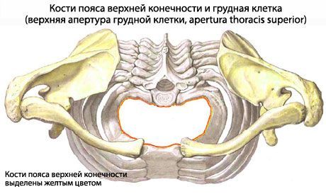
The upper limb girdle (unguium membri superiores), consisting of the paired scapula and clavicle, is attached to the rib cage by muscles and ligaments. In front, the clavicles are connected to the manubrium of the sternum on both sides. The skeleton of the free part of the upper limb (skeleton membri superioris liberi) consists of three sections: proximal (humerus), middle (radius and ulna of the forearm) and distal - the bones of the hand. The skeleton of the hand is divided into the bones of the wrist, metacarpal bones and phalanges of the fingers.
Bones of the girdle of the upper limbs
The scapula is a flat triangular bone. It is adjacent to the rib cage on its posterolateral side at the level of the 2nd to 7th rib. The scapula has three angles: lower (ingulus inferior), lateral (angulus lateralis) and upper (angulus superior). The scapula also has three edges: medial (margo medialis), facing the spinal column; lateral (margo lateralis), directed outward and slightly downward, and upper (margo superior), which has a scapular notch (incisure scapulae) for the passage of vessels and nerves.
The clavicle (clavicula) is a long, S-shaped tubular bone located between the clavicular notch of the sternum medially and the acromial process of the scapula laterally. The clavicle has a body (corpus claviculae) and two ends: the sternal end (extremitas sternalis) and the acromial end (extremitas acromiahs).
 [ 3 ]
[ 3 ]
Skeleton of the free part of the upper limb
The skeleton of the free part of the upper limb is formed mainly by tubular bones, which provide a wide range of motion.
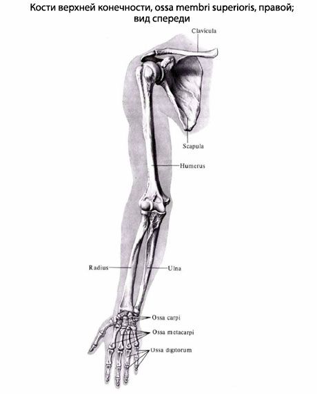
The humerus is a long tubular bone. There is a body of the humerus (corpus humeri) and two ends: upper and lower. The upper end (proximal) is thickened and forms the spherical head of the humerus (caput humeri). The head faces medially and slightly backward. Along the edge of the head there is a groove - the anatomical neck (collum anatomicum).
The bones of the forearm (ossa antebrachii) consist of two bones. The ulna is located medially, the radius is located laterally. These bones touch each other only at their ends, between their bodies there is an interosseous space of the forearm.
The ulna is thickened in its upper part. At this (proximal) end is a trochlear notch (incisura trochlearis), intended for articulation with the trochlea of the humerus.
The radius bone (radius) at the proximal end has the head of the radius (caput radu) with a flat depression - the glenoid fossa (fovea articuldris) for articulation with the head of the condyle of the humerus.
The hand (manus) has a skeleton, which includes the bones of the wrist (ossa carpi), metacarpal bones (ossa metacarpi) and bones of the fingers of the hand - the phalanges of the fingers (phalanges digitorum manus).
Joints of the bones of the upper limb
The bones and joints of the bones of the upper limbs in humans are adapted to grasping, holding and moving various objects (tools). The lower limbs have other functions. The lower limbs perform the functions of support and movement of the body in space. In connection with these functions, the bones of the lower limbs are larger and more massive than the bones of the upper limbs. The joints of the lower limbs are also larger, their mobility is less than that of the joints of the upper limbs.
In the intermetatarsal joints, the movements are most often combined: rotation of the calcaneus together with the navicular and the anterior end of the foot around the oblique sagittal axis. When the foot rotates inward (pronation), the lateral edge of the foot rises, and when it rotates outward (supination), the medial edge of the foot rises.
Rotation of the calcaneus together with the navicular and forefoot around the sagittal foot.
Slight rotation around the sagittal (front-back) axis.
Использованная литература