
All iLive content is medically reviewed or fact checked to ensure as much factual accuracy as possible.
We have strict sourcing guidelines and only link to reputable media sites, academic research institutions and, whenever possible, medically peer reviewed studies. Note that the numbers in parentheses ([1], [2], etc.) are clickable links to these studies.
If you feel that any of our content is inaccurate, out-of-date, or otherwise questionable, please select it and press Ctrl + Enter.
Hand
Medical expert of the article

Last reviewed: 07.07.2025
The hand (manus) has a skeleton, which includes the bones of the wrist (ossa carpi), metacarpal bones (ossa metacarpi) and bones of the fingers of the hand - the phalanges of the fingers (phalanges digitorum manus).
Bones of the wrist. The wrist (carpus) has 8 short (spongy) bones arranged in 2 rows. In the upper (proximal) row, if viewed in the medial direction (from the thumb to the little finger), are the following bones: scaphoid, lunate, triquetrum and pisiform. The lower (distal) row is formed by the polygonal (trapezium bone), trapezoid, capitate and hamate bones. The name of the bones reflects their shape. On the surfaces of each bone there are articular surfaces for articulation with adjacent bones.
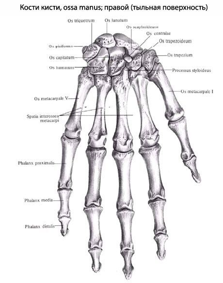
The scaphoid bone (os caphoideum) is large and has a convex surface that participates in the formation of the wrist joint. The lunate bone (os lunatum) also has a convex proximal surface. The triquetrum bone (os triquetrum) has a flat articular surface for articulation with the pisiform bone. The pisiform bone (os pisiforme) is the smallest of all the bones of the wrist. This bone is located in the thickness of the tendon of the ulnare flexor carpi ulnaris and is a sesamoid bone.
The three bones of the first row with their upper (proximal) surfaces face the bones of the forearm and form an ellipsoid articular head. The distal surfaces of these bones are directed toward the four carpal bones of the second row.
The trapezium bone (os traperium) has a saddle-shaped articular surface for articulation with the base of the first metacarpal bone. On the palmar surface of the trapezium bone there is a groove, which is limited by a tubercle on the lateral side. The trapezoid bone (os trapezoideum) resembles the trapezium in shape. The capitate bone (os capitdtum) is the largest of the carpal bones. It has a head directed proximally and slightly outward. The hook bone (os hamatum) on the palmar surface has a hook bent towards the radial side (hamulus ossis hamati).
The carpal bones form a bony arch, the convex side of which faces backwards, and the concave side faces forwards (towards the palm). As a result, a groove of the wrist (sulcus carpi) is formed on the palmar surface, limited on the radial side by the tubercle of the scaphoid bone and the tubercle of the trapezium bone, and on the ulnar side by the hook of the hamate bone and the pisiform bone.
Metacarpal bones. The metacarpus includes five (IV) short tubular bones - metacarpal bones (ossa metacarpalia). Each metacarpal bone consists of a base (basis), a body (corpus) and a head (caput). The bodies of the metacarpal bones have a triangular shape, their ends are thickened. Therefore, when the metacarpal bones are connected to each other, interosseous spaces remain between their bodies. On the palmar side, the bodies of the metacarpal bones are slightly concave, on the dorsal side - slightly convex. The bases of the II-V metacarpal bones at the proximal ends have flat articular surfaces for articulation with the bones of the second row of the wrist.
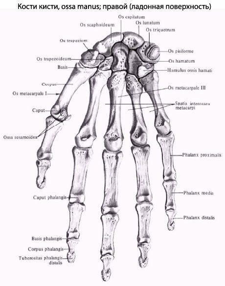
The first metacarpal bone (os metacarpale 1) is shorter and thicker than the others. At its base is a saddle-shaped surface for articulation with the polygonal bone. The second metacarpal bone is the longest. The bases of the second through fifth metacarpal bones have lateral articular surfaces for articulation with each other. The heads of the metacarpal bones are hemispherical, their convex articular surfaces serve for articulation with the proximal phalanges of the fingers.
Bones of the fingers. The hand is divided into the thumb (pollex, s.digitus primus); index finger (index, s.digitus secundus); middle finger (digitus medius, s.tertius) - the longest, ring finger (digitus anularis, s.quartus) and little finger (digitus minimus, s.quintus).
Phalanges of the fingers (phalanges digitorum). These are short tubular bones. Each finger, except for the first (thumb), has three phalanges: proximal (phalanx proximalis), middle (phalanx media) and distal (phalanx distalis). The thumb has only two phalanges - proximal and distal. The proximal phalanges are the longest, the distal are the shortest. A distinction is made between the base of the phalanx (basis phalangis), the body of the phalanx (corpus phalangis) and the head of the phalanx (caput phalangis). The bases of the proximal phalanges have articular fossae for articulation with the corresponding metacarpal bones. The bases of the middle and distal phalanges are provided with articular surfaces for articulation with the heads of the proximal phalanges. The end of each distal (nail) phalanx is flattened and forms the tuberosity of the distal phalanx (tuberositas phalangis distalis).
In the bones of the upper limb, as in other bones, there are nutrient holes. Through these holes penetrate the vessels that feed the bone and nerve fibers.
Movements of the hand in the radiocarpal, intercarpal and midcarpal joints around the frontal axis are possible in the range of 100°, abduction - adduction (around the sagittal axis) - 80°.
Flex the wrist: flexor carpi ulnaris, flexor carpi radialis, flexor digitorum superficialis, flexor digitorum profundus, flexor pollicis longus, palmaris longus.
Extend the wrist: muscles - long and short extensor of the wrist, ulnar extensor of the wrist, extensor of the fingers, long extensor of the thumb, extensor of the little finger.
Abduct the wrist: muscles - radial flexor of the wrist, long and short extensors of the wrist (with simultaneous contraction).
Adduct the hand: muscles - ulnar flexor of the wrist, ulnar extensor (with simultaneous contraction).
The movements of the fingers are carried out in the metacarpophalangeal joints around the frontal axis (flexion - extension), as well as abduction - adduction (around the sagittal axis), circular movements and passive rotation around the longitudinal axis. The thumb and little finger can be opposed to each other. The movements of the thumb are performed by the following muscles.
Flex the thumb: flexor pollicis longus, flexor pollicis brevis.
Extend the thumb: short and long extensors of the thumb.
Abduct the thumb: long and short muscles that abduct the thumb.
Adductor pollicis: muscle that adducts the thumb of the hand.
Opposition: The muscle that opposes the thumb.
The following muscles flex the II-V fingers of the hand: the superficial and deep flexors of the fingers (the phalanges of these fingers are also flexed by the interosseous and lumbrical muscles).
Extends the fingers: the extensor digitorum muscle.
Adduction to the middle finger - palmar interosseous muscles.
Abduction from the middle finger - dorsal interosseous muscles.
What do need to examine?
How to examine?