
All iLive content is medically reviewed or fact checked to ensure as much factual accuracy as possible.
We have strict sourcing guidelines and only link to reputable media sites, academic research institutions and, whenever possible, medically peer reviewed studies. Note that the numbers in parentheses ([1], [2], etc.) are clickable links to these studies.
If you feel that any of our content is inaccurate, out-of-date, or otherwise questionable, please select it and press Ctrl + Enter.
Fetal anencephaly of the brain
Medical expert of the article

Last reviewed: 12.07.2025
Among the defects of intrauterine development, such a type of irreversible disorder of embryonic morphogenesis of the fetus's brain as anencephaly stands out. In ICD-10, this defect is classified as a congenital anomaly of the nervous system with the code Q00.0.
Epidemiology
According to medical statistics, fetal anencephaly is one of the most common types of neural tube defects, and in the United States, approximately three pregnancies out of 10,000 are complicated by this anomaly each year. Although these figures do not take into account gestations that end in miscarriage.
In the UK, such defects were found in 2.8 babies per thousand live births and 5.3 per thousand spontaneously terminated pregnancies (at least eight weeks). [ 1 ]
According to EUROCAT (European Commission on the Epidemiological Monitoring of Congenital Anomalies), over 10 years (2000-2010) the overall prevalence of anencephaly was 3.52 per 10,000 live births. After prenatal diagnosis, 43% of all pregnancies were terminated for medical reasons. [ 2 ], [ 3 ]
Causes anencephaly
Already three weeks after fertilization – during the development of the human embryo – neurulation occurs, that is, the formation of the neural tube, which is the rudiment of the brain and spinal cord.
The key cause of anencephaly in neural tube dysraphism is a failure of its closure in the fourth to fifth week of embryonic development. This anomaly occurs when the rostral end of the embryo's neural tube, which forms the fetus's head and transforms into the brain, remains open. This causes the impossibility of further development of the main structures and tissues of the brain.
Anencephaly of the fetus is characterized by the absence of hemispheres and, accordingly, the cortex and neocortex of the brain that provides higher functions of the central nervous system, as well as the bones of the vault of the cranial part of the skull and the skin covering them. [ 4 ]
Risk factors
Embryonic morphogenesis is a complex process, therefore not all probable risk factors for its disruption leading to anencephaly have been identified yet.
It has been established that with a deficiency of folic acid – pteroylglutamic acid (or vitamin B9), necessary for the synthesis of purine and pyrimidine bases of some amino acids and DNA – neural tube defects, in particular, anencephaly and spina bifida, are observed much more often.
For more information, see What Causes Folate Deficiency?
In addition, the risk of impaired intrauterine brain development may be associated with:
- problems of a genetic nature (since the appearance of an anencephalic baby in a family increases the likelihood of this anomaly appearing in subsequent pregnancies to 4-7%);
- persistent maternal viral infections;
- uncontrolled diabetes mellitus, other endocrine pathologies and obesity;
- negative influence of the environment, in particular, ionizing radiation, which provokes spontaneous mutations;
- teratogenic effects of chemicals, including drugs, as well as alcohol and medications.
More information - Effects of toxic substances on pregnancy and fetus
Pathogenesis
The closure of the already formed neural tube occurs 28-32 days after conception, and researchers see the pathogenesis of anencephaly in the disruption of the formation of the rudiment of the central nervous system of the fetus even during the formation of the neural plate (between the 23rd and 26th day after conception), from which, in fact, the neurula stage begins, ending with the closure of the plate into the neural tube.
The essence of the violation of morphogenesis of the brain in the embryo with this anomaly is that the opening of the neural tube canal (neuropore) at the anterior end of the embryo remains open.
Next, the neural tube bends to form protrusions for the formation of differentiated neural stem cells of the forebrain, midbrain, and hindbrain. And to form the cerebral hemispheres (the endbrain), the pterygoid plate of the midbrain must expand. But since the anterior neuropore did not close in time, the formation of the skull becomes abnormal, and the morphology of the brain tissue changes, losing its functions.
Considering the leading role in embryonic development and control of the formation of organs and tissues of the fetus of homeotic or homeotic genes (HOX genes or morphogens), which are located on different chromosomes and contain DNA sequences encoding protein transcription factors, the possibility of a violation of neurulation at the gene level should be taken into account. [ 5 ]
Symptoms anencephaly
Despite the individual appearance of newborns, there are clear external signs of children with anencephaly.
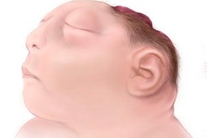
Immediately after birth, the first signs of this congenital defect are visible: a deformed skull of the infant with missing bones of its vault - the occipital or parietal; partial absence of the frontal bone or, less commonly, the temporal bones is also possible. There are no hemispheres of the brain and cerebellum, and in the uncovered defect of the skull there may be exposed tissue (glia), or the existing structures - the brain stem, the not fully formed basal nuclei of the forebrain - are covered by a thin membrane of connective tissue. [ 6 ]
Another external sign is the eyeballs protruding noticeably from the orbits, which is explained by the underdevelopment of the frontal bone of the skull, which forms the upper edge of the eye sockets.
In 80% of cases, anencephaly is not accompanied by other congenital anomalies, but a cleft soft palate (cleft palate) may be observed. [ 7 ]
Complications and consequences
Babies with this brain defect usually die during or shortly after birth, because the central nervous system does not function with this brain anomaly (only some basic reflexes are present, and not always). And experts note a 100% mortality rate among babies with anencephaly.
Rare cases of longer life are discussed at the end of the publication.
Diagnostics anencephaly
Prenatal diagnostics are carried out, and the diagnosis of fetal anencephaly can be made during pregnancy – in the early stages.
This can be done using visualization – instrumental diagnostics using ultrasonography.
Anencephaly is detected on ultrasound - as a defect of the open neural tube during an ultrasound examination of the fetus at 12 weeks of pregnancy. In this case, polyhydramnios (excess amniotic fluid) is often observed. Therefore, their analysis may be necessary - amnioscopy and amniocentesis.
Later, if the pregnancy has not terminated spontaneously, during an ultrasound examination of the fetus
Anencephaly, microcephaly, and hydrocephalus of the fetus are differentiated, since in microcephaly the skull is underdeveloped with the presence of convolutions of abnormal width. And in the case of hydrocephalus of the newborn or congenital hydrocephalus the head size is increased.
In addition, at 13-14 weeks, an analysis of alpha-fetoprotein in the blood of pregnant women is needed, since in the presence of anencephaly in the fetus, the level of this specific embryonic protein is always elevated.
Differential diagnosis
Differential diagnosis is carried out, first of all, with occipital brain hernia of newborns (encephalocele), which occurs due to a partially unclosed cranial vault; microhydranencephaly, alobar type of holoprosencephaly, schizocephaly.
Treatment anencephaly
Treatment of anencephaly in a child – in cases where the child is alive after birth – is palliative, since this defect is irreversible.
Prevention
Although not all etiological factors leading to anencephaly are known, numerous studies have proven the effectiveness of taking folic acid when planning pregnancy: with a daily dose of 0.8 mg.
See also: Folic acid during pregnancy
Forecast
A natural question arises: how long do people live with anencephaly? Regarding the life expectancy of infants with this congenital anomaly, the prognosis cannot be positive in the long term…
According to British obstetricians, just over 70% of newborns lived for a very short time after birth (from a few hours to two or three days), and only 7% lived for about four weeks. Then came death from cardiorespiratory collapse – cessation of breathing and cardiac arrest. [ 8 ]
However, there have been a few cases where surviving babies with anencephaly have lived for longer periods of time after birth.
For example, a girl named Angela Morales from Providence (Rhode Island, USA) lived for 3 years and 9 months; at the age of two months, she underwent surgery to close a hole in the back of her skull, as there was a constant leakage of cerebrospinal fluid.
A boy named Nicholas Cox, born in Pueblo (Colorado, USA), lived two months longer.
And Jackson Emmett Buell (born August 2014 in Orlando, Florida) with 80% of his brain (including his cerebral hemispheres) and most of his skull missing, is still alive. But he was diagnosed with microhydranencephaly, and his brainstem and thalamus are intact, and he still has some cranial nerves.