
All iLive content is medically reviewed or fact checked to ensure as much factual accuracy as possible.
We have strict sourcing guidelines and only link to reputable media sites, academic research institutions and, whenever possible, medically peer reviewed studies. Note that the numbers in parentheses ([1], [2], etc.) are clickable links to these studies.
If you feel that any of our content is inaccurate, out-of-date, or otherwise questionable, please select it and press Ctrl + Enter.
Anatomy and physiology of the female reproductive system
Medical expert of the article

Last reviewed: 04.07.2025

 [
[ External female genitalia
These include the labia majora and minora and the clitoris, which together form the vulva. It is bordered by two folds of skin - the labia majora. They consist of fatty tissue, saturated with blood vessels, and are located in the anteroposterior direction. The skin of the labia majora is covered with hair on the outside, and thin shiny skin on the inside, onto which numerous gland ducts open. The labia majora are connected in front and behind, forming the anterior and posterior commissures (adhesions). Inside them are the labia minora, which are located parallel to the labia majora and form the vestibule of the vagina. On the outside they are covered with thin skin, and on the inside they are lined with a mucous membrane. They are pink-red in color, connecting behind in front of the commissure of the labia majora, and in front - at the level of the clitoris. They are richly supplied with sensitive nerve endings and participate in achieving a voluptuous feeling.
In the vestibule of the vagina, the ducts of the Bartholin glands, located in the thickness of the labia majora, open. The secretion of the Bartholin glands is intensively secreted at the moment of sexual arousal and provides lubrication of the vagina to facilitate friction (periodic forward movements of the penis in the vagina) during sexual intercourse.
The bulbs of the cavernous bodies of the clitoris are located in the thickness of the labia majora, which increase in size during sexual arousal. The clitoris itself, which is a kind of greatly reduced semblance of the penis, also increases in size. It is located in front and above the entrance to the vagina, at the junction of the labia minora. The clitoris has many nerve endings and during sex it is the dominant, and sometimes the only organ, thanks to which a woman experiences orgasm.
Just below the clitoris is the opening of the urethra, and even lower is the entrance to the vagina. In women who have not had sex, it is covered by the hymen, which is a thin fold of mucous membrane. The hymen can have various shapes: a ring, a crescent, a fringe, etc. As a rule, it breaks during the first sexual intercourse, which can be accompanied by moderate pain and slight bleeding. In some women, the hymen is very dense and blocks the entrance to the vagina for the penis. In such cases, sexual intercourse becomes impossible and you have to resort to the help of a gynecologist, who cuts it. In other cases, the hymen is so elastic and pliable that it does not break during the first sexual intercourse.
Sometimes, during rough sexual intercourse, especially in combination with a large penis, a rupture of the hymen can be accompanied by quite severe bleeding, such that the help of a gynecologist is necessary.
It is extremely rare for the hymen to have no opening at all. During puberty, when a girl begins to menstruate, menstrual blood accumulates in the vagina. Gradually, the vagina becomes filled with blood and compresses the urethra, making urination impossible. In these cases, the help of a gynecologist is also necessary.
The area located between the posterior commissure of the labia majora and the anus is called the perineum. The perineum consists of muscles, fascia, vessels, and nerves. During childbirth, the perineum plays a very important role: due to its extensibility, on the one hand, and elasticity, on the other, it allows the fetus's head to pass through, increasing the diameter of the vagina. However, with a very large fetus or with rapid labor, the perineum cannot withstand excessive stretching and can rupture. Experienced obstetricians know how to prevent this situation. If all methods for protecting the perineum are ineffective, then they resort to an incision in the perineum (episiotomy or perineotomy), since a cut wound heals better and faster than a lacerated one.
Internal female genital organs
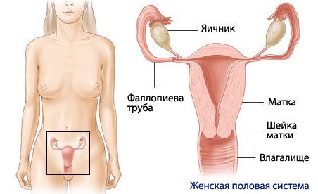
These include the vagina, uterus, ovaries, and fallopian tubes. All of these organs are located in the pelvis, a bony "shell" formed by the inner surfaces of the iliac, ischial, pubic bones, and sacrum. This is necessary to protect both the woman's reproductive system and the fetus developing in the uterus.
The uterus is a muscular organ consisting of smooth muscles, resembling a pear in shape. The average size of the uterus is 7-8 cm in length and about 5 cm in width. Despite its small size, during pregnancy the uterus can increase in size 7 times. Inside, the uterus is hollow. The thickness of the walls, as a rule, is about 3 cm. The body of the uterus - its widest part, faces upward, and the narrower one - the cervix - is directed downward and slightly forward (normally), falling into the vagina and dividing its back wall into the posterior and anterior fornices. The bladder is located in front of the uterus, and the rectum is located behind it.
There is an opening in the cervix (cervical canal) that connects the vaginal cavity with the uterine cavity.
The fallopian tubes, which extend from the lateral surfaces of the fundus of the uterus on both sides, are a paired organ 10-12 cm long. The sections of the fallopian tube are the uterine part, the isthmus, and the ampulla of the fallopian tube. The end of the tube is called the funnel, from the edges of which numerous processes of various shapes and lengths (fringes) extend. The tube is covered with a connective tissue membrane on the outside, and a muscular membrane underneath; the inner layer is the mucous membrane, lined with ciliated epithelium.
The ovaries are a paired organ, a sex gland. An oval body: length up to 2.5 cm, width 1.5 cm, thickness about 1 cm. One of its poles is connected to the uterus by its own ligament, the second is facing the side wall of the pelvis. The free edge is open to the abdominal cavity, the opposite edge is attached to the broad ligament of the uterus. It has a medulla and a cortex. The medulla contains vessels and nerves, and the cortex is where follicles mature.
The vagina is an elastic muscular-fibrous tube about 10 cm long. The upper edge of the vagina embraces the cervix, and the lower edge opens into the vestibule of the vagina. The cervix protrudes into the vagina, and a dome-shaped space is formed around the cervix - the anterior and posterior fornices. The vaginal wall consists of three layers: the outer layer is dense connective tissue, the middle layer is thin muscle fibers, and the inner layer is a mucous membrane. Some of the epithelial cells synthesize and store glycogen reserves. Normally, the vagina is dominated by Doderlein bacilli, which process the glycogen of dying cells, forming lactic acid. This causes the maintenance of an acidic environment in the vagina (pH = 4), which has a detrimental effect on other (non-acidophilic) bacteria. Additional protection against infection is provided by numerous neutrophils and leukocytes located in the vaginal epithelium.
The mammary glands are made up of glandular tissue: each contains about 20 individual tubuloalveolar glands, each of which has its own outlet on the nipple. In front of the nipple, each duct has an expansion (ampulla or sinus), which is surrounded by smooth muscle fibers. The walls of the ducts contain contractile cells that contract reflexively in response to sucking, expelling the milk contained in the ducts. The skin around the nipple is called the areola, it contains many glands similar to the milk glands, as well as sebaceous glands that produce an oily fluid that lubricates and protects the nipple during sucking.
Использованная литература