
All iLive content is medically reviewed or fact checked to ensure as much factual accuracy as possible.
We have strict sourcing guidelines and only link to reputable media sites, academic research institutions and, whenever possible, medically peer reviewed studies. Note that the numbers in parentheses ([1], [2], etc.) are clickable links to these studies.
If you feel that any of our content is inaccurate, out-of-date, or otherwise questionable, please select it and press Ctrl + Enter.
Fallopian tube
Medical expert of the article

Last reviewed: 04.07.2025

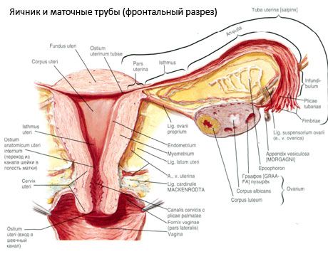
The fallopian tube (tuba uterina, s.salpinx) is a paired organ used to conduct the egg from the ovary (from the peritoneal cavity) to the uterine cavity. The fallopian tubes are located in the pelvic cavity and are cylindrical ducts running from the uterus to the ovaries. Each tube lies in the upper part of the broad ligament of the uterus, which is like a mesentery of the fallopian tube. The length of the fallopian tube is 10-12 cm, the lumen of the tube is from 2 to 4 mm. On one side, the fallopian tube communicates with the uterine cavity through a very narrow uterine opening of the tube (ostium uterinum tubae), on the other - it opens through the abdominal opening (ostium abdominale tubae uterinae) into the peritoneal cavity, near the ovary. Thus, in a woman, the peritoneal cavity through the lumen of the fallopian tubes, the cavity of the uterus and the vagina communicates with the external environment.
The fallopian tube initially has a horizontal position, then, having reached the wall of the small pelvis, it bends around the ovary at its tubular end and ends at its medial surface. The fallopian tube is divided into: the uterine part (pars uterina), which is enclosed in the thickness of the uterine wall, and the isthmus of the fallopian tube (isthmus tubae uterinae) - the part closest to the uterus. This is the narrowest and at the same time the thickest-walled part of the fallopian tube, which is located between the layers of the broad ligament of the uterus. The part following the isthmus is the ampulla of the fallopian tube (ampulla tubae uterinae), which accounts for almost half the length of the entire fallopian tube. The ampullar part gradually increases in diameter and passes into the next part - the funnel of the fallopian tube (infundibulum tubae uterinae), which ends in long and narrow fringes of the tube (fimbriae tubae). One of the fringes differs from the others in its greater length. It reaches the ovary and often adheres to it - this is the so-called ovarian fimbria (fimbria ovariса). The fimbria of the tube directs the movement of the egg towards the funnel of the fallopian tube. At the bottom of the funnel there is an abdominal opening of the fallopian tube, through which the egg released from the ovary enters the lumen of the fallopian tube.
 [
[ Structure of the wall of the fallopian tube
The wall of the fallopian tube is represented externally by the peritoneum - the serous membrane (tunica serosa), under which is the subserous base (tela subserosa). The next layer of the wall of the fallopian tube is formed by the muscular membrane (tunica muscularis), continuing into the muscles of the uterus and consisting of two layers. The outer layer is formed by longitudinally arranged bundles of smooth muscle (non-striated) cells. The inner layer, thicker, consists of circularly oriented bundles of muscle cells. Peristalsis of the muscular membrane ensures the movement of the egg towards the uterine cavity. The submucosa of the fallopian tube is absent, therefore under the muscular membrane there is a mucous membrane (tunica mucosa), forming longitudinal tubular folds (plicae tubariae) along the entire length of the fallopian tube. Closer to the abdominal opening of the fallopian tube, the mucous membrane becomes thicker and has more folds. They are especially numerous at the funnel of the fallopian tube. The mucous membrane is covered with epithelium, the cilia of which vibrate towards the uterus, facilitating the movement of the egg. Microvillous prismatic epithelial cells secrete a secretion that moistens the surface of the mucous membrane and ensures the development of the fertilized egg (embryo) as it moves through the lumen of the fallopian tube.
Vessels and nerves of the fallopian tubes
The blood supply to the fallopian tube comes from two sources: from the tubular branch of the uterine artery and the branch from the ovarian artery. Venous blood flows through the veins of the same name into the uterine venous plexus. The lymphatic vessels of the tube flow into the lumbar lymph nodes. The innervation of the fallopian tubes is carried out from the ovarian and uterovaginal plexuses.
On the radiograph, the fallopian tubes appear as long and narrow shadows, widened in the ampullar region.
Использованная литература