All iLive content is medically reviewed or fact checked to ensure as much factual accuracy as possible.
We have strict sourcing guidelines and only link to reputable media sites, academic research institutions and, whenever possible, medically peer reviewed studies. Note that the numbers in parentheses ([1], [2], etc.) are clickable links to these studies.
If you feel that any of our content is inaccurate, out-of-date, or otherwise questionable, please select it and press Ctrl + Enter.
Vagina
Medical expert of the article
Last reviewed: 04.07.2025

The vagina (vagina, s.colpos) is an unpaired hollow organ shaped like a tube, located in the pelvic cavity and extending from the uterus to the genital slit. At the bottom of the vagina it passes through the urogenital diaphragm.
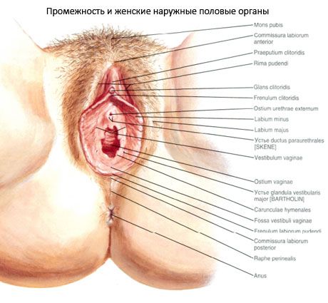
The length of the vagina is 8-10 cm, the thickness of the wall is about 3 mm. The vagina is slightly curved backwards, its longitudinal axis with the axis of the uterus forms an obtuse angle (slightly more than 90°), open to the front. The upper end of the vagina begins at the cervix, goes downwards, where the lower end opens into the vestibule with the opening of the vagina. In girls, the opening of the vagina is covered by the hymen, the place of attachment of which separates the vestibule from the vagina. The hymen is a crescent-shaped or perforated connective tissue plate. During the first sexual intercourse, the hymen ruptures and its remnants form hymen flaps (carunculae hymenales). In the collapsed state, the lumen of the vagina on the cross section has the appearance of a frontally located slit (cavity).
The vagina has an anterior wall (paries anterior), which in its upper third is adjacent to the fundus of the urinary bladder, and in the rest of its area is fused with the wall of the female urethra. The posterior wall (paries posterior) of the vagina in its upper part is covered by the peritoneum of the rectouterine depression, and the lower part of the wall is adjacent to the anterior wall of the rectum. The walls of the upper part of the vagina, covering the vaginal part of the cervix, form a narrow slit around it - the vaginal fornix (fornix vaginae). Due to the fact that the posterior wall of the vagina is longer than the anterior one, and is attached higher to the cervix, the posterior part of the fornix (pars posterior) is deeper than the anterior part (pars anterior).

Structure of the vaginal walls
The vaginal wall consists of three membranes. The outer adventitial membrane (tunica adventitia) is made of loose connective tissue containing a significant amount of elastic fibers, as well as bundles of smooth (non-striated) muscle cells. The middle muscular membrane (tunica muscularis) is represented mainly by longitudinally oriented bundles of muscle cells, as well as bundles with a circular direction. At the top, the muscular membrane of the vaginal wall passes into the muscles of the uterus, at the bottom it becomes more powerful and its bundles are woven into the muscles of the perineum. Bundles of striated (striated) muscle fibers, covering the lower end of the vagina and at the same time the urethra, form a kind of muscular sphincter.
The inner lining of the vaginal wall is represented by the mucous membrane (tunica mucosa). Due to the absence of a submucosa, it directly fuses with the muscular membrane. The surface of the mucous membrane is covered with multilayered squamous epithelium; the mucous membrane does not contain glands. The mucous membrane is quite thick (about 2 mm). The epithelial cells of its surface layer contain a significant amount of glycogen. The structure and thickness of the epithelium depend on the phase of the ovarian-menstrual cycle. By the time of ovulation, due to increased estrogen secretion, the glycogen content in the epithelial cells increases. Glycogen is used to maintain normal sperm function. The conversion of glycogen into lactic acid provides an acidic reaction in the vagina. The mucous membrane forms numerous transverse folds - vaginal folds (rugae vaginale) or wrinkles. On the anterior and posterior walls of the vagina, closer to the midline, the folds become higher, forming longitudinally oriented columns of folds (columnae rugarum). The anterior column of folds (columna rugarum anterior) located on the anterior wall of the vagina is better expressed than on the posterior wall. Below, it is a longitudinally oriented protrusion - the urethral keel of the vagina (carina urethritis vaginae), corresponding to the nearby urethra. The posterior column of folds (columna rugarum posterior) is located to the right or left of the anterior one, therefore, in a collapsed vagina, the anterior and posterior columns do not overlap each other. The basis of the columns of folds is the mucous membrane, which is thicker here than in other places and contains bundles of smooth muscle cells and numerous veins. In this regard, the columns of folds on the section have a spongy structure.
Vessels and nerves of the vagina
The blood supply to the vagina is provided by branches of the internal iliac artery: the vaginal artery, which is the descending branch of the uterine artery and supplies mainly its upper section; the inferior vesical artery, which supplies blood to the middle section of the vagina; the middle rectal artery; the internal pudendal artery, which supplies the lower section of the vagina; and the posterior branches of the labia.
Lymphatic drainage from the vaginal area occurs: from its lower third - to the superficial and deep inguinal lymph nodes, from the upper two thirds - to all three main groups of pelvic lymph nodes - iliac, internal iliac and sacral.
The vagina is innervated mainly by branches that extend from the general uterovaginal plexus. From the lower anterior parts of this plexus, the vaginal yervae extend, providing sympathetic and parasympathetic innervation.
The vagina receives sensory innervation from branches of the sacral plexus.
Использованная литература

