
All iLive content is medically reviewed or fact checked to ensure as much factual accuracy as possible.
We have strict sourcing guidelines and only link to reputable media sites, academic research institutions and, whenever possible, medically peer reviewed studies. Note that the numbers in parentheses ([1], [2], etc.) are clickable links to these studies.
If you feel that any of our content is inaccurate, out-of-date, or otherwise questionable, please select it and press Ctrl + Enter.
Ventriculomegaly of the brain: what it is, causes, consequences
Medical expert of the article

Last reviewed: 12.07.2025
In both hemispheres of the brain, the diencephalon and between the cerebellum and medulla oblongata there are four special cavities - cerebral ventricles (ventriculi cerebri), which produce cerebrospinal fluid. Their pathological expansion or enlargement is defined as ventriculomegaly.
Epidemiology
The statistics cited in various sources are as follows:
- The prevalence of ventriculomegaly, according to The Fetal Medicine Foundation, is one case per 100 fetuses at 22 weeks of gestation and one case per 1,000 live births;
- in more than 94% of cases, ventriculomegaly of the largest lateral ventricles (ventriculi laterales) is observed;
- craniocerebral anomalies are observed in 15-65% of cases of ventriculomegaly, and chromosomal defects – on average in 14.7%;
- The frequency of neurodestruction associated with this pathology in infancy is approximately 12% (according to other data, almost 60%).
Causes ventriculomegaly
When listing the possible causes of dilation of the cerebral ventricles, it is important to note that pathological dilation of the ventricular system of the brain – due to excess volume of cerebrospinal fluid (CSF) in the ventricles and subarachnoid spaces and disturbances in its circulation – is called hydrocephalus, often associated with increased intracranial pressure.
Since it is impossible to measure intracranial pressure during the period of intrauterine development (prenatally), the terms hydrocephalus and ventriculomegaly in the fetus are used as synonyms. Although hydrocephalus should be called an increase in the fetal ventricles to more than 15 mm.
Experts believe that the main causes of ventriculomegaly are brain dysgenesis, excess CSF in the ventricular system of the brain, and chromosomal defects.
Antenatal ventriculomegaly, i.e. ventriculomegaly in the fetus, may be the result of a neural tube defect in the embryo, as well as primary congenital anomalies of the brain: agenesis of the corpus callosum; subependymal heterotopia; colloid or arachnoid cyst of the brain; Arnold-Chiari malformation type 2, leading to Arnold-Chiari syndrome; cystic dilatation of the fourth ventricle - Dandy-Walker syndrome, etc.
Risk factors
Risk factors are associated with the mother’s age (over 35 years), intrauterine infections, gestosis and fetal oxygen starvation.
Pathogenesis
The pathogenesis of overproduction of CSF by the ventricles of the fetal brain may be due to:
- infections (bacterial, fungal, herpes meningitis);
- tumors (platelet glioma, endodermal sinus tumor, disseminated oligodendroglial tumor);
- hypertrophy, hyperplasia or tumor of the choroid (vascular) plexus of the ventricles of the brain.
An association of pathological dilation of the cerebral ventricles with trisomy of chromosomes 13, 18 and 21 has been noted – with Patau, Edwards and Down syndromes, respectively.
Ventriculomegaly in a newborn baby can be caused by:
- birth trauma with a sharp increase in venous pressure in the dural sinuses or in the internal jugular veins;
- cerebral ischemia in newborns;
- narrowing of the intraventricular (Monroe) openings connecting the third ventricle with the lateral ones;
- congenital stenosis of the aqueduct of Sylvius, a canal between the third ventricle of the brain (ventriculus tertius) and the fourth (ventriculus quartus).
In addition, rapidly or gradually developing ventriculomegaly in a child is possible with:
- traumatic brain injuries (especially with hemorrhage);
- brain damage caused by pork tapeworm - neurocysticercosis;
- meningioma, diffuse glial tumors or teratoma of the brain;
- choroid papilloma (with damage to the lateral ventricles of the brain).
Ventriculomegaly in adults
Secondary ventriculomegaly in adults may be caused by head trauma, inflammation of the meninges, cerebral neoplastic lesions, stenosis of the aqueduct of Sylvius, intracranial aneurysm, chronic subdural hematomas, and intraventricular or general cerebral hemorrhage, including hemorrhagic strokes.
In addition, as Canadian researchers have established, the development of ventriculomegaly, the pathogenesis of which is caused by a violation of CSF absorption or leakage of cerebrospinal fluid into the subdural space, occurs more often in older people than in young people.
The reason is brain atrophy – loss of volume of its parenchyma, as well as changes in glial cells and myelination of neurons, which leads to a decrease in the elasticity of cerebral tissue.
That is, changes can occur in the aging human brain that represent a compensatory expansion of the space filled with cerebrospinal fluid – hydrocephalus ex vacuo.
Symptoms ventriculomegaly
The first signs of this pathology in the fetus are the large size of the cerebral ventricles, reaching 12-20 mm. The optimal period for its detection is 24-25 weeks of gestation.
Symptoms of ventriculomegaly in newborns include lethargy, inactive sucking, and difficulty swallowing; the child often burps and cries; sleep periods are short; blood vessels often show through the skin on the head and face; a bulging fontanelle, periodic trembling of the lower jaw, and convulsive movements of the limbs are noted. Until the cranial sutures have fused, a rapid increase in its circumference (macrocephaly) is observed.
Children of the first two or three years have headaches; the pain intensifies with tension and sudden movements, jumping, bending. Nausea and vomiting occur spontaneously. In the presence of chromosomal syndromes, the symptoms acquire their characteristic features.
Ventriculomegaly in adults can manifest itself as headaches, nausea, vomiting, increased intracranial pressure, and visual impairment. The latter is caused by swelling of the optic disc – papillodema, which in the early stages can be asymptomatic or cause headaches. Over time, this swelling can lead to the appearance of a blind spot, blurred vision, and periodic limitation of the visual field. As a result, complete loss of vision is possible.
If the third ventricle located in the diencephalon enlarges, then due to pressure on the subcortical vegetative centers in the gray matter of its walls, gait disturbances, paresthesia, urinary incontinence, and deterioration of cognitive functions may occur.
Stages
The degree of enlargement of the cerebral ventricles in the neonatal period – based on the norm of its size up to 10 mm (along the atrium of the posterior or anterior horn of the lateral ventricle) – determines the degree of ventriculomegaly.
Their names are not unified, therefore, the expansion of the lateral ventricles (mainly assessed as ventriculi laterales) by 20% – up to 12 mm can be defined as grade 1 ventriculomegaly or mild ventriculomegaly.
If the expansion is 20-50% of the norm - from 12 to 15 mm, then this is moderate ventriculomegaly, and when the indicator is > 15 mm, ventriculomegaly can be classified as more severe, pronounced ventriculomegaly, as well as prethreshold or borderline ventriculomegaly.
If the fetal ventricle enlarges to ≥ 20 mm, intraventricular hydrocephalus is diagnosed.
Forms
Depending on the localization, the following are distinguished:
- ventriculomegaly of the lateral ventricles (ventriculi laterales) or lateral bilateral ventriculomegaly;
- unilateral ventriculomegaly, when only one lateral ventricle is enlarged. There may be ventriculomegaly on the left - left-sided lateral or ventriculomegaly on the right - right-sided lateral.
In cases where the ventricles are of different sizes (this difference should not exceed 2 mm), asymmetric ventriculomegaly is diagnosed.
If the ultrasound does not reveal any associated brain abnormalities, it is antenatal isolated ventriculomegaly. In many cases that appear to be isolated dilation of the cerebral ventricles in the fetus, other abnormalities are actually found after the birth of the child (especially when the ventriculomegaly exceeds 15 mm). According to observations, with this variant of pathology, there is a 4-fold increase in the risk of an abnormality of the 21st chromosome.
Finally, replacement ventriculomegaly (more commonly used for hydrocephalus) refers to an increase in CSF volume to replace lost brain parenchyma.
Complications and consequences
Naturally, the question arises: is ventriculomegaly dangerous?
Like all pathologies of the brain, the enlargement of its ventricles has serious consequences and complications. This includes macrocephaly, and a delay in the general development of the child, and destructive changes in the structures of the brain: the size of the cerebral cortex increases significantly, neuroglia grows in the periventricular and supratentorial areas, and the maturation of the cortical sulcus is delayed.
Setting sun syndrome or Graefe syndrome may develop.
Neurological disorders often occur, which negatively affect memory, learning ability, adaptive properties of the psyche and behavior.
According to some data, at the age of two, children with ventriculomegaly have neurological problems in almost 62.5% of cases.
 [ 26 ]
Diagnostics ventriculomegaly
The only method by which ventriculomegaly is diagnosed is instrumental diagnostics.
To detect X-anomalies, genetic analysis (karyotyping) of the fetus is required based on a sample of amniotic fluid. For more information on how it is taken, see – Invasive methods of prenatal diagnostics
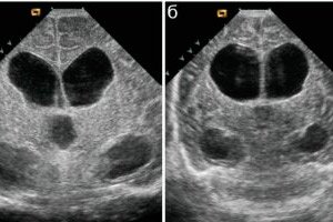
Ventriculomegaly in the fetus is detected during the mother's pregnancy - during an ultrasound examination after the 22nd week of gestation, when the size of the lateral ventricles can be visualized and measured.
Assessment of the fetal head includes determination of the shape of the skull and its bilar-articular diameter (the size of the head from one temple to the other). Ultrasound imaging allows for a clear definition of the medial border of the ventricles; echo signs of the choroid plexus - an echogenic structure occupying the central part of the lateral ventricle; cerebrospinal fluid may be visible.
If pathology is detected, screening is performed at a later stage (every 4 weeks) to monitor the condition of the ventricles.
Before the 18th week, and especially in the first trimester, ventriculomegaly is not examined by ultrasound: echo signs of pathology may simply be absent (the accuracy of the readings is less than 47%), since the spaces of the hemispheres are almost completely occupied by ventriculi laterales.
More information – Prenatal diagnostics of congenital diseases
Differential diagnosis
Clarification of the diagnosis and differential diagnostics require MRI of the brain. It is performed on adults and young children. Pregnant women undergo magnetic resonance imaging only in cases where the ventricular system cannot be assessed due to the position of the fetus in the uterus. According to diagnosticians, in half of the cases, MRI can detect additional, sonographically non-visualized CNS anomalies.
MRI signs of ventriculomegaly include: dark (low-intensity) signal from the ventricle of the brain (in the coronal plane) in T1 mode and bright (increased strength) - on images scanned in T2-weighted mode.
Adults may undergo CT or X-ray of the brain with radioisotope contrast - ventriculography.
Hydrocephalus is differentiated from ventriculomegaly based on the parameters of the enlarged cerebral ventricle and intracranial pressure; a spinal puncture is performed.
Among other pathologies, the most frequently identified are chronic syringomyelia with cavities in the spinal cord and inflammation of the ventriculi cerebri – ventriculitis.
Who to contact?
Treatment ventriculomegaly
Antenatal ventriculomegaly is not treated, and after the birth of the baby, treatment of ventriculomegaly is exclusively symptomatic - based on the recommendations of a pediatric neurologist.
The arsenal of tools available today includes:
- diuretics (used for hydrocephalus – Mannitol, Ethacrynic acid, etc.);
- drugs containing potassium (to avoid disruption of the balance of interstitial fluid, which is caused by long-term use of diuretics);
- antihypoxants;
- vitamins for the brain.
It is useful to do massage, physical activity is also welcomed for ventriculomegaly - moderate, without sudden movements.
Doctors do not hide the fact that managing this condition and consulting parents is not an easy task, since it is difficult to confidently name the exact cause of the pathology, predetermine the course of its development and predict the degree of risk of consequences.
Forecast
Ventriculomegaly associated with fetal anomalies and structural malformations often has a poor prognosis, ranging from disability (often moderate) to loss of the baby.
However, in cases of mild isolated ventriculomegaly there is a 90% chance of a normal outcome. In other cases there is a delay in the development of the nervous system - from mild to moderate.
[ 41 ]