
All iLive content is medically reviewed or fact checked to ensure as much factual accuracy as possible.
We have strict sourcing guidelines and only link to reputable media sites, academic research institutions and, whenever possible, medically peer reviewed studies. Note that the numbers in parentheses ([1], [2], etc.) are clickable links to these studies.
If you feel that any of our content is inaccurate, out-of-date, or otherwise questionable, please select it and press Ctrl + Enter.
Types of streptoderma: symptoms and peculiarities of the course of various forms of the disease
Medical expert of the article

Last reviewed: 04.07.2025
Streptoderma is a collective term that doctors use to describe a variety of types and forms of skin pathology caused by streptococcal infection. Different types of streptoderma have their own characteristic symptoms, features of the course and conditions of development. And since streptoderma is considered a skin disease, but representatives of different sexes have their own skin characteristics, then when considering the predisposition to the occurrence of certain types of streptococcal disease, gender differences must also be taken into account. Although, undoubtedly, the decisive role still belongs to human immunity.
Diseases united by the general term " streptoderma " may differ in the presence or absence of an exudative surface, characteristics of individual elements of the rash, the size of pathological foci and the speed of their spread, the duration of treatment, the presence or absence of factors complicating the course of the disease, etc. For this reason, in the classification of streptoderma, several groups can be considered, combining certain types and forms of streptococcal pathology.
Presence of exudate
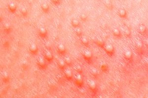
Considering various types of streptoderma and their manifestations, medical scientists came to the conclusion that streptococcal infection on the skin surface can behave differently. In most cases, this concept concealed a lesion of the skin with the characteristic appearance of blisters with a turbid yellowish liquid on the skin surface - phlyctena, around which a red rim of inflammation is observed.
This type of disease was called weeping streptoderma. The disease is more typical for people with delicate skin, i.e. for children and women, as well as for representatives of the stronger sex whose skin has not become roughened by the sun and wind.
Inflammatory elements formed by the accumulation of bacteria and their waste products, with wet streptoderma can be located on different parts of the body, including the nail folds, corners of the lips, perineum and genitals, and oral cavity.
This type of streptoderma is called weeping because of the appearance of blisters filled with liquid on the body, which subsequently burst, forming weeping. Subsequently, dense yellow crusts form in place of the bursting blisters.
In people with thick and hardened skin (most often in men), streptoderma may proceed differently, similar to white lichen. With this type of disease, whitish or slightly pinkish lesions of regular or irregular round shape, up to 5 cm in size, covered with peeling epidermis, appear on the skin. This is dry streptoderma.
It is called dry because there is no weeping surface. Apart from the greyish-white or greyish-yellow peeling films on the skin, there are no other manifestations of streptococcal infection. In other words, this is streptoderma without blisters and rough yellow crusts.
The foci of infection in dry streptoderma are localized mainly in the face and behind the ears, so the disease is sometimes called simple lichen of the face. But it should not be confused with white (vitiligo) or pityriasis versicolor, the causative agents of which are not streptococcus bacteria, but fungi. Despite some similarity in manifestations, the symptoms of the disease have some differences (for white and pityriasis versicolor of fungal origin, itching is not typical, moreover, the localization of the latter rarely affects the face or head). The causative agent of the disease can be easily identified by analyzing the scraping.
The medical name for the dry variety of skin disease caused by streptococcus is erythematous-squamous streptoderma. This type of pathology is considered a mild form of the disease, since it mainly affects the superficial layers of the epidermis, which is more likely if the skin is rough and thick.
However, statistics show that simple lichen of the face is often diagnosed in children, if the child's immunity is able to inhibit the penetration of infection into the deep layers of the epidermis or the disease was provoked by a small number of bacteria.
The development of the disease is facilitated by chapped or dry skin of the face, which can cause microcracks, insufficient hygiene, insufficient removal of moisture after washing, especially before going outside. Through microdamage, bacteria penetrate into the upper layers of the epidermis, where the pathological process develops.
The variety of types of weeping streptoderma
Looking at the statistics of streptoderma, one can see that the vast majority of patients with this diagnosis are children. The number of registered cases of streptococcal pyoderma in children under 15 years of age is estimated at 111 million. [ 1 ] Children's skin has its own structural features, so it is more delicate and thin. All kinds of damage easily appear on it, plus bacteria have the ability to multiply not only in the superficial layers. It is not surprising that in childhood, weeping forms of the disease are usually diagnosed.
In adults, streptococcal skin infection is diagnosed less frequently, but it is believed that women are more likely to have the same weeping form of the disease, while men with rougher skin are more likely to have a dry form.
Weeping streptoderma, also known as weeping streptococcal impetigo, is the most common type of streptoderma in people with delicate, sensitive skin. This category includes children and women, although sometimes this form of the disease can be diagnosed in men in the nail area, mucous membranes, and areas with less rough skin.
The disease manifests itself by the formation of separate small spots of bright pink or red color on the skin, which in a matter of hours turn into blisters with an inflammatory rim. Transparent exudate is initially visible inside the blisters, and the blisters themselves remain tense for some time. It seems that they can burst at any moment, but in reality, after some time, the blisters become softer, and the liquid inside them becomes cloudier and acquires a yellowish tint. [ 2 ], [ 3 ]
There are two options for resolving the problem. The blisters either dry up and form crusts, or spontaneously open (erosions with purulent contents are visible in their place). The erosions also subsequently tighten, become covered with a crust, which peels off over time, leaving behind a pink spot. After some time, the spot disappears without a trace.
Weeping streptoderma, as the most common type of streptococcal disease affecting the skin, can be divided into several subtypes depending on the localization of pathological foci with phlyctenas and the nature of the disease.
Let's consider the different types of weeping streptoderma from the point of view of their symptoms, localization and characteristics of the course of the disease:
Slit impetigo
This is a type of streptoderma localized in the corners of the mouth (other names: angular stomatitis). The disease develops like any other type of streptococcal impetigo. At first, redness and irritation are visible in the corners of the mouth, then small oval blisters form, after which painful cracks remain on the skin.
Crevice impetigo usually develops in patients who are used to sleeping with their mouth open, as a result of which the corners of the lips are constantly moistened with saliva, as well as in those who have a bad habit of frequently licking their lips. As a result, the structure of the epidermis is damaged, it becomes looser, microdamages easily appear on it, through which the infection penetrates.
The disease is difficult to treat because when the lips move, the crusts burst, leaving behind rather deep cracks that take a long time to heal. [ 4 ]
Crevice impetigo can also be localized at the base of the wings of the nose or in the corners of the eyes. Near the nose, the disease usually develops against the background of rhinitis (cold or allergic), in the corners of the eyes, inflammatory elements can appear in people with a tendency to lacrimation.
Streptoderma with erythema annulare
This type of streptoderma is distinguished by the behavior of phlyctema. Usually, the resolution of these formations on the skin is manifested by their drying up, after which the growth of the blister completely stops. With the annular form of pathology, after the resolution of the inner part of the blister, it continues to grow along the perimeter. A fairly large rounded inflamed focus is formed with a dry crust in the middle and small bubbles along the contour. [ 5 ]
The disease has a not entirely clear mechanism of development, a long recurring course (the lesions may disappear and then reappear after a while) and usually develops against the background of reduced immunity and endocrine disorders.
The ongoing inflammation is most likely a response of the immune system to the invasion of foreign microorganisms, that is, it is an allergic reaction, in which streptoderma develops somewhat differently with large ring-shaped lesions, somewhat reminiscent of lichen planus in the stage of crusting.
As for allergies, they do not cause streptoderma in themselves, but their skin manifestations in the form of rashes and peeling are a predisposing factor that opens the gates of infection deep into the skin. Streptoderma is an infectious disease, so without the presence of an infectious agent in the wound (in this case, active streptococcus bacteria), purulent inflammation does not develop.
Bullous streptoderma
This type of streptoderma is considered one of the most severe and dangerous. The fact is that any type of streptoderma is contagious, but with its bullous form the risk of infection is especially high, because the purulent elements are quite large. Cases of complications with toxic shock have been described. [ 6 ], [ 7 ]
If individual small blisters in classic impetigo do not particularly bother patients, then in bullous streptoderma the elements can reach 1-3 cm. Upon careful examination inside the phlyctema (or rather bullae), one can see not only yellow pus, but also red blood inclusions. Bullae tend to increase in size and spontaneously open with the release of purulent-bloody contents. In their place, rather large erosions remain, which are covered with brown crusts, while their growth does not stop, which makes this form similar to ring impetigo.
With bullous streptoderma, inflammatory elements appear mainly on the extremities: the hands are usually affected in the area of the hand, the legs - on the feet and the skin of the shins.
In this form of the disease with large open areas of damaged skin, there is a high risk of also developing a staphylococcal infection, which complicates the treatment of the disease by the formation of pus in the wounds. [ 8 ]
Streptococcal impetigo of the nail folds (tourniole)
Characterized by an infectious lesion of the skin around the nail plate. Most often diagnosed on the skin of the fingers, but can also occur on the feet, especially when they are constantly moisturized (sweating feet, working in rubber boots or in high humidity conditions), as well as when hangnails appear and are injured.
With this type of streptoderma, redness of the skin in the area of the nail fold and noticeable pain when pressing are first observed. Later, a dense blister with purulent-serous contents forms at the site of the redness, the size of which can vary. After opening the blister and removing the pus from it, a cavity remains, which has an arcuate or horseshoe-shaped cavity. Later, the cavity tightens, leaving behind a flaky area, which also subsequently disappears without a trace. [ 9 ]
Tournioles usually do not itch, but the pain can be quite noticeable until the blister bursts.
Intertriginous streptoderma
This type of weeping streptococcal infection is characterized by the formation of rash elements at the site of diaper rash. It is most often diagnosed in young children, but can also affect obese adults, so excess weight can be considered among the risk factors for the development of this type of streptoderma. This form of streptoderma is also possible in bedridden patients who suffer from diaper rash due to their forced position. [ 10 ]
The foci of the disease have a very specific localization - these are skin folds in the area of the arms and legs, on the abdomen, under the mammary glands, under the buttocks, in the armpits, in the groin. At the point of contact of skin areas, increased sweating and the development of prickly heat are usually noted. Under the influence of moisture, the skin becomes looser (maceration). Increased humidity and temperature against the background of high permeability of the above-mentioned areas of the skin can play a cruel joke. [ 11 ]
On the surface of such areas, irritation, hyperemia and the formation of small bubbles appear, which burst when rubbed and turn into painful, difficult-to-heal erosions.
Papuloerosive streptoderma
Another name: syphilitic impetigo. It has a certain similarity with the intertriginous form of streptoderma, but is diagnosed mainly in infants.
The disease occurs against the background of diaper dermatitis (diaper rash), the cause of which is considered to be the improper use of diapers and waterproof diapers. A child may not get out of diapers for days, despite the fact that some parents, in order to save money, even change them irregularly. This is convenient for the parents themselves, saving them from washing and unnecessary worries, but it can cause serious harm to the child. [ 12 ]
The situation is somewhat different with waterproof diapers. It is advisable to put an additional layer of breathable fabric between them and the baby's skin, and such diapers should be changed after each act of urination, and not when there is no dry spot left on them.
Diapers and waterproof nappies prevent the evaporation of liquid from the skin surface, as a result of which it becomes looser and more sensitive to irritants. And irritants can be sweat and natural excrements (urine and feces of the child, especially liquid). In this case, irritation occurs equally in both children who are breastfed and in "artificially fed".
Sometimes diaper dermatitis can occur even when using cloth diapers if they were poorly rinsed from synthetic detergents. In this case, irritation will be provoked by an allergic reaction to household chemicals.
It is worth noting that in children with exudative diathesis (inadequate reaction of the child's body to the slightest irritants, and sometimes even to ordinary influences) diaper rash can occur even when washing with gentle baby products. Predisposition to allergic reactions and their skin manifestations with the formation of erosive lesions at the site of the rash puts such children at risk for papuloerosive streptoderma, because streptococcus does not sleep and is always ready to penetrate into areas of delicate skin with weakened immune defense. [ 13 ]
Papulo-erosive streptoderma is often called syphilitic. The reason for this is the appearance on the skin in the area of the buttocks, inner and back of the thighs, in the perineum or scrotum in boys of separate seals that have a bluish-red tint and a size that sometimes reaches the size of a small pea. A clearly defined inflammatory halo of red color is noticeable around the papules. Such formations, hard to the touch, resemble a hard chancre that forms with syphilis.
Later, vesicles with purulent-serous contents appear on the surface of the papules. The phlyctenae spontaneously open up in a short time and painful erosions covered with crusts remain in their place. During the drying process, the crusts can burst, forming cracks. A border of exfoliated epidermis is visible around the drying elements.
The rapid opening of phlyctemas and the presence of resolved scaling elements on the periphery distinguishes streptoderma from syphilis. In addition, such rashes do not appear on the mucous membranes, as is typical for syphilitic infection.
Vulgar streptoderma
This is a type of skin infection characterized by a mixed infection, i.e. it is a combination of streptococcal and staphylococcal impetigo. [ 14 ]
The disease can initially be provoked by a mixed infection, since streptococci and staphylococci get along well together on our skin, being representatives of opportunistic microflora. But in some cases, a staphylococcal infection can join later, if the wound after opening the phlyctena is kept in antiseptic conditions.
In this case, the disease initially develops as a classic streptococcal impetigo, but subsequently pus appears at the site of the opened phlyctemas (especially characteristic of Staphylococcus aureus, which is why such streptoderma is called purulent), which also accumulates under the forming crusts, making the erosions deeper. Purulent streptoderma can leave behind rather large areas of altered skin with depressions, which only after a certain time will become level with the rest of the skin. [ 15 ]
Vulgar streptoderma can be considered as a complicated variant of infection, in which both the skin and hair follicles are affected. Staphylococcus usually penetrates into the foci of streptococcal infection when scratching the affected area, if the patient experiences itching (most often, children scratch pimples, not realizing the consequences of their actions). The addition of a secondary infection can be accompanied by increased itching and severe pain in the resulting erosions, an increased risk of infection spreading both along the skin and inside the body with an increase in lymph nodes. [ 16 ]
What does the severity of the disease depend on?
Streptoderma is a disease that can occur in mild, moderate and severe forms. It is important to consider not only the forms of streptoderma, but also the characteristics of the patient's body. The weaker the person's immunity, the more severe the course of the disease and the higher the likelihood of relapse.
Some types of streptoderma usually occur in a mild form. This applies to the simple form of streptococcal impetigo and its crevice variety, rarely accompanied by general malaise. But bullous and purulent forms of streptococcal infection tend to be severe with the appearance of new elements of the rash over several weeks and even months.
The situation is also worsened by a predisposition to allergic reactions, in which hyperemic foci of infection can be quite large in size and accompanied by additional allergic rashes.
In some cases, there is a coexistence of different forms of the disease. For example, a dry form of streptoderma is diagnosed on the face, and a wet form on the back, chest or arms.
Squamous (dry) streptoderma and the above-mentioned variants of weeping streptococcal infection are manifestations of simple streptoderma, which subsequently does not leave behind visible skin defects. Sooner or later, the wounds heal and become comparable to healthy skin.
Another matter is deep streptoderma, which is also called streptococcal ictim. Usually, streptococcal infection affects only the upper layers of the epidermis, but in the deep form of the disease, its lower layers are also affected (basal and spinous, the so-called germ layer, due to the division of cells of which the regeneration of the skin occurs).
The external manifestations of the disease are not much different from classic impetigo, except that small elements merge into larger blisters, which after opening leave behind large and deep erosions covered with purulent-serous yellow crusts with peeling along the periphery. After healing of such erosions, the skin does not fully recover, so the disease leaves behind an unsightly trace in the form of scars.
Course of streptoderma
Like most diseases, streptococcal skin infection can occur in two forms: acute and chronic. Streptoderma is an infectious disease, in the treatment of which systemic antimicrobial therapy comes to the fore. If, in parallel with antibiotic therapy, measures are not taken to strengthen the immune system or the disease is not taken seriously (maybe it will go away on its own), there is a chance that acute streptoderma, the duration of treatment of which is usually from 3 to 14 days, will become chronic.
Chronic streptoderma has a relapsing course. Inactive bacteria continue their hidden existence in the horny layers of the skin and on its surface, but with the slightest decrease in the body's defenses, they become active again with the formation of new lesions (sometimes on the site of old ones, sometimes nearby).
Depending on the number of pathogens that have entered the skin and the state of the immune defense, focal and diffuse streptoderma can be considered. The focal type of the disease is more characteristic of the acute course. In this case, individual elements or their groups appear on the body.
Diffuse streptoderma is a type of chronic infection, the provoking factors of which are vascular diseases of the legs (thrombophlebitis, varicose veins). A characteristic feature of this type of streptoderma is the presence of an infiltrate in the tissues and systemic damage to large areas of the skin. The mechanism of development of diffuse streptoderma is associated with long-term trophic disorders (impaired blood circulation in the skin, hypoxia of the dermis, metabolic disorders and innervation of the skin) caused by chronic vascular and endocrine diseases, hypothermia, after erysipelas, etc. [ 17 ]
The disease begins with the appearance of individual rash elements on the surface of hyperemic skin, which subsequently merge into larger lesions. The skin around them remains reddened and swollen with an unhealthy shine. On the surface of the swollen skin, after the blisters open, painful erosions of various sizes with purulent crusts appear.
The first elements that appear disappear within 10-12 days, but new ones appear in their place, so the acute stage can be quite long.
The disease has a recurrent course, so the rash and infiltrate of individual, fairly large areas of the body may disappear and then reappear. In this case, the lower extremities are mainly affected in the area of the shins and lower thighs.
Whatever the forms and types of streptoderma, their culprit is a streptococcal infection. And how the disease will progress depends on the state of the immune system and the treatment measures taken, which should include antimicrobial therapy and the use of immunostimulants that increase the functionality of the immune system, and therefore the body's defenses.