
All iLive content is medically reviewed or fact checked to ensure as much factual accuracy as possible.
We have strict sourcing guidelines and only link to reputable media sites, academic research institutions and, whenever possible, medically peer reviewed studies. Note that the numbers in parentheses ([1], [2], etc.) are clickable links to these studies.
If you feel that any of our content is inaccurate, out-of-date, or otherwise questionable, please select it and press Ctrl + Enter.
Treatment of astrocytoma of the brain and spinal cord
Medical expert of the article

Last reviewed: 04.07.2025
Astrocytoma of the brain is a collective name for several variants of the tumor process of glial tissue, which differ in their aggressiveness in terms of growth, and the likelihood of degeneration into a malignant tumor, and the prognosis of treatment. It is clear that we cannot talk about a general treatment plan. At the same time, not only approaches to the treatment of different types of astrocytomas differ, but also therapeutic schemes for each individual patient.
There are official specially developed protocols for the diagnosis and treatment of astrocytoma as one of the glial tumors, as well as recommended treatment regimens for individual types of tumors, taking into account the degree of their malignancy. Abroad, medicine works according to a common, proven protocol for the treatment of benign and malignant tumors, which gives good results. In our country, such unity is not observed. Treatment protocols are often drawn up by attending physicians with an emphasis on their own experience, although in fact they should be developed by specialists to help the practicing physician.
In Ukraine, the implementation of treatment protocols that familiarize doctors with effective methods of diagnosing and treating a specific disease (in this case, astrocytoma) and enable patients to control the justification of the doctor's actions is still in the development stage. Few specialists use international methods, and domestic ones are often compiled by the wrong people and answer the wrong questions (they put the cost of treatment at the forefront, which does not help save the patient's life, while the existing methods are not given the necessary attention).
It is clear that even the most highly scientific protocols offering treatment methods with proven effectiveness are not dogma. Medicine does not stand still, developing more and more new methods that allow saving a patient and prolonging his life as much as possible, therefore, existing protocols, which are essentially documented clinical recommendations, should be regularly amended to optimize the doctor's work.
Treatment with unproven effectiveness, based on the knowledge and experience of a particular doctor, is one of the reasons for a poor prognosis of the disease. In oncology, time is of the essence, and the doctor has no right to make a mistake, to test various treatment methods on the patient. Treatment protocols for tumors of different malignancies are designed to facilitate the doctor's work and make it as effective as possible. No one prohibits the use of new methods with unproven effectiveness with the consent of the patient or his relatives, but this must be done within the framework of the treatment protocol as auxiliary procedures.
Since the treatment of tumor diseases includes various types of care for the patient, the treatment protocol for such patients is based on the relevant protocols (for example, protocols for providing palliative care for pain syndrome and bleeding in terminally ill patients), which are used not only in the practice of oncology hospitals.
Today, astrocytoma treatment protocols include the use of standard methods such as surgery, chemotherapy, and radiotherapy, which has nothing to do with radio wave treatment and is essentially an effect on the lesion with ionizing radiation (radiotherapy). Let's consider these methods in more detail.
Surgical treatment of astrocytomas
If in the treatment of most somatic diseases, where surgical intervention is required, surgery is considered an extreme measure, then in the case of a tumor process, it is preferred first and foremost. The fact is that classical surgery among tumor treatment methods is considered the safest for humans, since its consequences cannot be compared with the consequences of chemotherapy and radiation. True, it is not always possible to remove a tumor surgically, so the decision to perform an operation is made taking into account such a concept as "operability".
The need for surgery for brain astrocytoma is due to the very fact of the presence of a brain tumor, because as the neoplasm grows (of any degree of malignancy), the mass effect increases (its consequences are compression of the brain vessels, deformation and displacement of its structures). When determining the possible scope of surgical intervention, the patient's age, general status (the patient's condition according to Karnovsky and the Glasgow scale), the presence of concomitant diseases, the location of the tumor and its surgical accessibility are taken into account. The surgeon's task is to remove as many components of the tumor as possible, minimizing the risks of functional complications and death, restore fluid outflow (cerebrospinal fluid circulation), and clarify the morphological diagnosis. The operation should be performed in such a way that it does not reduce the patient's quality of life, but helps him to live a more or less full life.
The choice of surgical tactics is based on the following points:
- location and surgical accessibility of the tumor, the possibility of its total removal,
- age, patient's condition according to Karnovsky, existing concomitant diseases,
- the possibility of reducing the consequences of the mass effect with the help of the chosen operation,
- interval between surgeries in case of recurrent tumor.
Surgical treatment options for brain tumors include open and stereotactic biopsy, complete or partial tumor resection. Removal of brain astrocytoma has various goals. On the one hand, it is an opportunity to reduce intracranial pressure and intensity of neurological symptoms by maximally reducing the tumor volume. On the other hand, it is the best option for taking the required amount of biomaterial for histological examination to accurately determine the degree of malignancy of the tumor. The tactics of further treatment depend on the latter factor.
If it is impossible to remove the entire neoplasm (total removal of astrocytoma means removal of the tumor within the visible healthy tissue, but not less than 90% of tumor cells), partial resection is used. This should help reduce the symptoms of intracranial hypertension, and also provides material for a more complete study of the tumor. According to research, the life expectancy of patients after total tumor resection is higher than that of patients with subtotal resection [ 1 ].
Tumor removal is usually performed using craniotomy, when an opening is made in the soft and bony coverings of the head, through which the tumor is surgically removed using microsurgical equipment, as well as navigation and control optics. After the operation, the meninges are hermetically sealed with an implant. An open biopsy is also performed in this way.
In a stereotactic biopsy, the material for examination is taken using a special needle. The minimally invasive operation is performed using a stereotactic frame and a navigation system (tomograph). The biomaterial is taken using a special needle without performing a craniotomy. [ 2 ] This method is used in certain cases:
- if differential diagnosis is difficult (it is not possible to differentiate the tumor from inflammatory and degenerative foci, metastases of another tumor, etc.),
- if it is not possible to remove the tumor surgically (for example, there are contraindications to surgery) or such removal is considered inappropriate.
For a highly accurate diagnosis, the material for histological examination should be a tissue area that intensively accumulates a contrast agent.
In elderly patients or those with severe somatic diseases, even the use of minimally invasive diagnostic methods may raise concerns. In this case, treatment tactics are based on clinical symptoms and tomogram data.
After removal of a brain astrocytoma, its histological examination is mandatory to determine the type of tumor and the degree of its malignancy. This is necessary to clarify the diagnosis and may affect the patient management tactics, since the probability of an erroneous diagnosis remains even after stereotactic and sometimes open biopsy, when an insignificant part of the tumor cells is taken for examination. [ 3 ] Tumor degeneration is a gradual process, therefore not all of its cells at the initial stage of malignancy may be atypical.
The final and reliable diagnosis is made based on the conclusion about the nature of the tumor by 3 pathomorphologists. If a malignant tumor is detected in a child under 5 years old, a genetic study is additionally prescribed (immunohistochemical method is used to study the deletion of the INI gene, which can lead to a change in the properties of cells and their uncontrolled division).
Immunohistochemical analysis of the tumor with the IDH1 antibody is also carried out in the case of glioblastoma. This allows for predictions regarding the treatment of this aggressive form of cancer, which causes the death of brain cells within 1 year (and only if the treatment is carried out).
Histological examination of tumor tissues allows an irrefutable diagnosis to be made only if there is a sufficient amount of biomaterial. If there is little of it, no signs of malignancy are found in it, and the focal proliferative activity index (Ki-67 marker) is no more than 8%, the diagnosis can sound twofold - "astrocytoma WHO grade 2 with a tendency to grade 3", where WHO is the international abbreviation of the World Health Organization. [ 4 ] Immunohistochemical analysis of proteins of the Bcl-2, Bcl-X, Mcl-1 family is also carried out [ 5 ]. A correlation of ATRX, IDH1 and p53 in glioblastoma with patient survival has been proven. [ 6 ]
If we are talking about a malignant astrocytoma without necrotic foci, with insufficient biopsy material, a diagnosis of "malignant astrocytoma WHO grade 3-4" can be made. This formulation once again confirms the fact that astrocytomas are predisposed to progression and degeneration into a malignant tumor, therefore even neoplasms of grade 1-2 malignancy are better treated without waiting for them to change their properties and behavior.
Modern technologies (radiosurgery)
Small tumors at early stages of the disease can be removed using minimally invasive techniques, if indicated. These include stereotactic radiosurgery methods, which remove benign and malignant tumors without tissue incisions and craniotomy using ionizing radiation.
Today, neurosurgeons and neuro-oncologists use two effective systems: a cyber knife based on photon irradiation and a gamma knife using gamma radiation. The latter is used only for intracranial surgeries. The cyber knife can be used to remove tumors of various locations without rigid traumatic fixation (when using a gamma knife, the patient's head is fixed with a metal frame screwed into the skull, for the cyber knife a thermoplastic mask is sufficient), pain and the use of anesthesia. [ 7 ], [ 8 ], [ 9 ], [ 10 ]
Astrocytoma of the brain can be found both in the head and in the spinal cord. With the help of the cyber knife, it is possible to remove such tumors without traumatic intervention on the spine.
When removing astrocytomas of the brain, the main requirements are:
- tumor verification, i.e. assessment of the morphological nature of the neoplasm, clarification of the diagnosis by biopsy,
- the tumor size is no more than 3 cm in diameter,
- absence of severe heart and vascular diseases (ECG is required),
- the patient's Karnofsky status is not lower than 60%,
- the patient's consent to the use of radiation therapy (this is what is used in radiosurgical systems).
It is clear that it is inappropriate to treat an advanced disease with this method. There is no point in locally irradiating a huge tumor without surgically removing it, since there is no certainty that all pathological cells will die. The effectiveness of radiosurgery in the treatment of diffuse tumors with blurred localization is also questionable, since ionizing radiation causes the death of not only cancerous but also healthy brain cells, which, given a large tumor, can make a person disabled in every sense of the word.
A disadvantage of radiosurgery is the impossibility of verifying the tumor after its removal, since there is no biological material for histological examination.
Radiation therapy for astrocytomas
The use of radiosurgical technologies for the treatment of benign and highly differentiated malignant astrocytomas at an early stage of their development gives a comprehensive answer to the question of whether astrocytomas are irradiated. Radiation therapy not only slows down tumor growth, it causes the death of cancer cells.
Radiological therapy is usually used in cases of malignant neoplasms; benign tumors can be removed surgically. But the insidiousness of glial tumors consisting of astrocytes lies in their predisposition to relapse. Both malignant and benign tumors can relapse. Relapse of benign astrocytomas of the brain is often accompanied by the degeneration of a generally safe tumor into a cancerous one. Therefore, doctors prefer to play it safe and consolidate the result of the operation with the help of radiotherapy. [ 11 ]
Indications for radiation therapy may include both a biopsy-confirmed diagnosis of a benign or malignant tumor, and a tumor relapse after treatment (including radiotherapy). The procedure may also be prescribed if tumor verification is impossible (without a biopsy) in the case of an astrocytoma located in the brainstem, at the base of the skull, in the optic chiasm area, and in some other areas that are difficult to access surgically.
Since most patients with brain astrocytomas are patients of oncology clinics (it so happens that the distribution of malignant and benign glial tumors is far from in favor of the latter), treatment of tumors by radiosurgical methods is less common than distant fractional radiotherapy. In case of malignant tumors, it is prescribed after removal of pathological cells. The interval between surgery and the first session of radiation therapy is usually 14-28 days. [ 12 ], [ 13 ]
In the case of particularly aggressive tumors with lightning-fast growth, radiation therapy, if the patient's condition is satisfactory, can be prescribed after 2-3 days. The lesion remaining after tumor removal (bed) is irradiated, with 2 cm of healthy tissue around it. According to the standard, radiation therapy involves the prescription of 25-30 fractions over 1-1.5 months.
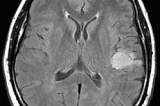
The irradiation zone is determined based on the MRI results. The total radiation dose to the lesion should not exceed 60 Gy, and if the spinal cord is irradiated, it should be even less, up to 35 Gy.
One of the complications of radiation therapy is the formation of a necrotic lesion in the brain after a couple of years. Dystrophic changes in the brain lead to a disruption of its functions, which is accompanied by corresponding symptoms similar to those of the tumor itself. In this case, the patient is examined and differential diagnostics are performed (PET with methionine, computer or magnetic resonance spectroscopy are prescribed) to distinguish radiation necrosis from tumor relapse. [ 14 ]
Along with remote radiotherapy, contact radiotherapy (brachytherapy) can also be used, but in the case of brain tumors it is used very rarely. In any case, the flow of ionizing radiation, affecting the pathological DNA of cells and destroying it, leads to the death of tumor cells, which are more sensitive to such an impact than healthy cells. Modernized linear accelerators make it possible to reduce the degree of destructive impact on healthy tissue, which is especially important when it comes to the brain.
Radiation therapy helps destroy the remaining hidden tumor cells and prevents their recurrence, but this treatment is not indicated for everyone. If doctors see that the risk of possible complications is high, radiation therapy is not performed.
Contraindications to radiation therapy include:
- location and infiltrative growth of the tumor in vital parts of the brain (stem, subcortical nerve centers, hypothalamus).
- swelling of brain tissue with symptoms of dislocation (displacement) of the brain
- presence of postoperative hematoma,
- purulent-inflammatory areas in the zone of exposure to ionizing radiation,
- inadequate patient behavior, increased psychomotor excitability.
Radiation therapy is not administered to terminally ill patients with serious somatic diseases, which can only worsen the patient's condition and hasten the inevitable end. Such patients are prescribed palliative therapy (according to the appropriate protocol) to reduce pain and prevent severe bleeding. In other words, doctors try to alleviate the patient's suffering in the last days and months of his life as much as possible.
Chemotherapy for astrocytoma
Chemotherapy is a method of systemic action on the body with the aim of destroying the remaining atypical cells and preventing their re-growth. The use of potent agents that have a detrimental effect on the liver and change the composition of the blood is justified only in the case of malignant tumors. [ 15 ] Early chemotherapy, parallel chemotherapy and short chemotherapy after radiotherapy are possible and well tolerated [ 16 ].
If we are talking about astrocytomas, then chemotherapy in some cases can be prescribed for a benign tumor, if there is a high risk of its degeneration into cancer. For example, in people with a hereditary predisposition (there were cases of confirmed oncology in the family), benign neoplasms, even after surgical removal and radiation therapy, can recur and turn into cancerous tumors.
An identical situation can be observed in dual diagnoses, when there is no certainty that the tumor has a low degree of malignancy or when there are contraindications to radiation therapy. In such cases, the lesser of two evils is chosen, i.e. chemotherapy.
Malignant astrocytoma of the brain is an aggressive tumor prone to rapid growth, so it is necessary to act against it with equally aggressive methods. Since astrocytomas are classified as primary brain tumors, drugs are selected for the treatment of this type of oncology, but taking into account the histological type of the tumor.
In chemotherapy of astrocytoma, cytostatic antitumor drugs with alkylating action are used. The alkyl groups of these drugs are capable of attaching to the DNA of atypical cells, destroying it and making the process of their division (mitosis) impossible. Such drugs include: "Temodal", "Temozolomide", "Lomustine", "Vincristine" (a drug based on the alkaloid of periwinkle), "Procarbazine", Dibromodulcitol [ 17 ] and others. It is possible to prescribe:
- platinum drugs (Cisplatin, Carboplatin), which inhibit DNA synthesis in atypical cells, [ 18 ]
- topoisomerase inhibitors (Etoposide, Irinotecan), which prevent cell division and the synthesis of hereditary information),
- monoclonal IgG1 antibodies (Bevacizumab), which disrupt the blood supply and nutrition of the tumor, preventing its growth and metastasis (they can be used independently, but more often in combination with topomerase inhibitors, for example, with the drug Irinotecan). [ 19 ]
For anaplastic tumors, the most effective are nitro derivatives (Lomustine, Fotemustine) or their combinations (Lomustine + drugs from another series: Procarbazine, Vincristine).
In case of relapses of anaplastic astrocytomas, the drug of choice is Temozolomide (Temodal). It is used alone or in combination with radiotherapy; combination treatment is usually prescribed for glioblastomas and recurrent anaplastic astrocytomas. [ 20 ]
Two-component regimens are often used to treat glioblastomas: Temozolomide + Vincristine, Temozolomide + Bevacizumab, Bevacizumab + Irinotecan. A course of treatment is prescribed for 4-6 cycles with intervals of 2-4 weeks. Temozolomide is prescribed daily for 5 days, the remaining drugs should be administered on certain days of treatment 1-2 times during the course.
This therapy is believed to increase the one-year survival rate of patients with malignant tumors by 6%. [ 21 ] Without chemotherapy, patients with glioblastoma rarely survive more than 1 year.
To assess the effectiveness of radiation and chemotherapy, a repeat MRI is performed. During the first 4-8 weeks, an atypical picture may be observed: the contrast increases, which may suggest the progression of the tumor process. Do not make hasty conclusions. It is more relevant to conduct a repeat MRI 4 weeks after the first one and, if necessary, a PET study.
WHO defines criteria by which the effectiveness of therapy can be assessed, but it is necessary to take into account the state of the patient's central nervous system and the concomitant treatment with corticosteroids. An acceptable goal of complex treatment is to increase the number of surviving patients and those who show no signs of disease progression within six months.
With a 100% disappearance of the tumor, they speak of complete regression, a decrease in the neoplasm by 50% or more is partial regression. Lower indicators indicate stabilization of the process, which is also considered a positive criterion, providing for a stop in tumor growth. But an increase in the tumor by more than a quarter indicates the progression of cancer, which is a poor prognostic symptom. Symptomatic treatment is also carried out.
Treatment of astrocytoma abroad
The state of our medicine is such that people are often afraid of dying not so much from the disease as from a surgical error, lack of necessary medicines. The life of a person with a brain tumor is not to be envied. What are the constant headaches and epileptic seizures worth? The psyche of patients is often at its limit, so not only professional diagnostics and the right approach to treatment are very important, but also the appropriate attitude towards the patient on the part of medical personnel.
In our country, disabled people and people with serious illnesses are still in an ambiguous position. Many people pity them in words, but in reality they do not receive the love and care they need. After all, pity is not the help that stimulates one to get back on one's feet after an illness. What is needed here is support and the instillation of confidence that there is almost always hope and that even the smallest opportunities must be used to live, because life is the highest value on Earth.
Even people with stage 4 cancer, who are given a cruel sentence, need hope and care. Even if a person is given only a couple of months, they can live through them in different ways. Doctors can ease the patient's suffering, and relatives can do everything so that their loved one dies happy.
Some people, with the support of others, do more in the allotted days and weeks than they do in their entire lives. But this requires the appropriate attitude. Cancer patients, more than anyone else, need the help of psychologists who help them change their attitude toward the disease. Unfortunately, such help is not always offered in domestic medical institutions.
We have oncology dispensaries and specialized departments, we treat cancer of various localizations, we have qualified specialists for this, but the equipment of our medical centers often leaves much to be desired, not all doctors undertake to perform operations on the brain, psychological assistance and the attitude of the staff usually leaves much to be desired. All this becomes the reason for looking for the possibility of treatment abroad, because reviews of foreign clinics are overwhelmingly positive, full of gratitude. This instills hope even in those who, it would seem, are doomed due to the diagnosis, which, moreover, may be inaccurate (poor equipment with diagnostic equipment increases the risk of error).
We have already become accustomed to the fact that foreign doctors undertake to treat patients who have been rejected by domestic specialists. Thus, many patients with malignant anaplastic astrocytoma have already been successfully treated in Israeli clinics. People have received the opportunity to continue living a full life. At the same time, the statistics of relapses in Israeli clinics are much lower than in our country.
Today, Israel, with its high-tech modern equipment of clinics and highly qualified personnel, is a leader in terms of treatment of oncological diseases, including brain astrocytoma. High assessment of the work of Israeli specialists is not accidental, because the success of the operations is facilitated by modern equipment, which is regularly updated and improved, and the development of scientifically proven effective schemes/methods of tumor treatment, and the attitude towards patients, whether they are citizens of the country or visitors.
Both state and private clinics care about their prestige, and their work is controlled by state bodies and relevant laws, which no one is in a hurry to violate (a different mentality). In hospitals and medical centers, the life and health of the patient come first, and both medical personnel and special international organizations take care of them. Patient support and assistance services help to settle in a foreign country, quickly and efficiently undergo the necessary examinations, and offer opportunities to reduce the cost of the services offered if financial difficulties arise.
The patient always has a choice. At the same time, a lower price for services does not mean their poor quality. In Israel, not only private but also state clinics can boast of their fame throughout the world. Moreover, this fame is deserved by many successful operations and many saved lives.
When considering the best Israeli clinics for the treatment of astrocytoma, it is worth noting the following government institutions:
- Hadassah University Hospital in Jerusalem. The clinic has a department for the treatment of CNS cancer tumors. The neurosurgical department provides patients with a full range of diagnostic services: examination by a neurologist, radiography, CT or MRI, electroencephalography, ultrasound (prescribed for children), PET-CT, angiography, spinal puncture, biopsy in combination with histological examination.
Operations to remove astrocytomas of varying degrees of malignancy are performed by world-renowned neurosurgeons who specialize in treating cancer patients. Treatment tactics and regimens are selected individually, which does not prevent doctors from adhering to scientifically based treatment protocols. The clinic has a neuro-oncological rehabilitation department.
- Sourasky Medical Center (Ichilov) in Tel Aviv. One of the largest public medical institutions in the country, which are ready to accept foreign patients. At the same time, the effectiveness of cancer treatment is simply amazing: 90% effectiveness of cancer treatment in combination with 98% of successful brain surgeries. Ichilov Hospital is included in the TOP-10 most popular clinics. It offers a comprehensive examination and management by several specialists at once, quick preparation of a treatment plan and calculation of its cost. All doctors working in the hospital are highly qualified, have completed internships in famous clinics in the USA and Canada, have a large stock of the latest knowledge and sufficient practical experience in treating cancer patients. Operations are carried out under the control of neuronavigation systems, which minimizes possible complications.
- Itzhak Rabin Medical Center. A multidisciplinary medical institution with the largest oncology center "Davidov" equipped with the latest technology. One fifth of cancer patients in Israel undergo treatment in this center, which is famous for its high accuracy of diagnosis (100%). About 34-35% of diagnoses made by hospitals in other countries are disputed here. People who considered themselves terminally ill receive a second chance and the most valuable thing - hope.
The latest developments, targeted and immunotherapy, and robotics are used in the treatment of cancer patients. During treatment, patients live in hotel-type wards.
- State Medical Center "Rambam". A modern well-equipped center, top-class specialists, extensive experience in treating patients with brain tumors, good attitude and care for patients regardless of their country of residence - this is an opportunity to receive quality treatment in a short time. It is possible to contact the hospital without intermediaries and fly out for treatment in 5 days. There is an opportunity to participate in experimental methods for patients with a poor prognosis for treatment with traditional methods.
- Sheba Medical Center. A renowned state university hospital that has been cooperating with the American MD Anderson Cancer Center for many years. The clinic's special feature, in addition to its good equipment, high diagnostic accuracy and successful operations to remove brain astrocytoma, is a special patient care program that includes ongoing psychological support.
As for private clinics where you can undergo qualified and safe treatment of brain astrocytoma, it is worth paying attention to such a multidisciplinary clinic "Assuta" in Tel Aviv, which was built on the basis of the institute. It is worth mentioning that this is one of the most famous and popular clinics, the cost of whose services is comparable to those in public hospitals and is controlled by the state. Accurate diagnostics, modern methods of treating brain cancer, a high percentage of recovery at stage 1 cancer (90%), the highest level of equipment of laboratories, diagnostic rooms, operating rooms, comfortable conditions for patients, professionalism of all doctors and junior medical staff involved in the treatment of the patient.
A special feature of almost all private and public clinics in Israel is the professionalism of doctors and a prudent, caring attitude towards patients. Today, there are no special problems in terms of communication and registration for treatment in Israel (except for financial ones, because foreign patients are treated there for a fee). As for payment for treatment, it is mainly made upon receipt, and in addition, there is the possibility of an installment plan.
High competition, government control and sufficient funding force Israeli clinics, as they say, to maintain their brand. We do not have such competition, as well as the ability to carry out accurate diagnostics and quality treatment. We have good doctors who are powerless against the disease not because of a lack of knowledge and experience, but because of the lack of necessary equipment. Patients would like to trust their domestic specialists, but they cannot, because their lives are at stake.
Today, treatment of brain tumors in Israel is the best option for taking care of yourself or your loved ones, whether it is brain cancer or there is a need to operate on other vital organs.