
All iLive content is medically reviewed or fact checked to ensure as much factual accuracy as possible.
We have strict sourcing guidelines and only link to reputable media sites, academic research institutions and, whenever possible, medically peer reviewed studies. Note that the numbers in parentheses ([1], [2], etc.) are clickable links to these studies.
If you feel that any of our content is inaccurate, out-of-date, or otherwise questionable, please select it and press Ctrl + Enter.
Tracheal extubation
Medical expert of the article

Last reviewed: 04.07.2025
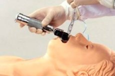
Anesthesiologists often use concepts such as intubation and extubation. The first term, intubation, actually means inserting a special tube into the trachea, which is necessary to ensure the patient's airway is clear. As for extubation, this is a procedure that is the opposite of intubation: the tube is removed from the trachea when it is no longer needed.
Extubation can be performed in a hospital setting or in an ambulance (outside a healthcare facility). [ 1 ]
Indications for the procedure
In cases where there is no longer a need to control the respiratory tract, the endotracheal tube inserted during intubation is removed. This is usually done when subjective and objective improvement in respiratory function is achieved. For a more comfortable and safe procedure, the doctor must ensure that the patient can breathe independently, that the respiratory tract is patent, and that the tidal volume is sufficient. In general, extubation is possible provided that the respiratory center has adequate ability to initiate inhalations with a normal frequency, depth, and rhythm. Additional conditions for the procedure are normal strength of the respiratory muscles, a “working” cough reflex, good nutritional status, adequate clearance of sedatives and muscle relaxants. [ 2 ]
In addition to normalizing the patient's condition and respiratory function, there are other indications. Extubation is performed in case of sudden blockage of the endotracheal tube by foreign agents - for example, mucous and sputum secretions, foreign objects. After removal, reintubation or tracheostomy is performed, at the discretion of the doctor.
Another indication for extubation may be a situation where the continued presence of the tube in the trachea becomes inappropriate – for example, if the patient is dying. [ 3 ]
Preparation
Preparation for extubation begins with careful planning of the procedure, namely, an assessment of the airway status and general risk factors.
The condition of the respiratory organs is assessed according to the following criteria:
- no difficulty breathing;
- absence of damage to the respiratory tract (swelling, injury, bleeding);
- no risk of aspiration and obstruction.
General factors are assessed based on cardiovascular, respiratory, neurological, and metabolic parameters, taking into account the characteristics of the surgical intervention and the patient's condition before extubation. [ 4 ]
In general, preparation consists of optimizing the general condition of the patient and other factors:
- check the quality of hemodynamics, respiration, measure temperature, assess metabolism and neurological status;
- prepare the necessary equipment and tools;
- monitor all vital functions of the body.
It is optimal if the extubation manipulation is performed on an empty stomach. Most often, the patient is fully conscious. [ 5 ]
Technique extubations
Extubation is the removal of the intubation tube when the patient has all the prerequisites for independent breathing. The manipulation is carried out in the following sequence of actions:
- If a gastric tube is present, the entire contents of the stomach are aspirated;
- carefully sanitize the nasal and oral cavity, pharynx, and tracheobronchial tree;
- deflate the cuff and gradually, slowly, preferably while inhaling, remove the endotracheal tube.
During extubation, the tube is removed in one clear but smooth movement. After this, a face mask is applied with 100% oxygen supply until the condition is normalized. [ 6 ]
Sometimes the extubation procedure is performed unplanned - for example, in patients with acute reactive psychosis, when the patient is poorly stabilized, or in conditions of insufficient sedation.
Emergency extubation in the following cases:
- at low or zero pressure in the airways;
- when the patient speaks;
- when the endotracheal tube extends several centimeters (depending on the age and initial depth of installation of the device).
The following are considered unreliable signs of the need for extubation:
- small tube outlet (up to 20 mm);
- expressed anxiety of the patient;
- paroxysmal cough, sudden cyanosis (it is necessary to check cardiovascular parameters).
If extubation occurs unplanned, the following step-by-step actions are followed:
- If there are clear signs of the need for extubation, the cuff is deflated and the endotracheal tube is removed. If necessary, the upper respiratory tract is sanitized, after which artificial ventilation of the lungs is performed using an Ambu bag (it is best to connect it to an oxygen source) or the mouth-to-mouth method. After the indicators have normalized, the need for reintubation is assessed.
- If unreliable signs are detected, an attempt is made to use an Ambu bag. Positive manifestations: the chest and abdomen change volume in time with respiratory movements, the skin turns pink, breathing noises are noted when listening to the lungs. If such signs are present, the endotracheal tube is brought to the required depth. If there are no positive manifestations, the cuff is deflated, the tube is removed. If there is a cough and cyanosis, the tracheobronchial tree is sanitized and artificial ventilation of the lungs is started using an Ambu bag.
If there is a need for re-intubation, it should not follow immediately after extubation. First, an attempt should be made to restore the patient's breathing using an Ambu bag for 3-5 minutes. Only after the condition has normalized is it determined whether re-intubation is necessary. Re-intubation is performed after preoxygenation. [ 7 ]
Extubation criteria
The endotracheal tube is removed when there is no longer a need for artificial patency of the respiratory tract. According to clinical characteristics, before extubation, there should be a softening of the signs of the initial cause of respiratory failure, and the patient himself should have all the prerequisites for normal spontaneous breathing and gas exchange processes. [ 8 ]
It is possible to determine that a person is ready for extubation based on the following criteria:
- is able to maintain a normal supply of oxygen to the blood, maintaining the ratio of PaO2 and FiO2 above 150 and 200 with the presence of O2 in the inhaled mixture no more than 40-50% and a PEEP value of no more than 5-8 mbar;
- is able to maintain the reaction of the arterial blood environment and the level of carbon dioxide on exhalation within acceptable values;
- successfully passes a spontaneous breathing test (30-120 minutes with a PEEP of 5 mbar, with a low support pressure of 5-7 mbar, with adequate gas exchange and stable hemodynamics);
- the spontaneous breathing rate during extubation does not exceed 35 per minute (in an adult);
- the norm of strength of the respiratory muscles is determined;
- the maximum negative inspiratory pressure exceeds 20-30 mbar;
- the vital capacity of the lungs exceeds 10 ml per kilogram (for newborns – 150 ml per kilogram);
- the transphrenic pressure index is less than 15% of the maximum during spontaneous breathing;
- the spontaneous minute ventilation rate for an adult at the moment of exhalation is 10 ml per kilogram;
- chest wall compliance exceeds 25 ml/cm;
- respiratory function less than 0.8 J/l;
- average blood pressure exceeds 80 mmHg.
The patient must be conscious and follow certain requests and commands from the doctor. As a test of readiness for extubation, a test such as the Gale tetrad is performed: the patient is asked to shake hands, lift and hold his head, touch the tip of his nose with his finger, and hold his breath. [ 9 ]
The extubation protocol is a set of diagnostic and tactical algorithms, including a full assessment of the patient's clinical condition, characteristics of the surgical operation, selection of the optimal ventilation scheme and drug support, determination of readiness for removal of the endotracheal tube, and optimization of spontaneous breathing.
The most justified indicators from a physiological point of view are those reflecting the respiratory rate and respiratory volume (frequency and volume index), as well as the values of the adaptability of the respiratory organs, maximum inspiratory effort and oxygenation. [ 10 ]
Contraindications to the procedure
Experts say there are no absolute contraindications to extubation. To achieve adequate gas exchange processes for some patients, the following may be required:
- non-invasive ventilation;
- prolonged air inflation (CPAP);
- inhaled mixture with increased oxygen concentration;
- reintubation.
It is necessary to be prepared for the fact that respiratory reflexes may be depressed immediately after extubation or a little later. Prevention of possible aspiration is mandatory. [ 11 ]
Extubation is the removal of the endotracheal tube in a conscious person, usually accompanied by coughing (or a motor reaction). The heart rate increases, central venous and arterial pressure increases, as well as intraocular and intracranial pressure. If the patient suffers from bronchial asthma, bronchospasm may develop. The development of complications can be prevented by administering lidocaine in an amount of 1.5 mg / kilogram one and a half minutes before extubation.
Removal of the tube under deep anesthesia is contraindicated if there is a risk of aspiration or airway obstruction.[ 12 ]
Consequences after the procedure
It is difficult to determine the outcome of extubation in advance, but it is necessary to take into account the fact that both premature and incorrectly performed manipulation can be fatal for the patient. The likelihood of developing certain consequences depends largely on the qualifications of the doctor, as well as on other background factors. Often, other pathologies in the patient's body, as well as secondary diseases, become the "culprits" of adverse consequences. [ 13 ]
To improve the prognosis, it is necessary to establish patient monitoring both before and after extubation. It is especially important to monitor the condition of patients in terminal states, when the probability of re-intubation remains high.
The clinical protocol for extubation should include careful monitoring of all vital signs and functions of the person after the manipulation, rapid identification and response to respiratory distress, and, if necessary, rapid reintubation or tracheostomy. [ 14 ]
Tracheal extubation is a key stage of recovery from general anesthesia. It is a complex procedure that can result in more complications than the initial intubation procedure. During the removal of the endotracheal tube, a controlled situation becomes uncontrollable: specialists are faced with physiological changes along with a limited time period and other compromising factors, which in general can be difficult even for a highly qualified anesthesiologist.
It should be noted that the vast majority of post-extubation complications are minor. However, in some cases, doctors have to deal with serious consequences, including cerebral hypoxia and death. [ 15 ]
Laryngospasm after extubation
Laryngospasm is the most common cause of upper airway obstruction after extubation. The clinical picture of laryngospasm can have varying degrees of severity and can be represented by both mild stridor breathing and complete respiratory obstruction. Most often, the complication is detected in childhood, against the background of surgical intervention on the respiratory system. [ 16 ]
The most common cause of laryngospasm after extubation is irritation by salivary secretions or blood, mainly when using light anesthesia. In such a situation, the patient is unable to either prevent the reflex response or cough well. The incidence of laryngospasm after extubation can be reduced by placing patients on their side and ensuring rest until they are fully awake. In addition, the complication can be prevented by intravenous administration of magnesium sulfate (dosage 15 mg/kg for 20 minutes) and lidocaine (dosage 1.5 mg/kg). [ 17 ]
Complications after the procedure
To prevent complications, the degree of risk for the patient must be determined before extubation. It is known that the easier the intubation, the lower the probability of post-extubation complications.
A special approach is necessary for long and traumatic operations with large blood loss. In obviously difficult cases, they resort to a step-by-step removal of the endotracheal tube.
One of the basic factors for the success of the procedure is the elimination of residual muscle relaxation. [ 18 ]
A high risk of complications is indicated in the following cases:
- there are difficulties with ventilation and intubation;
- limited mobility of the cervical spine, temporomandibular joints, or instability in these areas;
- the patient suffers from morbid obesity and has obstructive sleep apnea (from the anamnesis);
- there are risks of postoperative bleeding and compression of the larynx by a hematoma, or there are cases of damage to the nerve fibers of the larynx or pharynx;
- intubation was performed "blindly";
- There are massive bandages that can impair air access - for example, in the neck, head, and face area.
The most common potential complications after extubation are:
- hemodynamic disorders;
- laryngospasm;
- cough, wheezing, noisy (stridor) breathing;
- respiratory arrest (apnea);
- damage to the vocal cords;
- swelling of the laryngeal tissues;
- pulmonary edema;
- oxygen deficiency;
- aspiration.
The greatest risk is due to the lack of ability to quickly perform reintubation and ensure normal gas exchange during intubation attempts. [ 19 ]
Why does a child have difficulty breathing after extubation?
One of the complications of extubation may be laryngeal edema, which becomes a serious factor in the development of upper airway obstruction in young children: it manifests itself within six hours after the procedure. Supraglottic edema displaces the epiglottis backward, which leads to occlusion of the glottis during inspiration. If there is retroarytenoid edema behind the vocal cords, this leads to limitation of their abduction during inspiration. Subglottic edema narrows the cross-section of the laryngeal space. [ 20 ]
Additional risk factors for the development of post-extubation edema include:
- tightly fitted tube;
- intubation trauma;
- long intubation period (more than an hour);
- coughing, head and neck movements during intubation.
A similar condition is also typical for adult patients – after prolonged translaryngeal intubation.
In case of laryngeal edema, it is recommended to administer a humidified, heated, oxygen-enriched gas mixture. Epinephrine is administered through a nebulizer, dexamethasone, and Heliox are used. In difficult situations, reintubation is performed with a tube of a smaller diameter.
Difficulty breathing after extubation may be due to hematoma formation and tissue compression. In such cases, immediate re-intubation and final hemostasis are practiced. [ 21 ]
Another cause is trauma to the respiratory tract caused by rough manipulations, mechanical damage during insertion or removal of the endotracheal tube. Obstructive symptoms may arise acutely or manifest later in the form of swallowing pain or voice changes.
A less common cause of difficulty breathing after extubation is vocal cord paralysis due to damage to the vagus nerve during surgery. If paralysis is bilateral, there is a risk of post-extubation obstruction, so immediate reintubation is performed.
Care after the procedure
The risk of complications after extubation is present not only immediately after the endotracheal tube is removed, but also throughout the entire recovery period. Therefore, it is important to ensure maximum attention and monitoring of the patient's condition by the attending physician and anesthesiologist.
An oxygen mask is used during the patient's transportation to the postoperative ward. Medical personnel provide full care until all respiratory reflexes are restored and physiological parameters are normalized. Each patient is provided with constant monitoring by nurses and an anesthesiologist. [ 22 ]
After a person is brought out of anesthesia, specialists evaluate their level of consciousness, respiratory rate and heart activity, blood pressure, body temperature and peripheral oxygen saturation. The use of capnography allows for early detection of airway obstruction.
Warning signs after extubation:
- respiratory disorders in the form of stridor breathing, agitation;
- postoperative complications (pathological drainage secretions, transplant perfusion, bleeding and hematomas, respiratory tract edema);
- development of mediastinitis and other respiratory lesions. [ 23 ], [ 24 ]
Mediastinitis is a consequence of perforation of the airway - for example, after difficult insertion of a tube. The complication is manifested by pain in the chest and neck, difficulty swallowing, painful swallowing, fever, crepitus. [ 25 ]
Traumatic injuries are most often found in the larynx, pharynx and esophagus. In some cases, pneumothorax and emphysema are observed.
Patients with irritated airways are placed in a vertical position and prescribed inhalation of humidified oxygen with sufficient flow. Monitoring of exhaled carbon dioxide concentration is recommended. The patient is not fed due to possible laryngeal dysfunction (even with clear consciousness), factors capable of disrupting venous circulation are excluded. It is important to ensure deep breathing and free expectoration of sputum. If the patient has obstructive sleep apnea, respiratory patency is compensated by installing a nasopharyngeal airway.
To reduce inflammatory edema after extubation, glucocorticoids are prescribed (100 mg hydrocortisone every six hours, at least twice). If respiratory obstruction develops, 1 mg of adrenaline can be administered via a nebulizer. A mixture of helium in oxygen also has a positive effect. [ 26 ]
Additional drug support includes analgesic and antiemetic therapy.
Reviews
Resumption of spontaneous breathing after extubation is often achieved without any particular problems. However, in some patients, activation of respiratory function is difficult, which requires the use of intensive care measures.
Activation of spontaneous breathing is a combined process that requires a multi-stage assessment of the individual clinical case. The mechanics of respiratory capacity, adequacy of ventilation and tissue oxygen supply are assessed. The nature of the therapy used, the general and psychological state of the patient, and other existing problems are necessarily taken into account.
The success of extubation largely depends on the skills of the medical staff: it is important to correctly interpret the patient's reaction to an attempt to activate independent respiratory function.
The duration of a person's stay in the intensive care unit, as well as the frequency of complications caused by a long intubation period, depend on the timing of extubation. According to reviews, most patients are transferred to spontaneous breathing relatively quickly. Much fewer patients experience difficulties in activating independent respiratory function, which prolongs the hospital stay and increases the risk of developing adverse effects.
Early extubation has the following benefits: reduced need for nursing care, reduced risk of airway injury, increased cardiac output and renal perfusion during spontaneous breathing.