
All iLive content is medically reviewed or fact checked to ensure as much factual accuracy as possible.
We have strict sourcing guidelines and only link to reputable media sites, academic research institutions and, whenever possible, medically peer reviewed studies. Note that the numbers in parentheses ([1], [2], etc.) are clickable links to these studies.
If you feel that any of our content is inaccurate, out-of-date, or otherwise questionable, please select it and press Ctrl + Enter.
Adhesions in the left and right lung: pleural, fibrous
Medical expert of the article

Last reviewed: 04.07.2025
A serious complication after respiratory diseases is adhesions in the lungs. Let's consider the pathogenesis and causes of their occurrence, the main symptoms, methods of treatment and prevention.
The lungs are a paired organ in the chest that is responsible for the breathing process. The right lung is 10% larger than the left, since the human heart is shifted to the left. The organ's volume is about 3 liters. The lungs are covered with a pleural membrane on all sides. After extensive pneumonia and other inflammatory or infectious lesions, cords, i.e., peculiar internal scars, can form between the lobes.
- The appearance of adhesions depends on the organ where they are formed. They can be thin like a polyethylene film or thick fibrous neoplasms.
- Most often, the cords are localized between the serous membranes of the pleural cavity; they are also detected in the diaphragm area.
- In particularly severe cases, the growths occupy all parts of the pleura, causing adhesions of the pleural sheets and complete overgrowth of the cavities.
Adhesive disease can affect any organs where there is connective tissue. This pathology has a negative impact on the functioning of the entire body and especially the respiratory organs. Growing, adhesions block blood vessels, disrupting blood circulation and causing discomfort during breathing, respiratory failure.
Why are adhesions in the lungs dangerous?
In most cases, pulmonary adhesions are formed during inflammatory and infectious lesions. The danger of adhesions is that the pathological process is hidden. Very often, signs of adhesive disease are hidden under the symptoms of acute respiratory viral infections and other diseases of the respiratory system. As they grow, connective tissue strands disrupt the blood supply to the lungs and can cause the pleural cavities to fuse.
Another danger of shvart is pulmonary and cardiac insufficiency. These pathological processes lead to the development of pneumosclerosis, that is, the replacement of healthy organ tissues with connective tissues. The disease threatens the following complications:
- Deformation of the lungs and bronchi.
- Violation of gas exchange in the respiratory organ.
- Oxygen starvation.
- Pulmonary hypertension.
- Secondary infection.
All of the above factors have a negative impact on overall well-being and the functioning of the entire body. Without timely diagnosis and treatment, there is a risk of death.
Epidemiology
As statistics show, the appearance of adhesions in the lungs is most often associated with surgical interventions, injuries and inflammatory pathologies.
According to the conducted research, growths on the pleural membrane can progress for many years and not reveal themselves in any way. Only in 20% of cases, pulmonary synechiae lead to fusion of the pleural sheets, respiratory failure and other life-threatening complications.
Causes adhesions in the lungs
Adhesions are overgrown connective or fibrous tissue. They are most often a complication of pleurisy or severe pneumonia of any etiology.
The main causes of adhesions in the lungs include:
- Bronchitis: acute, chronic.
- Pneumonia.
- Parasitic infections: ascariasis, echinococcosis, amebiasis, paragonism.
- Infection of the lungs with Koch's bacillus.
- Malignant processes.
- Sarcoidosis.
- Congenital anomalies of the organ.
- Pulmonary infarction.
- Traumatic injuries.
- Internal bleeding.
- Occupational hazards (inhalation of dust and chemicals).
- Poor environmental situation in the place of residence.
- Bad habits.
- Surgical interventions on the chest.
- Allergic reactions and frequent inhalation of allergens.
If the growths are point or isolated, then there are no painful sensations, but if the adhesions are extensive, then this is accompanied by pronounced pathological symptoms. The presence of numerous adhesions leads to the exclusion of the lung from the gas exchange process. Because of this, oxygen starvation, respiratory failure and deterioration of general well-being develop.
Regardless of the cause of origin, connective tissue adhesions can become infected in advanced respiratory pathologies. Because of this, cicatricial changes tighten and deform the lungs, causing serious disruptions in their functioning.
Adhesions in the lung after surgery
Today, there are minimally invasive surgical techniques that allow lung operations to be performed through small incisions. But even laparoscopic intervention can cause postoperative adhesions.
All operations performed on the lungs are divided into two groups according to volume:
- Pulmonectomy (pneumonectomy) is the complete removal of a lung. It is prescribed for malignant lesions and multiple pathological lesions.
- Resection is the removal of a part of an organ.
Removal of a lung, its segment or lobe entails serious pathological changes in the structure of the lung tissue. If the postoperative process is complicated by inflammatory reactions, then synechiae are formed.
Adhesive disease leads to disruption of the body's oxygen supply. Shortness of breath, increased weakness, chest pain, cardiovascular problems, dizziness appear. The postoperative period leaves a negative imprint on the entire body. Internal organs are displaced, blood supply changes.
Pleural adhesions limit the linear dimensions of the remaining parts of the lung. This disrupts the breathing process. If the growths become infected, for example, due to a neglected cold, this leads to intoxication of the body. To prevent postoperative complications, patients are expected to undergo a long rehabilitation period with a course of physiotherapy procedures.
Risk factors
Enlarged connective tissue pulmonary cords are most often located between the serous membranes of the pleural cavity. They arise due to many reasons, and there are also a number of risk factors for the occurrence of this pathology:
- Chronic infectious and inflammatory diseases of the respiratory system.
- Mechanical injuries.
- Congenital and genetic pathologies.
- Radioactive exposure.
- Allergic reactions.
- Syphilis.
- Tuberculosis.
- Operations.
The cords can be of any localization, they arise in the place where there is connective tissue. The pleural lesion can be total, affecting all parts of the organ, and single flat. In especially severe cases, the pleural sheets fuse.
Pathogenesis
The mechanism of development of adhesive disease has a biochemical basis. Adhesions occur after inflammatory and infectious diseases, injuries, surgeries. Before considering the pathogenesis of the formation of cords in the lungs, it is necessary to familiarize yourself with the structural features of this respiratory organ:
- The lungs and chest cavity are covered with pleura. It is a serous membrane of mesothelial cells with a fibroelastic framework. The framework contains nerve endings, lymphatic and blood vessels.
- The pleura consists of two layers: parietal and visceral. The first is the outer shell of the inner surface of the chest cavity, provides free movement of the lungs in the chest.
- The visceral layer envelops each lung to allow them to slide normally relative to each other. The two parts of the pleura are connected to each other. The visceral part has a dual blood supply, receiving blood from the pulmonary and bronchial arteries.
- The pleural cavity and its sheets perform the act of breathing. The cavities contain fluid, which ensures the sliding of the sheets during inhalation and exhalation. The tightness of the organ is maintained by pressure.
Pleural adhesions are most often a complication of previous diseases: pneumonia, bronchitis, parasitic invasions. In this case, the adhesion process helps the body localize the inflamed area. The formation of strands occurs with mechanical damage, that is, chest injuries and after surgical interventions.
Connective tissue adhesions disrupt blood circulation in the affected tissues. This leads to changes in the structure of the lungs and provokes painful sensations and malfunctions of the organ. Adhesions can be single or multiple. Their appearance threatens air exchange disorders, oxygen starvation, and ongoing respiratory diseases.
Symptoms adhesions in the lungs
In most cases, the enlarged connective tissue strands on the lungs do not make themselves known in any way. Symptoms that allow one to suspect them are manifested by various complications. Also, the symptoms depend on the localization of the adhesions, so the disorders are quite diverse:
- Difficulty and rapid breathing.
- Respiratory failure and shortness of breath.
- Increased heart rate.
- Dizziness and loss of consciousness.
- Lowering blood pressure.
- Paradoxical movements of the chest with the participation of accessory respiratory muscles.
The painful condition is accompanied by a deterioration in general health and increased weakness. The appearance of the above symptoms is a reason to immediately seek medical help for diagnosis.
 [ 20 ]
First signs
At the first stages, the disease does not have any pronounced clinical signs. Discomfort occurs with increased physical activity, when compensatory mechanisms begin to deplete. Many patients note the appearance of such symptoms:
- Dyspnea.
- Chest pain.
- Dizziness.
- Difficulty restoring breathing.
- Increased heart rate.
If the appearance of adhesions occurs with a secondary infection, then purulent sputum, increased cough and shortness of breath, and elevated body temperature appear. Anemia with pronounced pallor of the skin may also develop.
As the connective tissue strands grow, the painful symptoms increase:
- Psychomotor agitation appears.
- The skin and mucous membranes take on a bluish tint.
- Blood pressure decreases.
- Loss of consciousness is possible.
If adhesions have caused the fusion of pleural sheets, the symptoms are acute and pronounced. Signs of terminal respiratory failure in combination with gas exchange disorders in the lungs and acute oxygen deficiency cause serious disruptions in the functioning of other organs and systems.
Adhesions in the left and right lungs
The left lung differs from the right in its smaller size and shape. It is slightly longer and narrower than the right. The organ consists of two branches: the upper and lower lobes, which have almost equal volume. The volume of the left bronchus is smaller than the right, while its length is twice as long as the right.
Adhesions in the left lung are formed for the same reasons as in the right. These may be previous and advanced inflammatory lesions, chronic obstructive disease, fibrous processes, injuries and a number of other pathologies. The symptoms of left-sided lesions are characterized by discomfort on the left side, which may affect the cardiovascular system.
The right lung is 10% larger than the left, but its linear dimensions are smaller. These differences are due to the displacement of the main muscle of the body (the heart) to the left, so there is more space on the right side. In addition, the liver is located on the right side of the abdominal cavity, which presses the sternum from below, thereby reducing its height.
The organ has three parts, or lobes, which are functionally equivalent. Each contains structural elements for gas exchange and proper functioning. The upper lobe differs from the others not only in location, but also in volume. The smallest size is in the middle lobe, and the largest is in the lower lobe. Adhesions in the right lung can occur on any of its branches.
Forms
Depending on the location and degree of prevalence, several types of connective tissue adhesions in the lungs are distinguished:
- Pleuroapical.
- Pereuropulmonary.
- Pleurodiaphragmatic.
Types of adhesions are also classified according to the etiology of their origin:
- Infectious/non-infectious.
- Traumatic.
- Postoperative.
- Exudative.
- Fibrous.
- Congenital.
Adhesions can be local, focal and multiple. Local ones represent a limited altered area of lung tissue. They do not cause painful symptoms. Focal ones are localized in several areas of the pleura, and multiple ones chaotically cover most of the lung.
The type of adhesive disease is determined during the diagnostic process. The form of treatment depends on the severity of the lesion and the severity of pathological symptoms.
Pleuroapical adhesions in the lungs
Thickening of the pleura of the pulmonary apices is pleuroapical adhesions. Such layers may indicate recent inflammatory processes. But most often, adhesions occur due to tuberculosis infection, i.e. they are its complication. Connective structures isolate the affected area from healthy tissues.
As a rule, tissue growth is detected during fluorography. The cords themselves are not a disease, so they do not require therapeutic intervention. If they cause chest pain, difficulty breathing, and other unpleasant symptoms, then medical assistance is needed.
[ 23 ], [ 24 ], [ 25 ], [ 26 ]
Pleuropulmonary adhesions
The appearance of pleuropulmonary cords in the lungs indicates previously suffered inflammatory processes with damage to the pleural tissues. Connective tissue adhesions are caused by impaired respiratory and circulatory functions. The cords localize the affected tissues from healthy ones, thereby restoring the normal functioning of the organ.
The lungs are surrounded by a pleural cavity. Normally, this area contains about 5 ml of synovial fluid, which acts as a shock absorber for the normal breathing process. If the organ is sick, then inflammatory exudate appears, which provokes the development of pleurisy. During the disease, fibrin is deposited on the walls of the organ. During recovery, the inflammation goes away, and the accumulated fluid is absorbed. But fibrin can remain in the pleura and cause adhesions. In especially severe cases, this leads to the fusion of the pleural sheets.
As a rule, pleuropulmonary formations are detected during fluorography. In most cases, this radiographic sign does not require special treatment. The need for therapy arises with the development of respiratory failure and other painful symptoms.
Pleurodiaphragmatic adhesions
Connective tissue at the borders of the serous membranes of the pleural cavity are pleurodiaphragmatic adhesions. In the lungs, they are formed due to inflammatory and infectious processes, as well as traumatic injuries and congenital anomalies. Adhesions can be total, growing throughout the entire volume of the pleura, and isolated.
If adhesions cause pain when breathing, shortness of breath and other unpleasant symptoms, you should consult a pulmonologist. The doctor will prescribe a comprehensive diagnosis.
- If the growths are associated with inflammatory processes, then an increased content of leukocytes in the sputum and a change in C-reactive protein in the blood are observed.
- When examining sputum, the level of alveolar macrophages, bronchial epithelium, the presence of erythrocytes and other indicators are determined. If the analysis reveals epithelium, then depending on which type of cells predominates, the doctor draws conclusions about the degree of damage to the respiratory tract.
- Spirometry with a bronchodilator is also performed to determine the functions of external respiration.
Treatment depends on the severity of the adhesive disease and the cause of its occurrence. If there is a high risk of pulmonary failure and other life-threatening pathologies, then surgical intervention is performed. In other cases, patients are prescribed drug therapy and a course of physiotherapy.
Diaphragmatic adhesions
The diaphragm is a flat muscle that consists of several muscle bundles. It is located between the sternum and the abdominal cavity, that is, directly under the lungs, in contact with the pleura. The appearance of diaphragmatic adhesions is most often associated with the involvement of the pleura in the inflammatory process. Thus, pleural layers isolate the affected areas.
As a rule, the adhesions in the lungs do not cause painful sensations. But in some cases, the following symptoms appear:
- Unproductive cough.
- Signs of respiratory failure.
- Discomfort when breathing and other painful sensations.
Such symptoms are a reason to seek medical help and undergo diagnostics. Tissue proliferation is detected using fluorography and X-ray examination. If diaphragmatic adhesions are calcified, this makes them easier to detect.
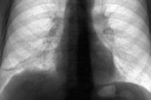
On the images, the cords appear as darkened areas of the pulmonary field with an enhanced vascular-connective tissue pattern. In case of multiple lesions, diffuse darkening is determined. It is also possible to reduce the height of the ribs, reduce the intercostal space and displace organs.
Treatment depends on the results of the diagnostics. If cicatricial changes quickly become denser and disrupt the normal functioning of the lung, then surgical intervention with a course of drug therapy is performed. The patient is also prescribed physiotherapy. Preventive measures are of particular importance for the prevention of adhesive disease. They consist of timely treatment of any diseases and increasing the protective properties of the immune system.
[ 27 ], [ 28 ], [ 29 ], [ 30 ], [ 31 ], [ 32 ], [ 33 ]
Basal adhesions
The enlarged connective tissue strands at the base of the lungs, i.e. in the root zone, are basal adhesions. The formation of adhesions in this area is extremely rare. The main reasons for the appearance of adhesions are:
- Chronic inflammatory processes.
- Bronchial obstruction.
- Mechanical trauma to the respiratory system.
- Genetic and congenital diseases.
- Long-term inhalation of dust and gases.
- Allergic alveolitis.
- Bacterial and viral diseases.
Formation of basal adhesions in the lungs is possible with thrombosis of the pulmonary arteries, left ventricular failure, and disruption of blood flow in the pulmonary circulation. That is, scarring of the pleura is the result of dystrophic changes. Connective tissues grow, deforming the structure of the organ.
The danger of the disease is that adhesions fill the intercellular space. Because of this, the lung tissue becomes denser and the volume of ventilated air decreases, the alveolar lumens narrow. Against this background, pneumosclerosis can develop. The main symptom of the pathological condition is respiratory failure. Without medical care, painful symptoms can progress, aggravating discomfort. Lack of oxygen negatively affects the functioning of the entire body.
[ 34 ], [ 35 ], [ 36 ], [ 37 ], [ 38 ], [ 39 ]
Fibrous adhesions in the lungs
Fibrous tissue is a type of connective tissue that replaces free space in the body. Fibrous adhesions on the pleura of the lungs most often appear in the following cases:
- After surgical interventions.
- For penetrating traumatic injuries.
- After acute infectious and inflammatory processes (pneumonia, tuberculosis).
With both single and multiple fibrous adhesions, symptoms arise that are similar to cardiac problems:
- Chest pain.
- Difficulty breathing.
- Increased weakness and shortness of breath.
- Tachycardia.
Gradually, nerve and blood vessels appear in the fibrous tissues. Adhesions can become saturated with calcium salts, i.e. ossify. This leads to limited lung movement, which disrupts their functioning. Excessive growth of adhesions is dangerous due to the gluing of pulmonary cavities and their overgrowing. The pathology is accompanied by severe symptoms: severe pain when breathing and acute respiratory failure. This condition requires urgent surgical treatment.
In the early stages, fibrous adhesions in the lungs do not cause painful sensations. But when the first signs of a painful condition appear and there is a suspicion of adhesive disease, it is necessary to consult a specialist.
Complications and consequences
Connective tissue growths in the lungs are dangerous due to serious consequences that negatively affect the functioning of the entire body. Pulmonary adhesions can cause the following complications:
- Respiratory failure.
- Oxygen starvation.
- Overgrowth of interlobar fissures and pleural cavities.
- Thickening of the pleural sheets due to multiple cicatricial changes.
- Pneumosclerosis.
- Limitation of mobility of the dome diaphragm.
Another rather serious complication of pulmonary adhesions is the appearance of cystic neoplasms. In the early stages, cystic fibrosis has vague symptoms:
- The body temperature gradually rises.
- The breathing rhythm is disturbed.
- The extremities and mucous membranes acquire a cyanotic tint.
- Breathing causes severe pain and is accompanied by wheezing.
In addition to the above problems, synechiae worsen the quality of life. Their appearance contributes to the development of not only pulmonary, but also cardiac insufficiency. It is also possible for a secondary infection to occur, which can be fatal.
Diagnostics adhesions in the lungs
Painful symptoms when breathing are the main reason for suspecting adhesions in the lungs. The doctor studies the patient's complaints, collects anamnesis and prescribes a set of diagnostic measures.
Diagnostic procedures are divided into two groups: to determine the general health of the patient and to identify complications of the adhesion process. The following studies are indicated to assess respiratory function:
- Physical examination – examination of the chest, palpation of tissues, axillary and subclavian lymph nodes. Percussion of the chest cavity and auscultation using a stethoscope. The doctor also measures the pulse, respiratory rate, body temperature and blood pressure. Based on the data obtained, a further diagnostic plan is drawn up.
- A set of laboratory tests – blood and urine analysis, blood gas composition, bacteriological composition of sputum.
- Instrumental methods – radiography, fluorography, MRI, spirography, CT, lung tissue biopsy.
The diagnostics are carried out by a therapist and a pulmonologist. Based on the results of the tests, a treatment plan is drawn up.
[ 51 ], [ 52 ], [ 53 ], [ 54 ], [ 55 ]
Tests
Laboratory diagnostics is a mandatory component of the examination of the body when adhesions in the lungs are suspected. Analyses are carried out not only at the diagnostic stage, but also during the treatment process.
- Blood test – if the proliferation of adhesions has caused respiratory failure, but changes in the blood composition are observed. An increase in leukocytes, erythrocytosis and an increase in the erythrocyte sedimentation rate are possible, which indicates inflammatory processes in the body. An increase in hemoglobin levels, an increase in hematocrit, and eosinophilia may also be observed.
- Urine analysis – allows you to assess the general condition of the body and the presence of complications of connective tissue growths. Urine may contain cylindrical epithelial cells, protein, and erythrocytes.
- Bacteriological analysis of sputum – is performed if cicatricial changes in the respiratory organs have led to acute or chronic respiratory failure. The formation of sputum with pus impurities indicates damage to the lungs by pathogenic microorganisms.
The test results make it possible to draw up a treatment plan or prescribe additional diagnostic tests. For example, after bacteriological tests, an antibiogram is drawn up to determine the sensitivity of bacteria to antibiotics and select an effective drug.
[ 56 ], [ 57 ], [ 58 ], [ 59 ], [ 60 ], [ 61 ]
Instrumental diagnostics
Very often, adhesions in the lungs are detected during fluorography, which is an instrumental diagnostic method. This type of examination is included in the complex of mandatory ones for any pathological symptoms from the respiratory organs.
Let's consider the main instrumental methods for detecting connective tissue growths in the lungs:
- Radiography – reveals single and multiple darkened foci that occur with pleurisy, extensive pneumonia, pulmonary infarction. With extensive pneumosclerosis, darkening of the entire organ volume is observed. This method does not show damage to the respiratory muscles and the respiratory center.
- Spirometry – assessment of external respiration, forced expiratory volume and peak air velocity. Allows to identify chronic respiratory failure and progressive pathological processes.
- Blood gas composition – to conduct the analysis, a device with a spectrophotometric sensor is placed on the patient’s finger. The device reads data on blood oxygen saturation and allows assessing the degree of respiratory failure. The procedure is painless and has no contraindications.
- Bronchoscopy is a complex diagnostic method in which a camera is inserted into the lumen of the bronchi. This allows one to examine the mucous membrane of the large bronchi and trachea, and identify adhesions. If there are signs of acute respiratory failure, the examination is not performed. The procedure is performed with preliminary anesthesia of the laryngeal mucosa.
- Electrocardiography – this method is necessary to assess the work of the cardiovascular system. If the adhesive disease is in an advanced form, it negatively affects the condition of the heart muscle. During the study, various cardiac pathologies can be detected: arrhythmia, infarction, pulmonary heart.
The complex of the above studies allows us to make a final diagnosis regarding the presence of adhesions in the pleural cavity and choose the tactics for their treatment.
What do need to examine?
Differential diagnosis
The symptoms of enlarged connective tissue strands resemble not only respiratory disorders, but also cardiovascular pathologies, as well as disorders of many other organs.
Adhesive disease is differentiated from pleurisy, pneumosclerosis, pulmonary infarction. Various diagnostic methods are used to identify the true disease: radiography, CT and MRI of the lungs, ultrasound of the heart, general clinical tests. In most cases, it is the results of fluorography that allow the final diagnosis to be made.
Who to contact?
Treatment adhesions in the lungs
The main reason for diagnosis and treatment of pulmonary adhesions is severe pain. A therapist or pulmonologist develops a treatment plan. Treatment is complex and depends on the severity of the adhesive disease, but in most cases it is symptomatic.
Prevention
All preventive measures for adhesive disease in pulmonary tissues are reduced to preventing respiratory diseases. The following measures are recommended for this:
- Sanitation of chronic foci of infection/inflammation in the body.
- Healthy lifestyle and balanced diet.
- Prevention of negative impacts on the body of biological, toxic and physical factors.
- Giving up bad habits.
- Taking vitamins.
- Physical activity and hardening of the body.
There are no other options for preventing connective tissue growths. No doctor can guarantee that adhesions will not form after completely cured inflammatory or infectious pathologies. Also, for timely detection of pleural adhesions and other pathologies of the respiratory organs, it is necessary to undergo fluorographic examination annually.
Forecast
The prognosis of adhesions in lung tissues depends on the severity of the pathological process, the volume of affected tissues and the presence of complications. If fibrous changes are focal, then provided that the patient undergoes the treatment prescribed by the doctor, the patient's life is not in danger. If adhesions are multiple, then the prognosis depends on the rate of development of respiratory and cardiac failure.
The worst prognosis is possible with the following complications:
- Secondary infection.
- Fusion of pleural sheets.
- Pneumosclerosis.
- Pulmonary heart.
- Oxygen starvation.
- Pulmonary hypertension.
The above-mentioned consequences significantly worsen the prognosis for recovery and threaten a fatal outcome. In any case, if the patient has adhesions in the lungs, and they cause painful symptoms, then an examination by a pulmonologist is recommended every 3-4 months. Timely diagnostics and regular preventive measures help to avoid the development of life-threatening complications.
[ 68 ]