
All iLive content is medically reviewed or fact checked to ensure as much factual accuracy as possible.
We have strict sourcing guidelines and only link to reputable media sites, academic research institutions and, whenever possible, medically peer reviewed studies. Note that the numbers in parentheses ([1], [2], etc.) are clickable links to these studies.
If you feel that any of our content is inaccurate, out-of-date, or otherwise questionable, please select it and press Ctrl + Enter.
Crucifix
Medical expert of the article

Last reviewed: 06.07.2025
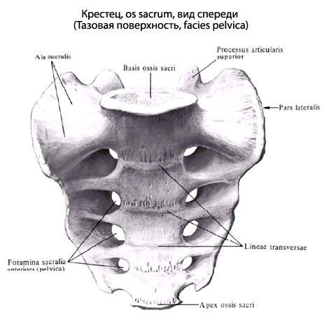
The sacrum (os sacrum) consists of five sacral vertebrae (vertebrae sacrales), which fuse into one bone in adolescence. The sacrum is triangular in shape. It is a massive bone, as it bears the weight of almost the entire body. There is a base of the sacrum, an apex of the sacrum, and two surfaces - the pelvic and dorsal.
The base of the sacrum (basis ossis sacri) is connected with the lower articular processes of the 5th lumbar vertebra by means of articular processes. In the area of the connection of the base with the 5th lumbar vertebra, a rounded angle protrudes forward - a cape (promontorium). On the concave pelvic surface (facies pelvica), facing forward, fourtransverse lines (lineae transversae) are clearly visible, traces of the fusion of the bodies of the sacral vertebrae with each other. On each side at the level of these lines there are pelvic sacral openings (foramina sacralia anteriora, s. pelvica).
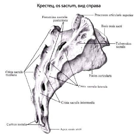
On the convex dorsal surface of the sacrum (facies dorsalis) the dorsal sacral openings (foramina sacralia posteriora, s.dorsalia) are visible on each side. Five longitudinal ridges were formed by the fusion of the processes of the sacral vertebrae. The unpaired median sacral crest (crista sacralis mediana) is the fused spinous processes. The paired intermediate ridge (crista sacralis intermedia) is the result of the fusion of the articular processes, and the paired lateral sacral crest (crista sacralis lateralis) was formed by the fusion of the transverse processes.
On the upper lateral sections of the sacrum are auricular surfaces (facies auriculares) for articulation with the surfaces of the iliac bones of the same name. On each side between the auricular surface and the lateral crest there is a sacral tuberosity (tuberositas sacralis), to which ligaments and muscles are attached. The vertebral openings of the fused sacral vertebrae form the sacral canal (canalis sacralis). This canal ends at the bottom in the sacral fissure (hiatus sacralis). On the sides, the fissure is limited by the sacral horns - a rudiment of the articular processes.
In addition to the joints and the ligaments that strengthen them, the pelvic bones are connected to the sacrum by two powerful extracapsular ligaments. The sacrotuberous ligament(lig. sacrotuberal) goes from the ischial tuberosity to the lateral edge of the sacrum and coccyx. The continuation of the sacrotuberous ligament downwards and forwards onto the branch of the ischium is the falciform process (processus falciformis) of this ligament. Sacrospinous ligament(lig. sacrospinal) connects the sciatic spine with the lateral surface of the sacrum and coccyx.
The sacrum, located between the two pelvic bones, is the "key" of the pelvic ring. The force of gravity of the trunk cannot displace the base of the sacrum forward and downward in the sacroiliac joints, since these joints are firmly strengthened by the interosseous sacroiliac, as well as the sacrotuberous and sacrospinous ligaments.
 [
[ Where does it hurt?
What do need to examine?
How to examine?