
All iLive content is medically reviewed or fact checked to ensure as much factual accuracy as possible.
We have strict sourcing guidelines and only link to reputable media sites, academic research institutions and, whenever possible, medically peer reviewed studies. Note that the numbers in parentheses ([1], [2], etc.) are clickable links to these studies.
If you feel that any of our content is inaccurate, out-of-date, or otherwise questionable, please select it and press Ctrl + Enter.
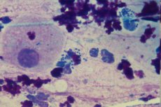
Flora smear results: leukocytes, erythrocytes, epithelium, mucus
Medical expert of the article

Last reviewed: 07.07.2025
A gynecological or urological smear is a study of not only the microflora that lives inside our body, but also other components of the internal environment, which can also carry important information for diagnostics. Before dealing with the specific composition of the flora in the smear, doctors (laboratory technicians) evaluate the presence and quantity of epithelial cells, blood and the immune system, as well as the quality and effectiveness of the processes occurring inside the body.
But let's look at the order of what the various entries and marks in the smear analysis forms for microflora mean. And let's start with leukocytes. As we know from school biology, leukocytes are white blood cells whose main area of activity is to protect the body. Penetrating the wall of blood vessels, they penetrate into infected tissues and begin to fight the infection.
Leukocytes in a smear on flora
They are present almost always, which ensures the health of its microflora. But in the absence of active reproduction of opportunistic microorganisms and the introduction of pathogenic microbes, the number of leukocytes is limited. Thus, in a normal smear in men, up to 5 units of leukocytes are found in the field of vision. In women, the number of leukocytes can vary depending on the area from which the smear is taken. The norm of leukocytes in a smear for flora taken from the urethra and vagina is from 0 to 10 in the field of vision. The analysis of the cervical canal of the uterus should show no more than 30 leukocytes in the field of vision.
The above number of leukocytes is not evidence of inflammation. These are normal values that are present in healthy men and women. An inflammatory process is indicated if the number of leukocytes is increased (leukocytosis). But it is important to understand that the number of leukocytes in the blood is not constant, it increases after eating, mental or physical overexertion, and simply in the evening, it is not surprising that blood donation for analysis involves the exclusion of the above factors.
However, when considering the issue of smear analysis, we are not talking about the total number of leukocytes in the blood, but only about those white cells that go into the tissues to fight pests. All leukocytes are capable of fighting infection, but among them there is a group of cells that reduces the number of bacteria by absorbing and digesting them. Such cells are called phagocytes, and the process of fighting pathogens is called phagocytosis.
Thus, phagocytosis in a smear on flora can be considered as a positive process of natural fight against infection, which is regulated by the immune system. That is, if local immunity cannot prevent the development of infection (for example, when skin or mucous tissue is damaged), phagocytes come into play. Absorbing bacterial cells, phagocytes increase in size and, eventually, are destroyed with the release of substances that provoke a local inflammatory reaction. That is, inflammation is provoked not only by the products of bacterial activity, but also by dying protective cells.
Inflammation is accompanied by hyperemia, edema, and an increase in temperature in the inflammatory focus, which is also carried out with the aim of destroying microbes and attracting other leukocytes to the focus. The pus secreted in the inflammation foci contains the "bodies" of leukocytes that died while performing their responsible function.
In phagocytosis, a smear analysis will show a large number of both active and dead leukocytes, which indicates an intensive fight of the body against the infection, but at the same time confirms the fact of the presence of an active infection. And, despite all the positive aspects of this process, there are situations when phagocytosis remains incomplete, i.e. not all bacteria or viruses are destroyed. Some remain undigested inside the phagocytes due to the weakening of these cells, while others, equipped with special protection from recognition, penetrate inside the cell and lead a hidden life. In this case, the inflammatory process becomes chronic or sluggish, gradually weakening the body and creating optimal conditions for the reproduction of other pathogens.
If phagocytosis is detected in the smear, doctors must determine its cause and, if necessary, provide medicinal assistance to the immune system so that the phagocytosis process is completed.
 [ 1 ], [ 2 ], [ 3 ], [ 4 ], [ 5 ]
Erythrocytes in a smear on flora
In addition to leukocytes, our blood also contains red blood cells, the number of which is much greater than white. These cells are not able to penetrate the walls of blood vessels, so normally the internal environment of the vagina or urethra should not contain erythrocytes. Erythrocytes in a smear on flora indicate damage to the walls of organs and the capillaries in them.
Individual red blood cells (1-3 cells) can be found in a smear taken from a woman on the eve of her period or shortly after it ends, when the vagina has not yet had time to completely cleanse itself. If the smear is taken immediately after the end of menstruation, the number of red blood cells can be 25-30 units or more, which prevents us from recreating a true picture of the internal environment.
The number of red blood cells in a smear taken in the middle of the cycle can change for several reasons:
- injury to the mucous membrane during a gynecological examination,
- recent injuries during hygiene procedures or sexual intercourse (which is why it is recommended to refrain from vaginal sex the day before taking a smear),
- injuries following the introduction of foreign bodies into the vagina, such as the installation of an intrauterine device, non-traditional methods of achieving sexual satisfaction, surgical interventions,
- hormonal disorders accompanied by spotting or fresh blood,
- erosions on the walls of the internal genital organs (for example, on the cervix),
- tumor diseases,
- an active inflammatory process in which microdamages are always detected in edematous tissues.
Erythrocytes in a smear from the urethra can also be detected in cases of urolithiasis and renal stone disease, when microdamage to the walls of organs is caused by hard crystals of urinary sediment, as well as in tumor processes.
In men, a small number of red blood cells in a urological smear may be due to trauma to the urethra during the collection of biomaterial. However, inflammatory pathologies caused by trauma, infection, allergic reaction, and tumor processes cannot be ruled out. In these cases, a noticeable increase in the level of leukocytes is striking.
As we can see, there are many reasons for the appearance of blood in a smear, and the doctor's task is to determine the one that caused the increase in red blood cells in the smear. The appearance of red blood cells in significant quantities indicates a hemorrhage of varying intensity. If we are talking about an infection, there will not be as many red blood cells as in trauma, but the level of white blood cells released to fight pathogens will be significantly increased. That is, the decisive role in diagnostics is played not by the number of red blood cells, but by the ratio of white and red blood cells in the biomaterial.
Epithelium in a smear for flora
This is also not a pathology, but an indicator of the condition of the vagina, which is regularly cleaned naturally. Flat epithelium is the surface layer of cells in the vagina or uterus. Its cells are renewed every 5-7 days, with old, dead cells exfoliating and excreted as part of normal discharge in women (3-15 cells). So, the detection of single particles of epithelium in a smear indicates good health of the female reproductive system. At the same time, doctors take into account the fact that in different phases of the menstrual cycle, the number of flat cells will be different.
As we can see, the requirements for preparation and timing of the analysis are not a simple whim of doctors, but conditions that determine the value of the analysis and the truth of its results.
But let's return to situations when the squamous epithelium in the smear is more or less than normal. In women of reproductive age, squamous epithelium is always present in the smear, and if it is not detected, we are talking about hormonal disorders, when metabolic processes in the epithelium occur irregularly and are not accompanied by the separation of dead, keratinized particles. In this case, there is a thinning or, conversely, thickening (colpohyperplasia) of the mucous membrane, which occurs with a deficiency of the female hormone estrogen.
If the analysis of the flora in the smear shows a deviation in the amount of flat epithelium towards its increase, this usually indicates inflammatory pathologies and their consequences. The fact is that during the inflammatory process, metabolic processes in the tissues change. Under the influence of infection and its metabolic products, many superficial cells of the mucous die and peel off from the surface, so they are easily removed during the smear. And inflammation of the vagina (vaginitis) in the overwhelming majority of cases is associated with an infection, so doctors primarily mean an infectious process, especially if an increase in the number of leukocytes is also noted. If key cells are detected in the smear, we are talking about infectious vaginitis caused by gardnerella.
Another reason for increased separation of epithelial cells is considered to be a disease such as leukoplakia, characterized by the appearance of individual keratinized foci on the mucous membrane. Leukoplakia is considered a precancerous condition, so the disease should not be ignored under any circumstances.
The true causes of this serious disease are unknown to doctors, however, there are a number of factors that contribute to the development of mucosal pathology: traumatic injuries, chronic inflammatory processes, atrophy of vaginal cells, hormonal abnormalities, vitamin A deficiency, heredity, etc.
In men, flat epithelium should be detected in quantities not exceeding 10 cells in the field of view, otherwise we are again talking about an inflammatory disease of the urethra (its nature is judged by the number of leukocytes) or leukoplakia.
In the vagina there is only flat epithelium, but the appearance of a large amount of cylindrical epithelium in a smear on the flora indicates deeper problems. The fact is that this type of epithelium, which borders on flat, is present in the cervical canal of the uterus.
It is clear that a woman's uterus, like her vagina, is regularly cleaned and renewed naturally, so the columnar epithelium is part of normal female discharge. But normally we are talking about the same 3-15 cells. If there are fewer, one can suspect a hormonal imbalance or abnormal changes, such as ectopia (or erosion - replacement of flat epithelium with columnar epithelium, which is easily damaged by the acidic environment of the vagina) or cervical dysplasia (changes in the structure of the epithelium and the appearance of atypical cells in it), which are considered precancerous conditions.
But an increase in the number of cylindrical cells in a smear most likely indicates inflammation in the uterus and cervical canal (less often in the vagina or urethra), but may also be evidence of uterine oncology, so additional studies are required, in particular a biopsy and histological examination of the biopsy. Other reasons for a shift in the level of cylindrical cells in a smear may be: a sharp increase in estrogen production, which is accompanied by the development of endometriosis, damage to the cervix during surgery.
During menopause, a similar situation is observed with benign mastopathy, which once again confirms the regulation of the mammary glands and reproductive organs by the same hormones.
[ 6 ], [ 7 ], [ 8 ], [ 9 ], [ 10 ], [ 11 ], [ 12 ], [ 13 ]
Mucus in a smear for flora
It is a normal component of vaginal discharge in women of reproductive age. It is not produced until the age of 12-14, and after menopause its amount decreases significantly. Mucus is secreted in the cervical canal and performs a protective function, helping to cleanse the uterus and vagina, protecting them from infection and moisturizing the mucous membrane.
Normally, mucus in women and girls who have reached puberty is secreted in an amount of no more than 4 ml per day. It has a viscous consistency, is translucent with a whitish tint, and has no odor. But depending on the physiological state of the woman, the amount of mucus can change. Most of it is in the first half of the menstrual cycle (especially during ovulation), and the minimum is recorded before menstruation, which should also be taken into account when deciphering the results of the smear.
Normally, mucus is detected in analyses of vaginal and cervical discharge, which is defined as a moderate amount. In smears from the urethra, if it is detected, then in insignificant quantities, but ideally it should not be there.
Mucus in the female reproductive system has a viscous consistency, so in a smear on the flora, or rather, its decoding, you can see the item "mucus strands". You should not be afraid of this expression, because it is not the presence of mucus that is indicative, but its quantity, which, moreover, can vary even in a healthy woman.
A large amount of mucus secreted most often indicates vaginal dysbacteriosis and an inflammatory process in the reproductive system. Urethral mucus is evidence of inflammation in the urinary system, which is possible in both men and women.
Decoding the analysis of a smear on flora may contain other points that are unclear to many women. For example, fibrin in a smear on flora is an insoluble protein that is usually present in the inflammation focus. But judging whether there is inflammation or not is necessary only in combination with studying the number and behavior of leukocytes. If single leukocytes are detected, then the detection of fibrin is not associated with inflammation, but with the peculiarities of taking the smear. In this case, the woman has nothing to worry about.
Detritus in a smear on flora is a substrate consisting of exfoliated cells of the mucous membrane and dead bacteria. Considering that cell renewal occurs regularly, and the composition of normal microflora contains various bacteria, both beneficial and opportunistic, the presence of detritus in vaginal discharge is quite understandable. Another matter is its quantity, which can change with various pathologies.
The vaginal microflora is famous for the diversity of life forms inhabiting it, which means that studying detritus gives doctors information about the flora contained in a smear, and therefore in a woman's vagina. The volume of detritus is a more or less constant value, so its increase can be considered as a deviation in the health of a representative of the fair sex. Most often, we are talking about inflammation of the vagina (vaginitis), but infectious and inflammatory processes of other localization should not be excluded: urethritis (inflammation of the urethra), cervicitis (inflammation in the cervical canal of the cervix) and endometritis (with localization of inflammation in the cavity of the organ), adnexitis (inflammation of the appendages). If the level of leukocytes is not increased or increased slightly, perhaps the cause lies in vaginal dysbacteriosis.
But the expression "cytolysis in a smear on flora" means a violation of the balance of lactobacilli and always means pathology. As we know, lactobacilli are the main mass of bacteria in the normal microflora of the female vagina. We are talking about 95-98% of the total number of bacteria.
Most often, smears show a decrease in the level of lactobacilli, but sometimes they begin to behave inappropriately, actively multiply, and the balance of microflora shifts towards an increase in lactobacilli. It would seem that there is nothing to worry about, because thanks to these beneficial bacteria, optimal acidity of the vagina is maintained, preventing pathogens penetrating there from multiplying.
But the optimal acidity is the one that does not destroy the body's own cells. But with an increase in the number of lactobacilli, the production of lactic acid also increases, which has an irritating effect on the delicate vaginal mucosa, which is accompanied by itching and burning. Irritation and destruction of the vaginal mucosa is called cytolysis, and the pathology is called cytolytic vaginosis.
Often this disease is combined with vaginal candidiasis, because a violation of the microflora is always accompanied by a struggle for territory of various microorganisms, and in this regard, the advantage remains with fungi, which get along well with lactobacilli.
The causes of cytolysis are still a mystery to scientists, although it has been possible to trace the relationship between the increase in the number of lactobacilli and the high level of glycogen in the second (luteal) phase of the menstrual cycle. This is explained by the fact that glycogen is a nutrient medium for lactobacilli, ensuring their activity and reproduction.
As we can see, even an experienced doctor cannot easily interpret the results of a urogenital smear. The diversity of flora in the smear and its relationship with various processes occurring in the body allow one to obtain only initial information, which, through analysis and comparison with the patient's medical history, complaints about health, and the results of instrumental studies, is transformed into a diagnosis.
It is very difficult for a non-specialist, even with a medical education, to judge possible disorders in the body or their absence based on the smear test. And what can we say about people who are far from medicine. Unfamiliar words and designations can be frightening, increasing the level of stress, which negatively affects the state of the immune system. Worrying about the test result, trying to find non-existent diseases in ourselves and not going to the doctor, we only prepare the ground for possible health problems, because a weakened immune system will no longer be able to resist infections. In such conditions, it is possible that a repeat test will indeed indicate a pathology.
On the other hand, timely visit to the doctor and detection of pathology at an early stage allows you to restore health faster and with fewer losses. And if the test result is normal, then you can save your nerves (and along with them, your immunity), which will be a good prevention of infectious diseases.