All iLive content is medically reviewed or fact checked to ensure as much factual accuracy as possible.
We have strict sourcing guidelines and only link to reputable media sites, academic research institutions and, whenever possible, medically peer reviewed studies. Note that the numbers in parentheses ([1], [2], etc.) are clickable links to these studies.
If you feel that any of our content is inaccurate, out-of-date, or otherwise questionable, please select it and press Ctrl + Enter.
Picornaviruses
Medical expert of the article
Last reviewed: 06.07.2025

Picornaviruses (picornaviridae, from Spanish pica - small) are a family of non-enveloped viruses containing single-stranded plus RNA.
The family has more than 230 representatives and consists of 9 genera: Enterovirus (11 serotypes), Rhinoviras (105 serotypes). Aphtovirus (7 serotypes), Heputoviras (2 serotypes: 1 human, 1 monkey), Cardiovirus (2 serotypes); Parechovinis, Erbovirus, Kobuvirus are the names of new genera. Genera consist of species, species - of serotypes. All these viruses are capable of infecting vertebrates.
Structure of picornaviruses
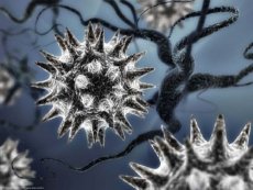
Picornaviruses are small, simply organized viruses. The virus diameter is about 30 nm. The virion consists of an icosahedral capsid surrounding infectious single-stranded plus RNA with the VPg protein. The capsid consists of 12 pentagons (pentamers), each of which, in turn, consists of 5 protein subunits - protomers. Protomers are formed by 4 viral polypeptides: VP1, VP2, VP3, VP4. Proteins VP1, VP2 and VP3 are located on the surface of the virion, and VP4 is inside the viral particle.
Reproduction of picornaviruses
The virus interacts with receptors on the cell surface. With the help of these receptors, the viral genome is transferred to the cytoplasm, accompanied by the loss of VP4 and the release of viral RNA from the protein shell. The viral genome can enter the cell by endocytosis with the subsequent release of nucleic acid from the vacuole or by injection of RNA through the cytoplasmic membrane of the cell. At the end of the RNA there is a viral protein - VPg. The genome is used, like RNA, for protein synthesis. One large polyprotein is translated from the viral genome. The polyprotein is then split into individual viral proteins, including RNA-dependent polymerase, which synthesizes the minus-strand matrix from the surface.
Structural proteins are assembled into a casing; the genome is included in it, forming a virion. The time required for the complete reproduction cycle - from infection to the end of virus assembly - is usually 5-10 hours. Their value depends on such factors as pH, temperature, type of virus and host cell, metabolic state of the cell, number of particles that have infected one cell. Virions are released from the cell by its lysis. Reproduction occurs in the cytoplasm of cells. In culture under an agar coating, viruses form plaques.


 [
[