
All iLive content is medically reviewed or fact checked to ensure as much factual accuracy as possible.
We have strict sourcing guidelines and only link to reputable media sites, academic research institutions and, whenever possible, medically peer reviewed studies. Note that the numbers in parentheses ([1], [2], etc.) are clickable links to these studies.
If you feel that any of our content is inaccurate, out-of-date, or otherwise questionable, please select it and press Ctrl + Enter.
Pericoronaritis
Medical expert of the article

Last reviewed: 12.07.2025
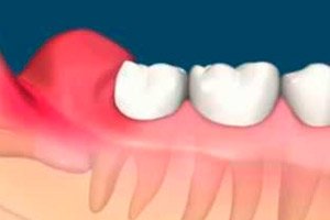
An odontogenic inflammatory disease, pericoronitis, occurs during teething. It mainly concerns third molars, which are the last to erupt – after 17 years of age, and many people experience various complications. Pericoronitis – inflammation and infection of the soft tissues around a partially erupted tooth – is often associated with affected third permanent molars. Other associated conditions include dental caries, root resorption of the adjacent tooth, and rarely the formation of cysts and tumors.
Epidemiology
The prevalence of pericoronitis in people aged 20-29, according to statistics from European dental surgeons, reaches 80%, and 67% of patients with this disease have deep infectious lesions of the periodontal tissues with spread to the cervical lymph nodes and even the paranasal sinuses. [ 1 ]
At least one wisdom tooth that has not erupted or has erupted partially is detected by dentists in 90% of 20-year-old patients. [ 2 ] By the way, in almost 2% of people, third molars do not erupt at all. Women (62.7%) suffer from pericoronitis more often than men (37.3%). [ 3 ], [ 4 ]
About 40% of all extracted teeth are wisdom teeth, the eruption of which led to pericoronitis.
Causes pericoronaryitis
The main causes of inflammation of the gum tissue around the crown of a partially erupted wisdom tooth – pericoronitis or pericoronitis – are an infection, the focus of which is formed in the pericoronary space during tooth retention (delayed eruption), its dystopia – when it is completely or partially covered by the mucous tissue of the gum (the so-called hood – dental operculum), and also if the tooth germ is initially incorrectly located inside the gum and cannot take its place in the dental row.
Pericoronitis of the wisdom tooth usually develops in adults with the eruption of the third molars of the lower jaw, which can grow at an acute and right angle to the second molar and other teeth, in the direction of the cheek or the back of the mouth. Very often, there is occlusion of the tissues surrounding the third lower molar by the chewing teeth of the upper jaw.
It is clear that pericoronitis in children cannot be associated with wisdom teeth, and, as clinical practice shows, inflammation of the tissues surrounding any erupting tooth in a child is a fairly rare phenomenon. Most often (about 36% of cases), inflammation accompanies the eruption of second lower permanent molars in children after 10-11 years.
Risk factors
The main risk factors are poor oral hygiene and difficulty cleaning partially erupted teeth. This leads to plaque, food debris and bacteria accumulating under the gum hood covering the tooth, creating conditions for the development of painful inflammation.
Predisposing factors include abnormalities in dental development, as well as the presence of acute or chronic upper respiratory tract infections, which, according to some data, are present in more than 40% of cases of pericoronitis. [ 5 ]
Pathogenesis
In all cases, the pathogenesis of inflammation of the gum tissue around the crown of an erupting tooth is caused by microbial flora, predominantly anaerobic, which develops in the distally located pericoronal space - an ideal place for the active growth and reproduction of bacteria. [ 6 ]
As a rule, the following bacteria (including obligate ones) are directly related to the inflammatory process and alteration of periodontal tissues: Prevotella melaninogenica, Capnocytophaga spp., Peptostreptococcus spp., Veillonella spp., Fusobacterium mucleatum, Streptococcus mitis. Bacteroides oralis, Propionibacterium spp., Actinomycetales odontolyticus and Actinomycetales pyogenes. [ 7 ], [ 8 ]
In this case, the morphological picture of pericoronitis does not depend on the type of infection, but on the characteristics of the inflammatory process, which can be either superficial (catarrhal) or deeper (involving soft tissues) - purulent, as well as ulcerative (with erosion of the mucous membrane).
Symptoms pericoronaryitis
The symptoms of periocoronitis do not all appear at once. The first signs are the development of inflammatory edema and the appearance of painful sensations in the jaw, which intensify quite quickly and can spread to the ear area and submandibular zone.
The spectrum of symptoms may vary from mild, aching to sharp or throbbing pain, redness, swelling, purulent discharge, limited mouth opening, fever, lymphadenopathy, halitosis, pharyngeal lesions and systemic toxemia.[ 9 ]
According to a study conducted by Jirapun and Aurasa, the symptoms associated with pericoronitis were classified as pain, 35.3%; swelling, 21.7%; discomfort with eating, 3.6%; purulent discharge, 3.0%; and other symptoms, 1.3% (such as trismus, sore throat, and lymphadenitis).
The swelling leads to partial blocking of the temporomandibular joint, causing difficulty in fully opening the mouth (trismus) and pain when chewing.
Inflammation can be acute, subacute and chronic; in many cases, the inflammatory process is accompanied by the formation of purulent-necrotic exudate secreted from under the mucous membrane covering the crown of the tooth, and this is purulent pericoronitis.
Acute pericoronitis is manifested by redness and swelling of the tissues surrounding the tooth, as well as the palate and part of the pharynx); fever; acute pulsating pain (increasing with chewing); dysphagia (difficulty swallowing). Acute purulent pericoronaritis is characterized by severe hyperthermia; bleeding of the mucous membrane of the affected area; halitosis (putrid odor from the mouth) and discharge of pus from the pericoronary sac; spread of pain to the entire jaw and pharynx. There may be enlargement and inflammation of the cervical lymph nodes.
Subacute pericoronitis differs from the acute form by the absence of trismus and more localized pain.
Chronic pericoronitis causes localized tissue edema and hyperemia; periodic dull (aching) pain; maceration of the buccal mucosa closest to the erupting tooth; halitosis and an unpleasant taste in the mouth; soreness of the submandibular lymph nodes (when palpated).
Complications and consequences
It is important to know that pericoronitis can cause serious consequences and complications, including:
- tonsillitis; [ 10 ]
- peripharyngeal abscess;
- peritonsillar abscess;
- gum flux;
- regional lymphadenopathy (inflammation of the submandibular and cervical lymph nodes);
- phlegmon of the retropharyngeal space and the floor of the oral cavity (Ludwig's angina);
- periodontal inflammation;
- spread of inflammation to the periosteum of the gum with the development of periostitis.
Diagnostics pericoronaryitis
For dentists, diagnosing periocoronitis is not difficult when examining the oral cavity: teeth and gums.
And to visualize unerupted teeth and determine treatment tactics, instrumental diagnostics are carried out: X-ray or orthopantomography to obtain an orthopantomogram - a panoramic image of all teeth and periodontal structures.
Differential diagnosis
Differential diagnostics helps to clarify the diagnosis in cases where the patient has a follicular cyst or exostosis of the jaw, a tumor of the gum or salivary gland.
Who to contact?
Treatment pericoronaryitis
The results of treatment of patients with pericoronitis depend on the form of the disease and the method of treatment. [ 11 ] Treatment of pericoronitis includes cleansing the pericoronary space, drainage of pus, drying the affected area, treatment with antiseptics, photodynamic therapy with methylene blue. [ 12 ]
To relieve inflammation, β-lactam antibiotics (Amoxicillin, Clavamitin, etc.) or Metronidazole are prescribed; NSAIDs, such as Ketonal or Ibuprofen, help with pain and inflammation.
The results of antibiotic susceptibility testing show that amoxicillin and pristinamycin are the most effective drugs against the strains tested and, in particular, against strains classified as aerobic. Metronidazole alone or in combination with spiramycin, amoxicillin at a dose of 4 mg/liter and pristinamycin are the most effective drugs against obligate anaerobic bacteria. The effectiveness of the latter drug confirms its value in acute cases and after failure of other antibiotics. [ 13 ], [ 14 ]
Dentists take into account not only the degree of inflammation and the severity of the infection, but also the position of the erupting tooth. And after the acute phase of the inflammatory process is over, one of the dental surgical procedures is performed. If the position of the tooth is normal, then to release its crown and complete eruption, excision of the pericoronitis is necessary, that is, operculectomy (regular or laser), in which a flap of the mucous tissue of the gum over the partially erupted tooth is removed.
Pericoronarotomy (pericoronarectomy) is also performed – excision of the hood in pericoronitis with antiseptic treatment of the wound and its drainage. In both cases, broad-spectrum antibiotics are prescribed in the postoperative period.
And when the position of the tooth is abnormal, they resort to extraction – removal of the wisdom tooth. [ 15 ]
Treatment of pericoronitis at home is carried out by rinsing the mouth with a warm solution of table salt, a decoction of sage, oak bark, peppermint, chamomile flowers, ginger root, as well as a solution with the addition of a few drops of 10% alcohol tincture of propolis. [ 16 ]
Prevention
Thorough cleaning of teeth and adherence to oral hygiene rules, as well as timely seeking medical help are key factors in preventing dental inflammatory diseases. [ 17 ]
Forecast
Pericoronitis is curable, but the prognosis regarding the duration of its treatment largely depends on the severity of the infectious inflammation and the state of the patient's immune system.
With minor inflammation and proper treatment, it may take several days or a week for it to completely stop. In severe cases or if complications of pericoronitis develop, recovery may take longer and require additional therapy.