
All iLive content is medically reviewed or fact checked to ensure as much factual accuracy as possible.
We have strict sourcing guidelines and only link to reputable media sites, academic research institutions and, whenever possible, medically peer reviewed studies. Note that the numbers in parentheses ([1], [2], etc.) are clickable links to these studies.
If you feel that any of our content is inaccurate, out-of-date, or otherwise questionable, please select it and press Ctrl + Enter.
Osteoarthritis: how are synovial joints organized?
Medical expert of the article

Last reviewed: 04.07.2025
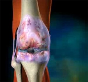 ">
">Osteoarthritis is a disease of the synovial joints (diarthroses). The main functions of diarthroses are motor (movement of the elements that make up the joint along certain axes) and support (load when standing, walking, jumping). The synovial joint consists of articulating bone surfaces covered with cartilage, a joint cavity containing synovial fluid, and a joint capsule. The inconstant anatomical elements of diarthrosis are ligaments located outside or, less commonly, inside the joint, and cartilaginous menisci.
According to the shape of the articulating bone surfaces, diarthroses are divided into the following types:
- flat joints (eg, some carpal and tarsal joints);
- ball-and-socket joints, in which one articular end is shaped like a ball or part of a ball, and the other is a concave surface congruent with the spherical articulating end; an example of a ball-and-socket joint is the shoulder joint, in which great freedom of movement of all kinds is possible - flexion, extension, abduction and adduction, circular movements;
- ellipsoid joints, in which one of the articulating ends has the form of an ellipse, and the other has the form of a congruent cavity; as a result of this anatomical structure, the range of motion in these joints is limited compared to spherical joints and, for example, circular movements are impossible in them; a distinction is made between simple ellipsoid joints and complex ones with several pairs of articular joints (for example, wrist joints);
- block joints, in which one articular end is shaped like a block, resembling a spool, bobbin, and the other concave articular end embraces part of the block and matches it in shape; a typical block joint is the interphalangeal joint of the hand and foot; movements in such joints can only be performed in one plane - flexion and extension; the elbow joint also belongs to block joints - it consists of three joints - the humeroulobronchial, humeroradial and proximal radioulnar, as a result of which in this complex joint, in addition to flexion and extension, supination and pronation are possible, i.e. rotational movements;
- rotational (wheel-shaped) joints, an example of which is the median atlantoaxial joint, consisting of a ring formed by the anterior arch of the atlas and the transverse ligament, and the odontoid process of the second cervical vertebra, which is included in the ring and serves as a kind of axis around which the ring of the atlas rotates; in the elbow joint, the radioulnar articulation should also be classified as a rotational type of joint, since the head of the radius rotates in the annular ligament, which encircles the head of the radius and is attached to the ulnar notch;
- saddle joints, an example of such joints is the carpometacarpal joint of the thumb; the trapezoid bone has an articulated surface in the form of a saddle, and the first metacarpal bone has a concave saddle shape; this anatomical structure allows for circular movements in the sagittal and frontal planes, circular movements along the axis are impossible in this joint;
- condylar joints, the anatomical feature of which is paired condyles - convex and concave, in which concomitant movements are possible; an example of a condylar joint is the knee, consisting of three components that form a single biomechanical system - the patellofemoral and internal and external tibiofemoral articulations; imperfect congruence of the condyles of the tibia is compensated for by the external and internal meniscus; powerful lateral ligaments prevent lateral and swinging movements of the tibia around the femur, and also protect the tibia from subluxation forward and backward during joint movements; flexion and extension, external and internal rotation in a semi-flexed position of the joint are possible in this condylar joint; during flexion-extension movements, the condyles of the femur rotate in relation to the condyles of the tibia and their simultaneous sliding occurs due to the movement of the axes of rotation; Thus, the knee joint is multiaxial or polycentric; during full extension, the lateral ligaments and tendons woven into the joint capsule are maximally tense, which creates conditions for the greatest stability and support capacity of the joint in this position.
The joint is surrounded by a fibrous capsule that attaches to the bone near the periphery of the articular cartilage and passes into the periosteum. The capsule of the synovial joint consists of two layers - the outer fibrous layer and the inner synovial layer. The fibrous layer consists of dense fibrous tissue, in some places the fibrous layer of the capsule becomes thinner with the formation of folds or bursae, in other places it is thickened, performing the function of a joint ligament. The thickness of the fibrous layer of the capsule is determined by the functional load on the joint.
The thickenings of the capsule form ligaments consisting of dense parallel bundles of collagen fibers that serve to stabilize and strengthen the joint and limit certain movements. Among the features of the capsule, in addition to its function as a support for the synovial membrane and connection with the ligaments, it should be noted that it contains a large number of nerve endings, unlike synovium, which has an insignificant number of such endings, and articular cartilage, which does not contain them at all. It is believed that, together with the nerves of the muscles, the nerves of the capsule participate in the control of position and also respond to pain.
The synovial membrane is the smallest in mass and volume, but the most important component of the synovial joint, since most rheumatic diseases occur with inflammation of the synovial membrane, which is generally called "synovitis". The synovial membrane lines all intra-articular structures except for the articular cartilage, its thickness is 25-35 μm. Histologically, it is a layer of connective tissue consisting of integumentary, collagenous and elastic layers. The synovial membrane normally has a certain number of folds and finger-like villi and forms a thin synovial layer (sometimes called the integumentary layer); it includes a layer of integumentary cells that form the lining of the non-articulated surfaces of the joint, and a subsynovial supporting layer consisting of fibrous-fatty connective tissue of varying thickness, which is connected to the capsule. The synovial layer often fuses with the subsynovial tissue by a smooth transition from an avascular inner lining containing many cells to a vascularized subsynovial connective tissue with fewer cells, which becomes increasingly saturated with collagen fibers as it approaches its junction with the fibrous capsule. Cells and nutrients exit the blood vessels of the subsynovial connective tissue into the synovial fluid due to the absence of morphological separation of the synovial and subsynovial layers (absence of a basement membrane, presence of spaces between the integumentary cells).
The synovial membrane is normally lined with 1-3 layers of synovocytes - synovial cells located in a matrix (ground substance) rich in microfibrils and proteoglycan aggregates. Synovocytes are divided into two groups - type A (macrophage-like) and type B (fibroblast-like). Type A synovocytes have an uneven cellular surface with a large number of outgrowths, they have a well-developed Golgi complex, many vacuoles and vesicles, but the ribosomal endoplasmic reticulum is poorly expressed. Macrophage synovocytes can also contain a large amount of phagocytized material. Type B synovocytes have a relatively smooth surface, a well-developed ribosomal endoplasmic reticulum, they contain only a small number of vacuoles. The classical division of synovocytes into A-cells, which perform a phagocytic function, and B-cells, whose main function is to produce components of the synovial fluid, primarily hyaluronic acid, does not reflect all the functions of synovocytes. Thus, synovocytes of type C have been described, which, according to their ultrastructural features, occupy an intermediate position between cells of type A and B. In addition, it has been established that macrophage-like cells are capable of synthesizing hyaluronic acid, and fibroblast-like cells have the ability to actively phagocytose.
 [
[