
All iLive content is medically reviewed or fact checked to ensure as much factual accuracy as possible.
We have strict sourcing guidelines and only link to reputable media sites, academic research institutions and, whenever possible, medically peer reviewed studies. Note that the numbers in parentheses ([1], [2], etc.) are clickable links to these studies.
If you feel that any of our content is inaccurate, out-of-date, or otherwise questionable, please select it and press Ctrl + Enter.
Open oval window in newborn children and adolescents
Medical expert of the article

Last reviewed: 12.07.2025
Quite often diagnosed pathology is an open oval window in a newborn. The child's cardiovascular system is very weak, but his life requires it to work hard. When the baby cries, pouts or coughs, the arterial pressure in the right atrium increases. To reduce it, the body opens the oval window.
Complete closure of the anomaly occurs before the age of two, but in some cases it is diagnosed at an older age. From a medical point of view, the danger lies not in the hole itself, but in such situations:
- As the child grows, his heart grows, but the valve does not enlarge. Because of this, it is not able to completely close the gap, causing blood to flow between the atria.
- The disorder entails a number of other pathologies of the cardiovascular system. Most often, this is increased pressure in the right atrium and opening of the valve.
The causes of the defect are related to the following factors: premature birth, diabetes in the mother, hereditary predisposition, infectious diseases suffered during pregnancy or severe intoxication. The problem also arises due to the bad habits of the expectant mother: smoking, alcoholism, drug addiction.
A painful condition in a newborn is characterized by the presence of the following symptoms:
- When coughing or crying, a blue color appears around the mouth, which goes away when the baby is calm.
- Extraneous noises are heard in the heart.
- During feeding, the heartbeat increases.
- Poor appetite.
- Insufficient weight gain.
- Slow physical development.
- Unexplained loss of consciousness.
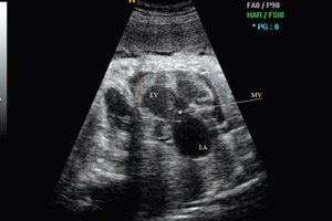
To confirm the diagnosis, the newborn must be examined by a cardiologist. The doctor will conduct an ultrasound of the heart, ECG and transthoracic Doppler echocardiography. These studies allow obtaining a two-dimensional image of the interatrial septum and valve movement, assessing the size of the PFO and excluding the presence of other defects. If the diagnosis is confirmed, then dispensary observation with annual ultrasound of the heart is indicated to assess the dynamics of the pathology.
If there are no hemodynamic disorders, then the treatment consists of general strengthening and health procedures: balanced nutrition, daily walks in the fresh air, hardening of the body. In case of minor deviations from the cardiovascular system, drug treatment and vitamin therapy can be prescribed to support the normal functioning of the heart and the body as a whole.
In rare cases, the atrial septal defect causes complications such as paradoxical embolism, infarction or stroke of the brain, etc. As a rule, this pathology does not cause serious problems up to two years and completely heals by the age of five.
Blood discharge with an open oval window
All newborns have an open oval window of the heart. As the child grows, the valve closes and is overgrown with connective tissue, causing the anomaly to disappear. But sometimes the opening closes partially or does not close at all. In this case, there is a risk of various serious consequences.
Blood flow through an open oval window may be impaired in the following cases:
- The oval opening stretches when the heart cavity is enlarged, but the valve size is insufficient to cover it.
- If the blood pressure in the right atrium is higher than in the left due to stretching of the interatrial septum, that is, valve insufficiency.
The valves open from right to left, causing blood to flow from left to right or from right to left. If the valve functions normally, then with increased blood pressure in the right atrium, a flow occurs from one atrium to the other, but in the opposite direction, that is, from right to left. Because of this, at rest and during normal life activity, the anomaly does not manifest itself.
If the pressure in the right atrium constantly exceeds the left atrium, then constant blood flow from right to left through the open window is possible. This is observed in patients with thrombophlebitis of the lower extremities and pelvic organs. If the defect reaches significant sizes and blood flows from the left atrium to the right, then emergency surgery is required.
Closure of patent foramen ovale
The valve communication between the atria formed during the intrauterine period is the open oval window. It provides blood supply to the brachiocephalic region of the fetus. The closure of the open oval window occurs immediately after birth:
- The pulmonary circulation starts, the lungs begin to function fully, providing gas exchange. Thanks to this, the open communication between the atrium is no longer needed.
- The valve closes because the pressure in the left atrium is higher than in the right. If this does not happen, pathology develops.
Normally, the gap closes completely in the first 2-3 months of life, but in some cases the process can take up to two years, which is also normal. Sometimes the window closes at 4-5 years or does not completely heal. The causes of this condition are related to various factors, such as hereditary predisposition, difficult pregnancy or poor ecology.
Most often, heart defects are detected in premature babies with intrauterine developmental pathologies. The problem is diagnosed using a phonendoscope based on heart noise or using ultrasound. The disorder may be associated with an atrial septal defect, when the valve between the atria does not perform its functions.
If the congenital communication does not cause pathological symptoms, then observation by a family doctor, cardiologist and regular echocardiography are recommended. If the anomaly is detected completely by chance during a routine ultrasound of the heart, then its closure is not performed. Treatment is necessary when pathological symptoms appear and there is a right-left direction of blood flow.
Patent foramen ovale in preterm infants
Premature birth is one of the reasons for incomplete closure of the septum of the heart in children. An open oval window is diagnosed in premature babies much more often than in those born on time. The open opening acts as a communication between the atria. Through it, venous blood participates in the blood circulation without affecting the lungs, which do not function until the moment of birth. Through the gap, the fetus receives oxygen and develops.
The heart has a septum that divides it into atria. In the center of the septum there is a depression, under which there is a passage with a valve that opens towards the left atrium. The diameter of the open slit is about 2 mm. When a baby is born, the window closes with the first breath. But in medicine, its gradual closure during the first year of life is considered normal. In some cases, the condition normalizes by 3-5 years.
The main reasons for the development of heart pathology in premature babies:
- Hereditary predisposition on the maternal line.
- Bad habits of women during pregnancy.
- Underdevelopment of the child's cardiovascular system.
- Unfavorable ecological living environment.
- Poor and unbalanced nutrition during pregnancy.
- Central nervous system disorders.
- Stressful situations and nervous tension.
- Intoxication during pregnancy.
An open gap in the wall between the right and left atrium has a number of characteristic signs: poor weight gain in the child, cyanosis of the perilabial triangle, frequent colds of a bronchopulmonary nature. As the baby grows, shortness of breath and rapid heartbeat appear.
The presence of a functioning window in the heart of a premature baby can cause unwanted complications. So, during the period of active growth, when the heart muscle increases in size, the valve remains the same. This leads to the fact that increased blood flow through the window provokes mixing of blood from different atria. Because of this, the load on the cardiovascular system increases significantly.
In some cases, the disorder is even useful. With the initial signs of pulmonary hypertension, blood from the pulmonary circulation moves into the left atrium through the open window. Against this background, the pressure decreases, which has a beneficial effect on the general condition of the body.
If the window is small and not accompanied by additional pathologies, then drug therapy is not carried out. The baby is registered with a cardiologist and his condition is monitored. If moderate sizes of the hole cause discomfort, then anticoagulants and disaggregants can be prescribed. Large sizes of the defect are accompanied by a decompensated condition, which requires surgical treatment.
Patent foramen ovale in adolescents
Such a pathology as an open oval window in adolescents is most often detected accidentally during an ultrasound examination of the cardiovascular system. All children are born with this anomaly, but as they grow older, the physiological feature is eliminated on its own. If closure does not occur, the disease proceeds in a latent form with rather scanty symptoms. The disorder is characterized by:
- Developmental delay.
- Anxiety and fatigue.
- Dizziness and headaches.
- Reduced stamina.
- Paleness of the skin.
- Blue discoloration of the nasolabial triangle during physical exertion.
- Frequent fainting.
- Shortness of breath.
- Tendency to colds.
To diagnose a disease condition, a set of hardware examinations should be performed. The doctor, usually a cardiologist, collects anamnesis of complaints and signs of the defect, evaluates the general condition of the patient. Particular attention is paid to laboratory and instrumental research methods.
Treatment depends entirely on the nature of the disorder. If there are no significant disturbances in the functioning of the heart, then a set of therapeutic measures is indicated to maintain the normal functioning of the body. In case of severe symptoms, surgical treatment may be required.
There are no methods for preventing PFO. In order to prevent the disease, it is recommended to stick to moderate physical activity, that is, not to overexert the body. It is also necessary to promptly treat all emerging diseases and prevent their complications.
Patent ductus arteriosus and patent foramen ovale
The functional pathological communication between the aorta and the pulmonary trunk is the patent ductus arteriosus. The patent foramen ovale provides embryonic circulation but is obliterated immediately after birth, unlike the ductus.
Patent arterial or Botallo's duct is a non-closure of the accessory vessel that connects the aorta and pulmonary artery. The duct is an important anatomical structure, but after birth and pulmonary breathing, there is no need for it. Normally, it closes in the first 2-8 weeks of life. In cardiology, this defect accounts for about 10% of all congenital heart defects and is most often diagnosed in women.
There are several reasons for the anomaly:
- Premature birth.
- Newborns weighing less than 1750 g.
- Respiratory distress syndrome.
- Asphyxia during childbirth.
- Persistent metabolic acidosis.
- Infectious diseases suffered by the mother in the first trimester of pregnancy.
Stages of patent ductus arteriosus:
- Pulmonary artery pressure is within 40% of arterial pressure.
- I – stage of primary adaptation (first 2-3 years of life).
- II – stage of relative compensation (from 2-3 years to 20 years).
- III – stage of sclerotic changes in the pulmonary vessels.
- Moderate pulmonary hypertension – pressure 40-75%.
- Severe pulmonary hypertension – pressure above 75%, left-to-right blood flow is preserved.
The following symptoms are characteristic of patent ductus arteriosus:
- The child's developmental delay.
- Increased fatigue.
- Pronounced pallor of the skin.
- Rapid and irregular heartbeat.
- Interruptions in cardiac activity.
- Tachypnea.
Diagnosis of the disease condition consists of chest X-ray, ultrasound of the heart, ECG and phonocardiography. In case of high pulmonary hypertension, MRI, aortography and right heart probing are indicated.
To treat this anomaly in premature infants, drug therapy is administered with the introduction of prostaglandin synthesis inhibitors to stimulate duct obliteration. In severe cases, surgical intervention is indicated. Patients undergo open endovascular operations.
Even a small arterial duct is associated with an increased risk of premature death. The problem leads to a decrease in the compensatory reserves of the myocardium and pulmonary vessels. According to statistics, the average life expectancy with the natural course of the duct is about 25 years. At the same time, spontaneous closure of the defect occurs extremely rarely.
Patent foramen ovale and atrial septal defect
Congenital heart defects make up a significant percentage of other diseases. Patent foramen magnum and atrial septal defect are cardiovascular anomalies. According to medical statistics, about 5 children out of 1000 are born with such problems. Moreover, the incidence of pathology is much higher among premature babies.
An atrial septal defect is a congenital anomaly in which there is a hole in the septum between the right and left atrium without a valve. Its presence leads to an increased load on the right ventricle and increased pressure in the pulmonary vessels.
Reasons for violation:
- Genetic factors.
- Diseases suffered during pregnancy: rubella, Coxsackie virus, mumps.
- Diabetes mellitus.
- Mother's bad habits: alcoholism, smoking, drug addiction.
Forms of atrial septal defect:
- Patent oval window.
- Primary defect of the inferior septum.
- Secondary defect of the superior septum.
Symptoms of the disease manifest themselves during the first life of the child. The pathology manifests itself as a bluish tint of the skin at the moment of birth. The tissues may turn pale when crying and restless. The main signs of the defect are:
- The child is lethargic and refuses to play.
- During physical exertion or crying, the heartbeat increases sharply.
- The baby sucks the breast weakly and has a poor appetite.
- Delay in physical development.
- Pronounced pallor of the skin.
To identify the disease state, a comprehensive diagnosis is indicated. Most often, patients are prescribed a course of drug treatment and physiotherapy. In particularly severe cases, surgical treatment is performed.
Atrial septal aneurysm and patent foramen ovale
The complex of congenital defects and anomalies of the heart includes many diseases. Atrial septal aneurysm and patent foramen ovale are components of MARS syndrome. They can be diagnosed in both adults and children. Most often, the disorder is detected in premature infants.
Atrial septal aneurysm (ASA) and PFO are minor cardiac anomalies. Bulging of the arterial wall is a rare defect, it occurs in 1% of children and is usually asymptomatic. The pathology can be isolated, but most often occurs with other anomalies and PFO. Because of this, the symptoms are very blurred, which complicates the diagnostic process.
- The main causes of myocardial wall protrusion are related to the impact of both external and internal factors. These may include stress, unfavorable environmental conditions of living, or infections suffered during pregnancy.
- In school-age children, the disease is associated with the rapid overgrowth of the open window with fibrous-muscular tissue, which leads to sagging of the thin walls in one direction or another.
- In adults, an aneurysm occurs after a major heart attack.
There are several types of aneurysm, which depend on the deflection of the interatrial septum:
- Sagging into the right atrium.
- Sag into the left atrium.
- An S-shaped bulge affecting both sides.
In this case, the direction of the sagging does not affect the symptoms and severity of the defect. Most often, a right-sided deviation is detected, since the pressure in the left atrium is higher than the right, so the heart wall deviates in the opposite direction.
The main manifestations of the disorder:
- Blueness of the nasolabial triangle.
- Tachycardia.
- Severe shortness of breath during physical activity.
- Increased fatigue.
- Headaches and dizziness.
- Heart rhythm disturbance.
- Pain in the heart.
- Inflammatory diseases of the bronchopulmonary system.
- Increased sweating and nausea.
- Subfebrile fever.
The pathological condition may be accompanied by complications, the main ones being: intracardiac thrombosis, embolism and blockage of other vessels.
Diagnosis of an aneurysm of the interatrial septum consists of ultrasound and Dopplerography. If necessary, echocardiography is performed, as well as a set of laboratory and instrumental studies. A set of drug and physiotherapeutic methods is used for treatment.
Norm of open oval window
There are several conditions in which the presence of a minor cardiac anomaly is physiological. The norm of an open oval window is characteristic of:
- For proper blood circulation during the embryonic period, blood is pumped through a through hole between the atria. After birth, there is no longer a need for it, as the lungs begin to work, so the hole gradually closes. This is a physiological norm. If the window closes before birth, it will cause many serious complications, the most dangerous of which is right ventricular failure.
- The FAO remains unclosed immediately after birth and this is also normal. Immediately after birth, the baby takes the first breath and screams, straightening the lungs. Due to the strong load in the left atrium, the pressure increases, closing the valve. According to medical statistics, about 50% of healthy premature babies have such an anatomical feature up to two years, and sometimes more.
- The residual element of the oval opening of the fetal heart closes gradually. This process occurs due to the valve growing into the fossa. The duration of this phenomenon is individual for each organism. In some cases, it is 3-5 years.
If other concomitant cardiovascular disorders are detected during the body diagnostics, this can also be considered normal, since MARS syndrome is a multifactorial pathology. In particularly severe cases, the holes do not close tightly, then the PFO remains open throughout life.
Read more about methods of treating patent foramen ovale in this article.
 [ 9 ]
[ 9 ]