
All iLive content is medically reviewed or fact checked to ensure as much factual accuracy as possible.
We have strict sourcing guidelines and only link to reputable media sites, academic research institutions and, whenever possible, medically peer reviewed studies. Note that the numbers in parentheses ([1], [2], etc.) are clickable links to these studies.
If you feel that any of our content is inaccurate, out-of-date, or otherwise questionable, please select it and press Ctrl + Enter.
Multifocal electroretinography
Medical expert of the article

Last reviewed: 06.07.2025
 [
[ When is multifocal electroretinography used?
Although most responses in electroretinography originate from the outer layers of the retina (photoreceptors, bipolar cells), multifocal electroretinography is also used to objectively assess ganglion cell function. Part of the response signals comes from ganglion cell fibers located near the optic disc. This component is underestimated in patients with glaucoma. This method does not require pupil dilation. Special systems have been developed to study the amplification, isolation, and mapping of this component of the response.
How does multifocal electroretinography work?
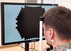
When an electroretinographic signal is received from the cornea through the contact lens of the electrode, all focal zones are independently and simultaneously excited. A special mathematical scheme of multifocal stimulation allows the delivered focal responses to be accurately extracted from a single electroretinographic signal. Patients do not need to answer questions. Using the Visual Evoked Response Imaging System (VERIS; Electro-Diagnostic Imaging, San Mateo, CA), the stimulus can consist of several hundred focal stimuli. Typically, 103 hexagonal areas, imaged on a video monitor, stimulate the central 50° of the patient's visual field. In most cases, focal stimulation consists of a pseudorandom presentation of flashes. Local electrophysiological responses are topographically collected and recorded, forming functional retinal maps similar to visual field maps.
Restrictions
Currently, multifocal electroretinography is used experimentally and is not included in routine clinical examination.
Visual evoked cortical potentials
Visual evoked cortical potentials (VECPs) are electrical signals generated by the visual cortex of the occipital lobe of the brain in response to stimulation of the retina by flashes of light or pattern stimuli. To assess the state of the visual pathways, pattern VECPs are preferred over flash VECPs due to their increased sensitivity in detecting disturbances of axonal conduction.
How Visual Evoked Cortical Potentials Work
The VEP method measures the electrical response of the visual cortex to a pattern or flash stimulus. Visual evoked response potentials are measured between electrodes on the scalp. One electrode, which measures the response itself, is placed above or lateral to the lateral occipital tuberosity (or inion), close to the primary visual cortex. Another electrode is placed at the control point. The final electrode is used for grounding.
When visual evoked cortical potentials are used
Initially, the VVS was used to determine secondary vision loss in diseases of the optic nerve and damage to the anterior visual pathways.
The multifocal method described in the previous section is also used to record cortical responses (multifocal VSEPs). In this case, the stimulus order is usually formed as a "dart" pattern, where each sector contains contrast reversal stimuli in a checkerboard pattern. The difficulty with this method is that local responses are reduced or absent, partly due to the anatomical tortuosity of the cerebral cortex. This method does not always reflect dysfunction. Unilateral local dysfunction is detected by comparing the response maps of the two eyes. Recent studies have found correlations between VSEPs and visual field defects.
Restrictions
Similar to the limitations of multifocal electroretinography, much work needs to be done with multifocal electroretinography before this method can be generally clinically adapted.