
All iLive content is medically reviewed or fact checked to ensure as much factual accuracy as possible.
We have strict sourcing guidelines and only link to reputable media sites, academic research institutions and, whenever possible, medically peer reviewed studies. Note that the numbers in parentheses ([1], [2], etc.) are clickable links to these studies.
If you feel that any of our content is inaccurate, out-of-date, or otherwise questionable, please select it and press Ctrl + Enter.
Lymph nodes
Medical expert of the article

Last reviewed: 07.07.2025
Lymph nodes (nodi lymphatici) are usually located near blood vessels, more often near large veins, usually in groups - from several nodes to ten or more. Taking into account the peculiarities of the position (anatomical and topographic principle), as well as the direction of lymph flow from the organs (the principle of regionality), there are about 150 regional groups of lymph nodes (from the Latin regio - region, area) in the human body. Accordingly, the areas of location are: lumbar lymph nodes (nodi lymphatici lumbales), axillary lymph nodes (nodi lymphatici axillaris), etc. A group of lymph nodes can have the name of the blood vessel next to which it is located: celiac lymph nodes (nodi lymphatici coeliaci), iliac lymph nodes (nodi lymphatici iliaci).
In some areas of the human body, groups of lymph nodes are arranged in two layers, one group above the other. Between these groups there is usually fascia. In such cases, the nodes lying on the fascia are called superficial, and those lying under the fascia are called deep: for example, on the broad fascia of the thigh are located the superficial inguinal lymph nodes (nodi lymphatici inguinales superficiales), and under the fascia are the deep inguinal lymph nodes (nodi lymphatici inguinales profundi).
In the body cavities: thoracic, abdominal, pelvic - the lymph nodes are located near the internal organs and on the walls of the cavities. Given the position of the nodes, the first of them are usually called visceral (visceral) lymph nodes (nodi lymphatici viscerales). These are such groups of lymph nodes as mediastinal, bronchopulmonary, tracheobronchial in the thoracic cavity; pararectal, paravesical, parauterine - in the pelvic cavity. On the walls of the cavities are parietal (wall) lymph nodes (nodi lymphatici parietales). These include the parasternal, intercostal, upper diaphragmatic lymph nodes in the thoracic cavity; lumbar, lower epigastric, lower diaphragmatic - in the abdominal cavity; iliac: common, external and internal lymph nodes - in the pelvic cavity.
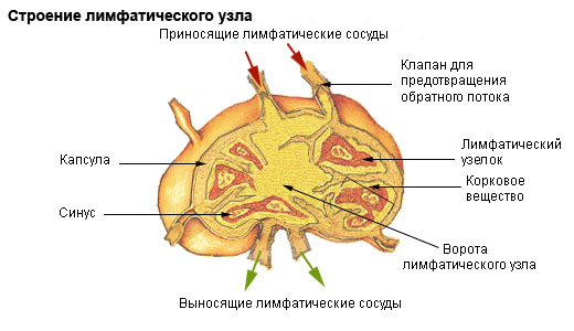
The lymph nodes are pinkish-gray in color, round, ovoid, bean-shaped, and even ribbon-shaped, ranging in size from a pinhead (0.5-1.0 mm) to a large bean (30-50 mm or more in length). Each lymph node is covered by a connective tissue capsule on the outside. Inside the lymph node, there is a connective tissue (reticular) stroma and parenchyma, represented by lymphoid tissue. There is also a system of interconnected channels - lymphatic sinuses, through which lymph flows through the lymph node. Under the capsule is the subcapsular (marginal) sinus, which goes with its ends directly to the gate of the node. From it, intermediate (first cortical, then cerebral) sinuses go into the parenchyma of the lymph node, in the area of the gate of the organ they pass into the portal sinus. The subcapsular sinus also opens into this sinus.
Lymph enters the lymph node through the afferent lymphatic vessels (vdsa afferentia). These vessels, 2-4 in number, approach the convex side of the node, pierce the capsule and flow into the subcapsular (marginal) sinus. Then, through this sinus and the intermediate sinuses, which are located in the parenchyma of the node and communicate with each other, the lymph enters the portal sinus. From the portal sinus, 1-2 efferent lymphatic vessels (vasa efferentia) exit, through which the lymph flows out of the lymph node. In the lumen of the sinuses of the medulla there is a fine-mesh network formed by reticular fibers and reticular cells. When lymph passes through the sinus system of the lymph node, foreign particles that have entered the lymphatic vessels from tissues (microbial bodies, dead and tumor cells, dust particles) are retained in the loops of the network. Lymphocytes enter the lymph from the parenchyma of the lymph node.
Through the efferent lymphatic vessels, lymph from some nodes is directed to the next lymph nodes lying on its path or to the collecting vessels - lymphatic trunks and ducts. In each regional group, the lymph nodes are connected to each other by internodal lymphatic vessels. Through these vessels, lymph flows from one node to another in the direction of the general flow, towards the venous angle. On its way from each organ, lymph passes through at least one lymph node, and more often through several. For example, on the path of lymph flow from the stomach there are 6-8 nodes, from the kidney, lymph passes through 6-10 lymph nodes. Only the esophagus is an exception. From its middle part, some lymphatic vessels flow directly into the nearby thoracic duct, bypassing the lymph nodes. Therefore, in esophageal cancer, tumor cells with lymph enter the thoracic duct and then into the blood, without passing through the lymph nodes. In rare cases, individual lymphatic vessels of the liver also flow directly into the thoracic duct.
What do need to examine?
How to examine?