
All iLive content is medically reviewed or fact checked to ensure as much factual accuracy as possible.
We have strict sourcing guidelines and only link to reputable media sites, academic research institutions and, whenever possible, medically peer reviewed studies. Note that the numbers in parentheses ([1], [2], etc.) are clickable links to these studies.
If you feel that any of our content is inaccurate, out-of-date, or otherwise questionable, please select it and press Ctrl + Enter.
Examination of lymph nodes
Medical expert of the article

Last reviewed: 04.07.2025

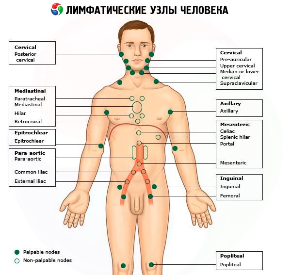
It is generally believed that in a healthy person the lymph nodes are not visible and not accessible to palpation. This rule, which is true in most cases, should be accepted only with certain reservations. Thus, given the wide prevalence of various dental diseases among the population (caries, periodontitis, periodontal disease, etc.), we must take into account the fact that in many people the submandibular lymph nodes can be palpated without much difficulty. In practically healthy people, due to minor, sometimes unnoticeable injuries to the skin of the lower extremities, small (pea-sized) inguinal lymph nodes can be palpated. According to a number of authors, the detection of single small axillary nodes by palpation may also not be a serious diagnostic sign. However, it should be emphasized once again that a more significant increase in lymph nodes, especially in cases where it is detected already during examination, is always a symptom of one or another disease, sometimes a very serious one.
When examining different groups of lymph nodes, the data obtained must be compared with the results of examination and palpation of the same (symmetrical) group of lymph nodes on the other side.
Palpation of the lymph nodes
During palpation, the size of the lymph nodes is determined first of all, which is usually compared with the size of some round objects (the size of “a millet grain”, “a lentil”, “a small (medium, large) pea”, “a hazelnut”, “a pigeon’s egg”, “a walnut”, “a chicken egg”, etc.).
The number of enlarged lymph nodes, their consistency (doughy, soft elastic, dense) are specified; attention is paid to the mobility of the lymph nodes, pain upon palpation (a sign of inflammatory processes), fusion with each other in conglomerates and fusion with surrounding tissues, the presence of edema of the surrounding subcutaneous tissue and hyperemia of the corresponding area of the skin, the formation of fistulas and cicatricial changes (for example, with tuberculous lymphadenitis ). In this case, the lesion may concern individual lymph nodes, their regional group (with inflammation, malignant tumors) or can be systemic, manifested by a generalized increase in the lymph nodes of various groups (for example, with leukemia, lymphogranulomatosis ).
Palpation of the lymph nodes is performed with the tips of slightly bent fingers (usually the second to fifth fingers of both hands), carefully, gently, with light, sliding movements (as if "rolling" over the lymph nodes). In this case, a certain sequence is observed in the examination of the lymph nodes.
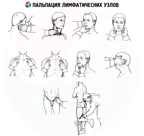
First, palpate the occipital lymph nodes, which are located in the area where the muscles of the head and neck are attached to the occipital bone. Then move on to palpating the postauricular lymph nodes, which are located behind the auricle on the mastoid process of the temporal bone. In the area of the parotid salivary gland, palpate the parotid lymph nodes. The mandibular (submandibular) lymph nodes, which enlarge with various inflammatory processes in the oral cavity, are palpated in the subcutaneous tissue on the body of the lower jaw behind the masticatory muscles (during palpation, these lymph nodes are pressed against the lower jaw). The submental lymph nodes are determined by moving the fingers from back to front near the midline of the chin area.
The superficial cervical lymph nodes are palpated in the lateral and anterior regions of the neck, respectively along the posterior and anterior edges of the sternocleidomastoid muscles. A prolonged increase in the cervical lymph nodes, sometimes reaching significant sizes, is observed in tuberculous lymphadenitis, lymphogranulomatosis. However, in patients with chronic tonsillitis, chains of small dense lymph nodes can often be found along the anterior edges of the sternocleidomastoid muscles.
In gastric cancer, a dense lymph node (the “Virchow gland” or “Virchow-Troisier gland”) can be found in the supraclavicular region (in the triangle between the legs of the sternocleidomastoid muscle and the upper edge of the clavicle), which is a tumor metastasis.
When palpating the axillary lymph nodes, the patient's arms are slightly moved aside. The fingers of the palpating hand are inserted as deeply as possible into the armpit (for hygienic reasons, the patient's T-shirt or shirt is taken into the palpating hand). The patient's moved arm is returned to its original position; the patient should not press it tightly to the body. Palpation of the axillary lymph nodes is performed by moving the palpating fingers in a top-down direction, sliding along the lateral surface of the patient's chest. Enlargement of the axillary lymph nodes is observed in metastases of breast cancer, as well as in any inflammatory processes in the upper limbs.
When palpating the ulnar lymph nodes, grasp the lower third of the forearm of the patient's arm being examined with your own hand and bend it at the elbow joint at a right or obtuse angle. Then, using sliding longitudinal movements, palpate the sulci bicipitales lateralis et medialis just above the epicondyle of the humerus with the index and middle fingers of the other hand (the latter are the medial and lateral grooves formed by the tendon of the biceps muscle).
The inguinal lymph nodes are palpated in the area of the inguinal triangle (fossa inguinalis) in a direction transverse to the inguinal ligament. Enlargement of the inguinal lymph nodes may be observed with various inflammatory processes in the area of the lower extremities, anus, external genitalia. Finally, the popliteal lymph nodes are palpated in the popliteal fossa with the shin slightly bent at the knee joint.
Enlargement of regional lymph nodes, such as in the neck, as well as in other areas, is sometimes the main complaint of patients, leading them to the doctor. In this case, it is rarely possible to see enlarged lymph nodes that deform the corresponding part of the body. The main method of examining lymph nodes is palpation. It is advisable to palpate the lymph nodes in a certain order, starting with the occipital, parotid, submandibular, submental, then supraclavicular, subclavian, axillary, cubital, inguinal.
Enlarged lymph nodes are observed in lymphoproliferative diseases (lymphogranulomatosis), systemic connective tissue diseases, and tumors (metastases). To clarify the cause of enlarged lymph nodes, in addition to general clinical and laboratory studies, a biopsy (or removal) of the node is performed for its morphological examination. The musculoskeletal system (joints, muscles, bones) is examined after the lymph nodes. In this case, the examination begins with identifying complaints, most often pain or limited movement in the joints, then an examination and palpation are performed.