
All iLive content is medically reviewed or fact checked to ensure as much factual accuracy as possible.
We have strict sourcing guidelines and only link to reputable media sites, academic research institutions and, whenever possible, medically peer reviewed studies. Note that the numbers in parentheses ([1], [2], etc.) are clickable links to these studies.
If you feel that any of our content is inaccurate, out-of-date, or otherwise questionable, please select it and press Ctrl + Enter.
Keratoderma: causes, symptoms, diagnosis, treatment
Medical expert of the article

Last reviewed: 07.07.2025
Keratoderma is a group of dermatoses characterized by a disruption of the keratinization process - excessive horny formation mainly on the palms and soles.
The causes and pathogenesis of the disease have not been fully elucidated. Research has established that keratodermas are caused by mutations in the genes encoding keratin 6, 9, 16. Vitamin A deficiency, hormonal dysfunctions, primarily of the sex glands, bacterial and viral infections are of great importance in the pathogenesis. They are one of the symptoms of hereditary diseases and tumors of internal organs (parapsoriatic keratodermas).
Symptoms. A distinction is made between diffuse (Unna-Tost keratoderma, Meleda keratoderma, Papillon-Lefevre keratoderma, mutilating keratoderma and syndromes that include diffuse keratoderma as one of the main symptoms) and focal (disseminated spotted keratoderma of Fischer-Buschke, acrokeratoelastoidosis of Kosti, limited keratoderma of Bruhauer-Franzesthesti, linear keratoderma of Fuchs, etc.) keratoderma.
Winy-Tost keratoderma (synonyms: congenital ichthyosis of the palms and soles, Winy-Tost syndrome) is transmitted in an autosomal dominant manner. There is a diffuse excessive keratinization of the skin of the palms and soles (sometimes only the soles), which develops in the first two years of life. The skin pathological process begins with a slight thickening of the skin of the palms and soles in the form of a strip of erythema of a livid color on the border with healthy skin. Over time, smooth, yellowish horny layers appear on their surface. The lesion rarely spreads to the dorsum of the wrists or fingers. In some patients, superficial or deep cracks may form and local hyperhidrosis is noted. In the patient observed by the author, the uncle on the mother's side, brother and son suffered from Winy-Tost keratoderma.
Cases of damage to nails (thickening), teeth, and hair in Winy-Tost keratoderma in combination with various skeletal anomalies and pathologies of internal organs, nervous and endocrine systems are described.
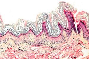
Histopathology. Histological examination reveals marked hyperkeratosis, granulosis, acanthosis, and small inflammatory infiltrates in the upper dermis. Differential diagnosis. The disease must be differentiated from other types of keratoderma.
Meleda keratoderma (synonyms: Meleda disease, congenital progressive acrokeratoma, Siemens' palmoplantar transgradient keratosis, Kogoy's hereditary palmoplantar progressive keratosis) is inherited in an autosomal recessive manner. This form of keratoderma is characterized by thick, yellow-brown horny layers with deep cracks. A violet-purple border several millimeters wide is visible along the edges of the lesion. The process typically spreads to the back of the hands and feet, forearms, and shins. Most patients experience local hyperhidrosis. In this regard, the surface of the palms and soles becomes slightly moist and covered with black dots (sweat gland ducts).
The disease can develop by the age of 15-20. Nails thicken and become deformed.
Histopathology. Histological examination reveals hyperkeratosis, sometimes acanthosis, and a chronic inflammatory infiltrate in the papillary dermis.
Differential diagnosis. Melela keratoderma must be distinguished from Unna-Tost keratoderma.
Keratoderma Papillon-Lefevre (synonym: palmoplantar hyperkeratosis with periodontitis) is inherited in an autosomal recessive manner.
The disease manifests itself in the 2nd-3rd year of life. The clinical picture of the disease is similar to Melela's disease. In addition, changes in the teeth are characteristic (abnormalities in the eruption of milk and permanent teeth with the development of caries, gingivitis, rapidly progressing periodontosis with premature tooth loss).
Histopathology. Histological examination reveals thickening of all layers of the epidermis, especially the horny layer, and insignificant cellular clusters of lymphocytes and histiocytes in the dermis.
Differential diagnosis. The disease should be distinguished from other keratodermas. An important distinguishing feature is the characteristic dental pathology, which is not found in other forms of hereditary diffuse keratodermas.
Keratoderma mutilans (synonyms: Fonwinkel syndrome, hereditary mutilating keratoma) is a type of diffuse keratoderma inherited in an autosomal dominant manner. It develops in the 2nd year of life and is characterized by diffuse horny deposits on the skin of the palms and soles with hyperhidrosis. Over time, cord-like grooves form on the fingers, which leads to contractures and spontaneous amputation of the fingers. Follicular keratosis is expressed on the back of the hands, as well as in the area of the elbow and knee joints. The nail plates are changed (often like watch glasses). Cases of hypogonadism, ruby alopecia, hearing loss, pachyonychia have been described.
Histopathology. Histological examination reveals severe hyperkeratosis, granulosis, acanthosis, and small inflammatory infiltrates in the dermis, consisting of lymphocytes and histiocytes.
Differential diagnosis. When differentiating mutilating keratoderma from other forms of diffuse keratoderma, the mutilation effect, which is not typical for other forms, should be taken into account first of all. When performing differential diagnostics of all forms of diffuse keratoderma, it is necessary to remember that it can be one of the main symptoms of a number of hereditary syndromes.
Treatment. Neotigazone is indicated in the general therapy of keratoderma. The dose of the drug depends on the severity of the process and is 0.3-1 mg/kg of the patient's weight. In the absence of neotigazone, vitamin A is recommended in a dose of 100 to 300,000 mg per day for a long time. External therapy consists of using ointments with aromatic retinoids, keratolytic and steroid agents.
 [
[ What's bothering you?
What do need to examine?
How to examine?