
All iLive content is medically reviewed or fact checked to ensure as much factual accuracy as possible.
We have strict sourcing guidelines and only link to reputable media sites, academic research institutions and, whenever possible, medically peer reviewed studies. Note that the numbers in parentheses ([1], [2], etc.) are clickable links to these studies.
If you feel that any of our content is inaccurate, out-of-date, or otherwise questionable, please select it and press Ctrl + Enter.
Karyotype test
Medical expert of the article

Last reviewed: 04.07.2025
One of the methods of cytogenetic research aimed at studying chromosomes is karyotyping. The analysis has a number of indications for implementation, as well as several types.
The karyotype is a set of human chromosomes. It describes all the features of genes: size, quantity, shape. Normally, the genome consists of 46 chromosomes, 44 of which are autosomal, that is, they are responsible for hereditary traits (hair and eye color, ear shape, etc.). The last pair is the sex chromosomes, which determine the karyotype: women 46XX and men 46XY.
During the diagnostic process, any genomic abnormalities are identified:
- Changes in quantitative composition.
- Violation of structure.
- Violation of quality.
Karyotyping is usually performed on newborns to determine genetic abnormalities. The analysis is also indicated for married couples who are planning a pregnancy. In this case, the study allows identifying a mismatch of genes, which can cause the birth of a child with hereditary pathologies.
Types of molecular karyotyping:
- Targeted
It is prescribed to confirm various anomalies and syndromes. It allows to determine the causes of pregnancy loss: frozen fetus, miscarriage, termination for medical reasons. It determines the etiology of an additional set of chromosomes in triploidy. The analysis is performed on micromatrices with 350 thousand markers concentrated in clinically significant areas of chromosomes. The resolution of this study is from 1 million bp.
- Standard
Detects abnormalities in the genome of clinical significance. Diagnoses microdeletion syndromes and pathologies associated with autosomal dominant diseases. Determines the causes of chromosomal abnormalities in undifferentiated syndromes in patients with developmental abnormalities, congenital defects, delayed psychomotor development, autism.
Allows to detect chromosomal abnormalities in the prenatal period. The method determines aneuploidies, pathological microdeletions in the fetus. The study is conducted on a micromatrix with 750 thousand high-density markers, which cover all significant areas of the genome. The resolution of the standard karyotype analysis is from 200 thousand bp.
- Extended
Allows to establish the causes of chromosomal abnormalities in undifferentiated syndromes in children. Reveals pathogenic deletions, i.e. the disappearance of chromosome sections and duplications – additional copies of genes. Diagnoses sections with loss of heterozygosity, causes of autosomal recessive pathologies.
Expanded chromosome microarray analysis is performed using a high-density microarray, which contains more than 2.6 million individual high-density markers. The resolution of this study allows for coverage of the entire genome and ranges from 50,000 bp to 10,000 bp. This allows all sections of the gene code to be studied with extreme precision, making it possible to identify the smallest structural abnormalities.
As a rule, a karyotype analysis is carried out as prescribed by a geneticist. Depending on the doctor's indications, one of the above types may be prescribed. Standard testing is cheaper, but is prescribed extremely rarely, since it does not reveal many chromosomal abnormalities. Targeted karyotyping is a more expensive analysis, so it is prescribed in the presence of clinical signs of syndromes and other anomalies. Extended diagnostics is the most expensive and most informative, since it allows for a complete study of all 23 sets of chromosomes.
Where can I get a karyotype test?
Chromosomal microarray analysis is done by order of a geneticist. The study is aimed at studying the patient's genome and identifying any anomalies in its structure.
Chromosomes are DNA strands, their number and structure is specific for each species. The human body contains 23 pairs of chromosomes. One pair determines gender: women have 46XX chromosomes, and men have 46XY. The remaining genes are autosomes, i.e. non-sexual.
Features of karyotyping:
- The analysis is carried out once, since the chromosome set does not change throughout life.
- Allows you to determine the causes of reproductive problems in spouses.
- Diagnoses multiple developmental defects in children.
- Detects genetic abnormalities.
The karyotype is taken in a specialized medical laboratory or genetic center. The study is conducted by a qualified doctor. As a rule, the tests are ready within 1-2 weeks. The results are deciphered by a geneticist.
Indications for the procedure karyotype test
The karyotyping procedure is prescribed to newborn babies to identify genetic abnormalities and hereditary pathologies, as well as to men and women at the stage of pregnancy planning. There are also a number of other indications for the analysis:
- Male and female infertility of unknown origin.
- Male infertility: severe and non-obstructive oligozoospermia, teratozoospermia.
- Spontaneous termination of pregnancy: miscarriages, frozen fetus, premature birth.
- Primary amenorrhea.
- History of early neonatal deaths.
- Children with chromosomal abnormalities.
- Children with multiple congenital malformations.
- Parents are over 35 years old.
- Multiple unsuccessful attempts at artificial insemination (IVF).
- Hereditary disease in one of the future parents.
- Hormonal disorders in women.
- Spermatogenesis of unknown etiology.
- Consanguineous marriages.
- Unfavorable ecological living environment.
- Long-term contact with chemicals, radiation.
- Bad habits: smoking, alcohol, drugs, drug addiction.
Karyotyping of children is performed in the following cases:
- Congenital malformations.
- Mental retardation.
- Delayed psychomotor development.
- Microanomalies and delayed psycho-speech development.
- Sexual anomalies.
- Disruption or delay of sexual development.
- Growth retardation.
- Child health prognosis.
Diagnostics is recommended for all spouses at the stage of pregnancy planning. The analysis can also be carried out during pregnancy, that is, prenatal chromosomal research.
What does a karyotype test look like?
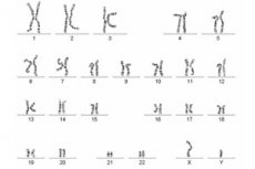
The set of features of a complete set of chromosomes is a karyotype. To systematize chromosome analyses, the International Cytogenetic Nomenclature is used, which is based on differential staining of the genome for a detailed description of all sections of DNA strands.
The study allows us to identify:
- Trisomy – there is a third extra chromosome in the pair.
- Monosomy – one chromosome is missing from a pair.
- Inversion is a reversal of a region of the genome.
- Translocation is the movement of sections.
- Deletion is the loss of a region.
- Duplication is the doubling of a fragment.
The results of the analysis are recorded according to the following system:
- The total number of chromosomes and the set of sex chromosomes are 46, XX; 46, XY.
- Extra and missing chromosomes are indicated, for example 47, XY, + 21; 46, XY -18.
- The short arm of the genome is designated by the symbol p, and the long arm by q.
- Translocation is t, and deletion is del, for example 46,XX,del(6)(p12.3)
The finished karyotype analysis looks like this:
- 46, XX – normal woman.
- 46, XY – normal male.
- 45, X – Shereshevsky-Turner syndrome.
- 47 XXY – Klinefelter syndrome.
- 47, XXX – trisomy of the X chromosome.
- 47, XX (XY), + 21 – Down syndrome.
- 47, XY (XX), + 18 – Edwards syndrome.
- 47, XX (XY), + 13 – Patau syndrome.
Cytogenetic research reveals various anomalies in the structure of DNA strands. The analysis also diagnoses predispositions to many diseases: endocrine pathologies, hypertension, joint damage, myocardial infarction, and others.
Preparation
Karyotype analysis uses blood cells, so it is very important to properly prepare for the diagnosis.
Preparation for a chromosome test begins 2 weeks before it is taken and consists of eliminating the impact of the following factors on the body:
- Acute and chronic diseases.
- Taking medications.
- Use of alcohol and drugs, smoking.
For analysis, 4 ml of venous blood is used. Blood is collected on an empty stomach.
Technique karyotype test
The human genome cannot be seen with the naked eye, chromosomes are visible only under a microscope at certain phases of cell division. To determine the karyotype, mononuclear leukocytes, skin fibroblasts or bone marrow cells are used. Cells in the metaphase of mitosis are suitable for the study. The biological fluid is placed in a test tube with lithium and heparin. The blood is cultured for 72 hours.
The culture is then enriched with special substances that stop cell division at the phase necessary for diagnostics. The culture is used to make slides that are subject to examination. Additional information about the state of the genome is obtained by staining it. Each chromosome has a striation that is clearly visible after staining.
In a classic chromosome study, staining is performed with different dyes and their mixtures. The dye binds differently to individual parts of the genome, making the staining uneven. Due to this, a complex of transverse marks is formed, which reflect the linear heterogeneity of the chromosome.
Basic dyeing methods:
- Q – provides highly detailed images. This method is called Kaspersson staining with quinacrine-yperite with diagnostics under a fluorescent microscope. It is used to analyze genetic sex, identify translocations between X and Y, Y and autosomes, and to screen for mosaicism with Y chromosomes.
- G – modified Romanovsky-Giemsa method. Has higher sensitivity compared to Q. Used as a standard method of cytogenetic analysis. Detects small aberrations, marker chromosomes.
- R – is used to detect homologous G and Q negative regions. The genome is treated with acridine orange dye.
- C – analyzes centromeric regions of chromosomes with constitutive heterochromatin and variable distal part of Y.
- T – used to analyze telomeric regions of DNA strands.
The stained and fixed cells are photographed under a microscope. From the resulting set of photographs, a numbered set of autosome pairs is formed, i.e. a systematized karyotype. The image of the DNA strands is oriented vertically, the numbering depends on the size, with the pair of sex chromosomes closing the set.
Blood preparations are analyzed under a microscope in 20-100 metaphase plates to identify quantitative and structural aberrations.
- Quantitative aberrations are changes in the number of genes. This is observed in Down syndrome, when there is an extra 21 chromosome.
- Structural aberrations are changes in the chromosomes themselves. This can be a loss of a section of the genome, the transfer of one part to another, a 180-degree rotation, etc.
The technique of karyotyping is a labor-intensive process. The study is carried out by highly qualified specialists. It may take a whole working day to diagnose the genome of one person.
Karyotype analysis of spouses
When entering into marriage, many couples face the problem of conception. Cytogenetic analysis is indicated to solve reproductive problems. Karyotyping of spouses allows to identify anomalies in the structure of the genome that prevent having children or disrupt the process of bearing children. It is impossible to change the karyotype, but thanks to diagnostics, it is possible to establish the true causes of infertility and termination of pregnancy, and find ways to solve them.
Chromosomal microarray analysis is performed to identify abnormalities in the structure and number of DNA strands that may be the cause of hereditary diseases in the future child or infertility of the spouses. There are international standards for conducting analysis in future parents:
- Chromosomal pathologies in the family.
- History of miscarriage.
- The pregnant woman is over 35 years old.
- Long-term mutagenic effects on the body.
The following karyotyping methods are used today:
- Analysis of chromosomes in blood cells.
Allows to identify cases of infertility, when the chance of having a child is significantly reduced or completely absent for one of the spouses. The examination also determines the risk of genome instability. To treat deviations, patients can be prescribed antioxidants and immunomodulators, which reduce conception failures.
For the study, venous blood is collected. Lymphocytes are isolated from the biological fluid, stimulated in a test tube, treated with a special substance, stained and studied. For example, in Klinefelter syndrome, which manifests itself as male infertility, the karyotype contains an extra chromosome 47 XX. Structural changes in the genome can also be detected: inversion, deletion, translocation.
- Prenatal research.
Determines chromosomal pathologies of the fetus in the early stages of pregnancy. Such research is necessary for the diagnosis of genetic diseases or developmental defects that lead to intrauterine fetal death.
The following methods can be used to conduct the research:
- Non-invasive – safe for mother and fetus. Diagnosis is made using ultrasound of the child and a detailed biochemical analysis of the woman’s blood.
- Invasive – chorion biopsy, cordocentesis, placentocentesis, amniocentesis. For analysis, placental or chorionic cells, amniotic fluid or umbilical cord blood are collected. Despite the high diagnostic accuracy, invasive methods have an increased risk of complications, so they are carried out only according to strict medical indications: fetal pathologies detected during ultrasound, the mother is over 35 years old, parents have chromosomal abnormalities, changes in blood biochemical markers.
Not only blood, but also ejaculate can be used for cytogenetic research. This method is called Tunel and allows one of the most common causes of male infertility to be determined, provided the karyotype is normal – sperm DNA fragmentation.
If gene mutations or chromosomal aberrations are detected in one of the spouses, the doctor talks about the possible risks and the probability of having a child with abnormalities. Since gene pathologies are incurable, the spouses make their own decision: to use donor material (sperm, egg), to risk giving birth or to remain childless.
If deviations in the genome are detected during the gestation process, both in the woman and in the embryo, then doctors recommend terminating such pregnancies. This is due to the increased risk of the birth of a baby with serious, and in some cases, incompatible with life deviations. A geneticist is involved in conducting tests and deciphering their results.
 [ 12 ], [ 13 ], [ 14 ], [ 15 ], [ 16 ]
Blood test for karyotype
Most often, karyotyping is performed by analyzing venous blood using cell culture. However, other biological material can also be used to conduct cytogenetic research:
- Cells from amniotic fluid.
- Placenta.
- Embryonic cells.
- Abortion material.
- Bone marrow.
The material that will be taken for diagnostics depends on the reason and purpose of the analysis. Approximate blood test algorithm:
- A small volume of liquid is placed in a nutrient medium at a temperature of 37˚C for 72 hours.
- Since chromosomes are visible at the metaphase stage of cell division, a reagent is added to the biological environment that stops the division process at the required phase.
- The cell culture is stained, fixed and analyzed under a microscope.
Blood analysis for karyotype provides highly accurate detection of any anomalies in the structure of DNA strands: intrachromosomal and interchromosomal rearrangements, changes in the order of genome fragments, etc. The main goal of diagnostics is to identify genetic diseases.
[ 17 ], [ 18 ], [ 19 ], [ 20 ]
Genetic analysis for karyotype
Cytogenetic diagnostics aimed at studying the size, number and shape of chromosomes is genetic karyotyping. The analysis has the following indications for implementation:
- Detection of congenital defects.
- Risk of having a child with hereditary pathologies.
- Suspected infertility.
- Spermogram abnormality.
- Miscarriage.
- Drawing up a treatment plan for certain types of tumors.
Also, genetic analysis for karyotype is included in the list of mandatory tests for spouses who plan to have children.
Most often, the study reveals the following pathologies:
- Aneuploidy is a change in the number of chromosomes, either increasing or decreasing. Imbalance leads to miscarriages, the birth of babies with severe congenital pathologies. Mosaic aneuploidy causes Down syndrome, Edwards syndrome and other diseases that are often incompatible with life.
- Karyotype reorganization – if the changes are balanced, then the chromosome set is not broken, but simply ordered differently. With unbalanced changes, there is a threat of gene mutations, which is especially dangerous for future generations.
- Translocation is an unusual structure of DNA strands, i.e. the replacement of one fragment of the genome with another. In most cases, it is inherited.
- Sex differentiation disorder is an extremely rare chromosomal disorder that does not always manifest itself with external symptoms. Inconsistency with the phenotypic sex can be one of the causes of infertility.
Karyotype analysis is performed in genetic laboratories by qualified geneticists.
Karyotype analysis with aberrations
Aberrations are disturbances in the structure of chromosomes caused by their breaks and redistribution with loss or duplication of genetic material. Karyotyping with aberrations is a study aimed at identifying any changes in the structure of the genome.
Types of aberrations:
- Quantitative – violation of the number of chromosomes.
- Structural – disruption of the genome structure.
- Regular – are found in most or all cells of the body.
- Irregular – arise due to the impact of various unfavorable factors on the body (viruses, radiation, chemical exposure).
The analysis establishes the karyotype, its features, signs of the impact of various negative factors. Chromosomal research with aberrations is carried out in the following cases:
- Infertility in marriage.
- Spontaneous miscarriages.
- History of stillbirth.
- Early infant mortality.
- Frozen pregnancy.
- Congenital malformations.
- Disorder of sexual differentiation.
- Suspected chromosomal abnormalities.
- Delayed mental and physical development.
- Examination before IVF, ICSI and other reproductive procedures.
Unlike classical karyotyping, this analysis takes longer to perform and is more expensive.
Karyotype analysis for a child
According to medical statistics, congenital pathologies are a significant cause of death in young children. To detect genetic abnormalities and hereditary diseases in a timely manner, the child undergoes a karyotype analysis.
- Most often, children are diagnosed with trisomy - Down syndrome. This pathology occurs in 1 out of 750 babies and manifests itself in various kinds of deviations in both physical and intellectual development.
- The second most common disorder is Klinefelter syndrome, which is characterized by delayed sexual development in adolescence and occurs in 1 in 600 male newborns.
- Another genetic pathology diagnosed in 1 in 2,500 female children is Shereshevsky-Turner syndrome. In childhood, this disease makes itself known by increased pigmentation of the skin, swelling of the feet, hands and shins. During puberty, there is a lack of menstruation, hair under the arms and on the pubis, and the mammary glands are also not developed,
Karyotyping is necessary not only for babies with visible abnormalities, as it allows one to suspect genetic problems and begin their correction. The analysis is done at a medical genetic center. Depending on the age of the child, blood can be taken from the heel or from a vein. If necessary, the geneticist may require the parents to undergo a karyotype analysis.
Karyotype analysis of a newborn
Neonatal screening is the first test performed on newborns. The test is performed in the maternity hospital on the 3rd-4th day of life, for premature babies on the 7th day. Early karyotyping allows identifying genetic abnormalities and DNA structure disorders before visible pathological symptoms appear.
Blood from the infant's heel is used for early diagnostics. Cytogenetic testing is aimed at identifying such common pathologies among babies as:
- Phenylketonuria is a hereditary disease characterized by decreased activity or absence of the enzyme that breaks down the amino acid phenylalanine. As it progresses, it leads to disturbances in the functioning of the brain and mental retardation.
- Cystic fibrosis – affects the glands that produce secretions, digestive juices, sweat, saliva, mucus. Causes problems with the lungs and gastrointestinal tract. The disease is inherited.
- Congenital hypothyroidism is a disorder of the thyroid gland with insufficient production of its hormones. It leads to delayed physical and mental development.
- Adrenogenital syndrome is a pathological condition in which the adrenal cortex produces insufficient amounts of hormones. This causes disruption of the development of the genitals.
- Galactesemia is a pathology in which the transformation of galactose into glucose is disrupted. Treatment consists of giving up dairy products. Without timely diagnosis, it can cause blindness and death.
If the results of the karyotype analysis reveal any deviations or anomalies in the newborn, then a set of additional studies is carried out to clarify the diagnosis. Such early diagnostics will allow timely detection of any problems in the child's body and the beginning of their treatment.
[ 21 ], [ 22 ], [ 23 ], [ 24 ], [ 25 ]
How long does it take to do a karyotype test?
The duration of the chromosome study takes from 10 to 21 days. When the results are ready depends on the type of analysis, i.e. with aberrations or classical karyotyping.
The completed karyotype analysis contains the following information:
- Number of chromosomes.
- Are there any changes in the chromosome structure?
- Are there any disturbances in the ordering of the genome?
The results are deciphered and interpreted by a geneticist. If any abnormalities are detected, the doctor provides medical recommendations for further diagnostics or treatment instructions.
Normal performance
Normal karyotypes for humans are 46, XX or 46, XY. As a rule, their change occurs at the early stages of the organism's development:
- Most often, the disorder occurs during gametogenesis (pre-embryonic development), when the parental germ cells produce the zygote karyotype. Further development of such a zygote leads to all the cells of the embryo containing an abnormal genome.
- The disorder may occur at the early stages of zygote division. In this case, the embryo contains several cell clones with different karyotypes. That is, mosaicism develops - a multiplicity of karyotypes of the entire organism and its organs
Changes in the genome manifest themselves in various pathologies and defects. Let's consider common karyotype anomalies:
- 47,XXY; 48,XXXY – Klinefelter syndrome, polysomy of the X chromosome in males.
- 45X0; 45X0/46XX; 45.X/46.XY; 46,X iso (Xq) – Shereshevsky-Turner syndrome, monosomy on the X chromosome, mosaicism.
- 47,XXX; 48,XXXX; 49,ХХХХХ – polysomy on the X chromosome, trisomy.
- 47,XX,+18; 47,ХY,+18 – Edwards syndrome, trisomy of chromosome 18.
- 46,XX, 5p- – Cri du chat syndrome, deletion of the short arm of the 5th pair of the genome.
- 47,XX,+21; 47,ХY,+21 – Down syndrome, trisomy of chromosome 21.
- 47,XX,+13; 47,ХY,+13 – Patau syndrome, trisomy of chromosome 13.
Cytogenetic research is aimed at determining the state of DNA strands, identifying defects and anomalies. Any deviations from normal indicators are a reason for a comprehensive examination of the body.
[ 26 ], [ 27 ], [ 28 ], [ 29 ], [ 30 ], [ 31 ], [ 32 ], [ 33 ], [ 34 ]
The device for analysis
To decipher the karyotype, the sequencing method is used. This method was developed in 1970 and is based on determining the sequence of amino acids in DNA. Sequencing devices use interactive cyclic enzymatic reactions with subsequent processing and comparison of the results obtained.
The main functions of sequencers:
- Primary complete study of unknown genomes, exomes, transcriptomes.
- Karyotyping.
- Paleogenetics.
- Metagenomics and microbial diversity.
- Resequencing and mapping.
- DNA methylation analysis.
- Transcriptome analysis.
In the first stage, the device creates a library of random DNA strand sequences. It then creates PCR amplicons, which are used as samples. In the final stage, the primary structure of all fragments is determined.
The latest generation of sequencers are fully automated and are widely used in genomic analysis, minimizing the possibility of erroneous results due to human error.
Deciphering the results of a karyotype analysis
A geneticist interprets the results of a cytogenetic study. As a rule, the analysis is ready in 1-2 weeks and may look like this:
- 46XX(XY), grouped into 22 pairs and 1 pair of sex genes. The genome has a normal size and structure. No anomalies were detected.
- The genome is disrupted, more/less than 46 chromosomes are detected. The shapes and sizes of one/several chromosomes are abnormal. The genome pairs are disrupted/incorrectly grouped.
As for pathological deviations in the karyotype, the following common disorders are distinguished:
- Trisomy - an extra somatic chromosome. Down syndrome, Edwards syndrome.
- Monosomy is the loss of one chromosome.
- Deletion - absence of a genomic region. -46, xx, 5p - Cri du chat syndrome.
- Translocation is the movement of one part of the genome to another.
- Duplication is the doubling of a fragment.
- Inversion is a rotation of a chromosome fragment.
Based on the results of the karyotype analysis, the doctor draws a conclusion about the state of the genotype and the degree of genetic risk. At the slightest changes in the structure of DNA strands, a set of additional studies is prescribed. The identified aberrations may not manifest themselves in any way, but they increase the risk of having babies with genetic abnormalities.