
All iLive content is medically reviewed or fact checked to ensure as much factual accuracy as possible.
We have strict sourcing guidelines and only link to reputable media sites, academic research institutions and, whenever possible, medically peer reviewed studies. Note that the numbers in parentheses ([1], [2], etc.) are clickable links to these studies.
If you feel that any of our content is inaccurate, out-of-date, or otherwise questionable, please select it and press Ctrl + Enter.
Ectodermal dysplasia
Medical expert of the article

Last reviewed: 04.07.2025
A relatively rare disease, ectodermal dysplasia, is a genetic disorder accompanied by a disorder of the functionality and structure of the derivative elements of the outer layer of the skin. Hair, nail plates, teeth, glandular system (mucous, sweat and sebaceous) are usually affected. The disease is complex and can occur in several forms. Treatment is mainly corrective, symptomatic: unfortunately, there is no talk of a complete recovery. [ 1 ]
Epidemiology
Variations of ectodermal dysplasia include Christ-Siemens-Touraine syndrome, Clouston syndrome, Rapp-Hodgkin syndrome, and EEC syndrome. The pathology was first described in the mid-19th century by Dr. Touraine. In 1913 and 1929, this description was supplemented by dentist Christ and dermatologist Siemens, in 1968 by Rapp and Hodgkin, and in 1970 by Rudiger.
In medical literature, the disease is more often found under the name ectodermal dysplasia and corresponds to the international coding Q82.4 (ICD-10).
To date, doctors cannot give an exact incidence rate. However, it is believed that the syndrome occurs in approximately one case out of 5-10 thousand. [ 2 ]
The etiologic heterogeneity of the disease is well known, with three genetic types of hereditary transmission: autosomal recessive, dominant, and X-linked recessive (the latter is the most common). [ 3 ]
At present, three genes have been identified in different chromosomes, which make it possible to detect this disease using genetic-molecular methods. The number of possible mutations is more than sixty.
Ectodermal dysplasia most often affects boys, which is associated with sex-linked inheritance. Girls often have a mild form of the pathology, or an asymptomatic one.
The pathology is registered in different countries of the world among representatives of any race. It can appear sporadically in clinically healthy couples, or manifest itself in a familial form (especially often - if the parents are closely related).
Causes ectodermal dysplasia
The only cause of ectodermal dysplasia is a mutation of a certain hereditary gene factor. In particular, the most common disorder is that of the EDA gene, localized on the X chromosome. This gene is responsible for encoding the protein substance ectodysplasin-A, the abnormal structure of which entails a violation of the formation of ectoderm elements. At present, the exact characteristics of the protein substance and the mechanism of development of mutational disorders have not been clarified.
The X-linked disease has its own characteristics: the problem is more often found in men, but women can also be not only carriers, but also have individual signs of the syndrome, albeit to a mild degree. For example, patients with ectodermal dysplasia may experience excessive dryness of the skin, wrinkling, thinning and dry hair, and dental deformation. Problems with the mammary glands and nipples are possible. Such signs indicate the possibility of incomplete dominance of EDA gene mutations.
Among other types of mutations, changes in the EDAR gene, which is responsible for encoding the receptor to the tumor necrosis factor, can be distinguished. This gene is localized on chromosome II, and is inherited in an autosomal recessive manner. The exact process of pathology development has not been clarified.
If we are talking about rare variants of ectodermal dysplasia, they arise under the influence of gene mutations in TDARADD, responsible for encoding the protein receptor to exodysplasin-A, localized on chromosome I. Pathogenetic mechanisms have not been fully studied. [ 4 ]
Risk factors
The most significant risk factors leading to the birth of a child with ectodermal dysplasia are defects:
- the EDA gene encoding ectodysplasin A, mapped to chromosome Xq12-q13.1;
- EDAR gene encoding tumor necrosis factor receptor, a member of the EDAR superfamily, mapped to chromosome 2q11-q13;
- TDARADD gene encoding ectodysplasin-A, a receptor-associated protein, mapped to chromosome 1q42.2-q43.
A hereditary predisposition to ectodermal dysplasia can be suspected by examining the family history.
Comprehensive genetic and molecular diagnostics allows us to assess the genetic risk of a child developing this syndrome.
Pathogenesis
Pathogenetic features of the development of this disease are poorly understood. It is known that ectodermal dysplasia appears as a result of mutational changes in certain genes. The cause of the most common form of pathology is damage to the EDA gene, localized on the X chromosome. This gene is responsible for encoding a protein agent called ectodysplasin-A. Pathological changes in its structure cause abnormal development of ectodermal derivatives. Unfortunately, to date, both the functional side of this protein agent and the pathogenesis of changes in the EDA gene mutation have not been sufficiently studied.
The main distinguishing feature of ectodermal dysplasia is that clinical disorders are found not only in male patients, but also in women: the carrier state is manifested by milder pathological changes. In particular, dry hair and skin, early wrinkling, curvature and other dental disorders are noted.
In addition, typical Christ-Siemens-Touraine syndrome is caused by mutational changes in the EDAR gene, responsible for encoding one of the receptors for tumor necrosis factor. This gene is localized on chromosome II, and is inherited in an autosomal recessive manner. Pathogenetic features have not been studied in this case either. [ 5 ]
A rarer variant of the anhidrotic type of ectodermal dysplasia is also known, with an autosomal dominant mode of inheritance. The cause is mutational changes in the TDARADD gene, which codes for the protein substance-receptor to exodysplasin-A and is localized on chromosome I. Most likely, the pathogenetic characteristics in this case are identical to the more common type of the disease, linked to sex.
For your information: the ectoderm is one of the three germ layers (the other two are the mesoderm and endoderm). The ectoderm is the outer layer that forms during the third week of embryonic development and causes the formation of the skin and appendages (hair, nails), rectal and oral epithelium, tooth enamel, lens and cornea, and sweat glands. In people with ectodermal dysplasia, some or all of the ectoderm structures are either absent or do not function properly.
Symptoms ectodermal dysplasia
The clinical picture of ectodermal dysplasia is determined by a number of numerous disorders affecting the ectoderm and sweat glands. The sebaceous and apocrine glands are also affected, but these defects are less pronounced. Other glandular systems - in particular, the lacrimal, digestive, nasal, bronchial - have signs of atrophy. Typical signs: atrophic processes, hypoplasia of the skin, hypoplasia of the mammary glands and nipples.
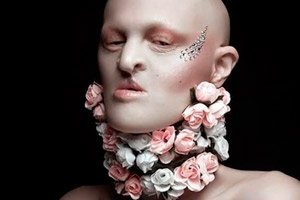
In the facial area, wrinkles, thinning of the eyelids, periorbital pigmentation disorders, papules, eczematous rashes, and palmar hyperkeratosis are found. The frontal tubercles and superciliary arches are clearly protruding forward, the bridge of the nose is smoothed, the nose is small and saddle-shaped, the nasal wings are hypoplastic, the lips are full and protruding, and the cheeks are sunken.
The hair is sparse, often with bald spots, and is characterized by dryness and light shades.
The teeth are irregularly shaped, often conical and pointed. Some teeth are completely absent (canines are always present).
The auricles are also deformed: they are usually small, set high, and the ear curl has an irregular shape.
In terms of the visual organs, clouding of the lens, myopia, blepharoconjunctivitis, decreased lacrimation, and liquid vitreous body may be observed.
Some patients suffer from complete hearing loss. There is a tendency to infectious diseases and thermoregulation disorders.
First signs
The first manifestations of ectodermal dysplasia are often detected already in the neonatal period. However, this may also occur later, since the clinical picture in young children is not always pronounced and worsens over the years.
The basic symptoms that can be used to suspect the presence of pathology are often the following:
- growth retardation against the background of a relatively large head;
- dry, thin hair, predominantly “vellus” hair with slow growth and low pigmentation, short and sparse eyelashes and eyebrows, or their complete absence;
- early alopecia, up to complete loss of hair;
- typical appearance of the "old man's face" type, protruding frontal region, brow ridges and tubercles, widened cheekbones, sunken bridge of the nose, small saddle-shaped nose and hypoplastic wings, sunken cheeks, protruding "fish-like" lips, "heavy" chin, irregular shape of the auricles;
- delayed eruption of teeth (from one to three years), disruption of the usual sequence of eruption, long period of retention of milk teeth, absence of some teeth; [ 6 ]
- conical dental configuration, pointed cutting edges, smoothed surface of molars;
- dental and bite disorders;
- underdeveloped salivary glands, weak salivation, dry mouth, hoarseness;
- excessive dryness of the skin, early wrinkling, which is especially noticeable on the face;
- pigmentation disorders, improper functioning of the sebaceous glands, papular rash;
- conjunctivitis, photophobia;
- underdeveloped mammary glands, or their absence;
- insufficiently developed mucous glands in the respiratory and digestive systems, which causes frequent bronchitis, rhinitis, sinusitis, and gastrointestinal pathologies;
- periodic sharp increase in temperature associated with improper heat transfer due to disruption of the sweat glands;
- less often – mental retardation, oligophrenia (more often, the development of intelligence corresponds to the norm);
- impaired social adaptation and orientation, stiffness and isolation;
- speech problems associated with abnormal tooth growth and dry mucous membranes of the oropharynx;
- impaired vision;
- little or no sweating.
Triad in anhidrotic ectodermal dysplasia
The anhidrotic variant of ectodermal dysplasia is manifested by a triad of basic signs:
- sparse hair growth of the atrichosis or hypotrichosis type; [ 7 ], [ 8 ]
- incorrect configuration of teeth (often conical, pointed), or underdevelopment and absence of teeth;
- disorders of sweat secretion such as hypohidrosis and anhidrosis, which is often caused by the absence of sweat glands as such.
Due to the presence of anhidrosis, the patient has such additional symptoms as high-temperature hypersensitivity and regular relapses of hyperthermia, which poses a real danger to human life. The skin is thinned and dry. Many patients suffer from chronic blepharoconjunctivitis, "dry eye syndrome", and asthma-like conditions. [ 9 ]
Forms
Various combined manifestations and their intensity determine the division of ectodermal dysplasia into several types, which can be called independent forms of pathology. The main types are: Christ-Siemens-Touraine syndrome, Clouston syndrome, Rapp-Hodgkin syndrome and EEC syndrome.
Christ-Siemens-Touraine syndrome, or anhidrotic ectodermal dysplasia, is characterized by complete dysfunction of the sweat glands, as well as a unique phenotype of facial structure: the child has a protruding forehead, thin and sparse eyebrows, rare short eyelashes, wrinkles. Periorbital pigmentation disorder, saddle-shaped nasal bridge, and jaw hypoplasia are typical. Hair may be depigmented or weakly pigmented.
Some specialists believed that full-blown anhidrosis is still rarely detected in patients, and in most patients the sweat excretory system is weak, but still functions. This opinion was taken into account and led to the fact that over time, doctors began to use a more correct name: hypohidrotic form of the disease. Hypohidrotic ectodermal dysplasia is a genetic disorder of the formation of the ectodermal layer. The pathology is characterized by disturbances in the formation of such elements of the ectoderm as skin and hair, glands (sweat, sebaceous) and teeth. The disease consists of three subtypes, which are practically indistinguishable symptomatically, since the main clinical sign is impaired sweating (mainly hypohidrosis). We are talking about the Christ-Siemens-Touraine syndrome with an X-linked type of inheritance, as well as autosomal recessive and autosomal dominant ectodermal dysplasia. There are also several less common subtypes that are accompanied by severe immune deficiency – the so-called congenital anhidrotic ectodermal dysplasia with immunodeficiency state.
Clouston syndrome is a hydrotic type of ectodermal dysplasia. The defining symptoms of the pathology are the same lesions of the teeth, hair and sweat system, but to a slightly lesser extent. Hypodontia is found in the area of the lower incisors, second molars and upper canines. Nail lesions are manifested in the form of hypoplasia, dystrophy, aplasia with paronychia. The number of sweat glands is reduced, with unchanged sebaceous glands. Hypotrichosis and baldness are possible. The mode of inheritance is autosomal and autosomal dominant.
Rapp-Hodgkin syndrome is also called hypohidrotic ectodermal dysplasia, accompanied by cleft lip, alveolar process, soft and hard palate. Distinctive manifestations are considered to be the following: hypohidrosis and hypotrichosis, pathological changes in the nails, hypodontia or oligodontia in combination with cleft upper lip, alveolar process, soft and hard palate. Common symptoms are also a sunken bridge of the nose, narrowing of the nose, micrognathia of the upper jaw, small mouth, reduced genitals. The syndrome is inherited in an autosomal dominant manner.
EEC syndrome has only recently been identified as a separate disorder, better known as the combined syndrome of ectrodactyly, ectodermal dysplasia with cleft palate and lip. Distinctive symptoms include defects of the feet and hands, cleft lip, and sometimes cleft tongue. These signs are present against the background of impaired sweating, hypotrichosis and alopecia, nail hypoplasia, dry and hypopigmented skin, conjunctivitis, photophobia, etc. Dental anomalies, late eruption, and multiple caries are also typical. Against the background of physical defects, mental development is usually adequate. The mode of inheritance is autosomal dominant, but recessively inherited variants also occur.
Ectodermal dysplasia in children
Despite the fact that ectodermal dysplasia is a congenital disease, it is not always possible to diagnose it in a newborn baby: often the diagnosis is made only after several years (often by 2-3 years). Practicing doctors note the need for early diagnosis, since not only the future lifestyle, but sometimes the life of the patient directly may depend on it. The symptoms of the pathology are varied, but not always noticeable. Some of them are more common, while others are less common. [ 10 ] Both parents and doctors should be alerted by the following signs:
- hypoplasia of sweat glands with hypo or anhidrosis, thermoregulatory disorders, frequent episodes of hyperthermia, causeless fever, regular overheating;
- hypotrichosis, sparseness, depigmentation and thinness of hair, shortening of eyebrows and eyelashes (or their absence);
- persistent or transient baldness (total or patchy);
- late teething with disruption of its sequence;
- insufficient number of teeth, abnormal configuration of teeth (often conical, spiky with a pointed edge), or absence of teeth;
- malocclusion, sometimes tooth mobility, large interdental spaces;
- low attachment of the upper labial frenulum, sharply pronounced buccal cords, shallow oral vestibule;
- insufficiently developed maxillary alveolar process;
- the radiograph shows shortened dental roots, widened periodontal spaces, flattened condylar processes of the mandible;
- hypoplasia of the mucous glands in the mouth, resulting in insufficient salivation and hoarseness;
- tendency to fungal stomatitis, cheilitis;
- typical "old man's face" with a prominent frontal area, a sunken bridge of the nose, a small saddle-shaped nose, sunken cheeks, full, indistinct, convex lips, and irregularly shaped ears;
- thin, dry, wrinkled skin, sometimes with papular rash;
- insufficient function of the lacrimal glands, frequent inflammatory diseases (keratitis, blepharitis, etc.);
- defects of the lip and palate;
- nail lesions, paronychia;
- defects of the feet and/or hands, hyperkeratosis of the palms and feet;
- insufficient development of the mammary glands and nipples (sometimes their absence);
- immunodeficiency, eczema;
- a tendency to respiratory and digestive diseases, as well as nosebleeds.
Different combinations of signs and their intensity determine individual variants of the course of ectodermal dysplasia.
Complications and consequences
Patients with ectodermal dysplasia should avoid any uncontrolled thermal effects. Infants require constant monitoring of body temperature. Older children should be provided with the necessary preventive and cooling measures - in particular, regularly drinking cool drinks, moistening clothes, using air conditioners.
It is necessary to start caring for the oral cavity and teeth as early as possible – to maintain their functionality and optimize their appearance. Orthodontist’s help often consists of installing corrective plates and sinus lifting with subsequent dental implantation. Supportive dental prosthetics are possible. [ 11 ]
For the hypohidrotic form of ectodermal dysplasia with immunodeficiency, medications to support the immune system are required, as well as intensive therapy for infectious diseases, or a hematopoietic stem cell transplant.
If the pathological syndrome is not detected in early childhood, then impaired thermoregulation can cause damage to the brain, which will ultimately lead to death. With adequate and timely diagnosis and competent treatment, patients have the opportunity to lead a normal life without negative impact on its duration. [ 12 ]
Diagnostics ectodermal dysplasia
The diagnosis of ectodermal dysplasia is often made after periodic cases of fever or late teething. Dysfunction and absence of sweat glands is confirmed by skin biopsy or noninvasive confocal microscopy. It is also possible to study graphite prints of the palmar surfaces and feet.
The quality of the sweat system function is assessed by numerical evaluation of pilocarpine-induced sweating. To confirm the diagnosis, genetic tests are performed and the hereditary anamnesis is examined.
Genetic evaluation involves direct sequencing of the EDA gene sequence to detect mutations.[ 13 ]
The assessment of the hereditary anamnesis is carried out along with the determination of the objective status of the mother. Often, she has certain signs indicating the carriage of the pathology. Such signs include dry skin, thinned weakened hair, underdeveloped mammary glands.
Genetic studies of carriage of the disrupted form of the EDA gene are often problematic due to frequent false negative results. Therefore, other methods of genetic research are used to prove carriage, in particular, the multiplex ligase reaction.
Instrumental diagnostics performed on patients with suspected ectodermal dysplasia may include the following procedures:
- ultrasound examination and electrocardiography;
- skin biopsy to assess the condition of the sweat glands;
- microscopy of hair structure;
- X-ray of the jaws to determine the quality of the dental rudiments.
Laboratory tests such as a complete blood count may indicate a shift in eosinophil levels and anemia, but such changes are not specific for ectodermal dysplasia.
Differential diagnosis
Anhidrotic ectodermal dysplasia is distinguished, first of all, from the hydrotic variant of the disease (Clouston syndrome). The symptoms of the two pathologies have much in common, but with the hydrotic form, the sweat glands function, so xeroderma and hyperthermia may be absent. Differentiation is also made between all existing types of ectodermal dysplasia and certain forms of ichthyosis. [ 14 ]
Who to contact?
Treatment ectodermal dysplasia
The treatment regimen for ectodermal dysplasia is determined depending on the existing disorders and involves the use of symptomatic agents against the background of lifelong special care, which is prescribed to the patient as a way of life and includes avoiding overheating and physical exertion. Treatment tactics also depend on the patient's age. [ 15 ]
The basic direction of systemic therapy is the use of second-generation H1-antihistamine blockers, since they are not able to penetrate the blood-brain membrane, are suitable for long-term use, and are convenient to use (once a day). In early childhood, first-generation H1-antihistamine blockers can be used, which is due to the slight sedative property of these drugs. [ 16 ]
In case of ectodermal dysplasia, external therapeutic agents are used without fail – to soften and protect the skin. The drugs of choice may be:
- emollients with a hydrophilic base and 5% urea;
- creams based on cyramides or petroleum jelly with a frequency of application of at least 2 times a day (with the onset of the remission period, switch to use once every 1-2 days);
- medical and cosmetic products intended for the care of dry and irritated skin.
Patients with an elevated SCORAD index (from 20 to 40 and more than 40) are prescribed active corticosteroid agents externally:
- mometasone furoate ointment 0.1% daily at night for 21 days;
- can be replaced with fluticasone propionate 0.005%.
If there is no response to topical corticosteroids, topical calcineurin inhibitors are prescribed - for example, tacrolimus ointment 0.1% twice a day for three months, or until symptoms disappear.
Among vitamin preparations, it is appropriate to take only vitamin D 3 (cholecalciferol), and only after assessing the content of calcidiol in the blood. Cholecalciferol is prescribed at 1000-1600 IU daily for 1-2 months.
Physiotherapy involves the use of phototherapy:
- UVA1 (340-400 nm) in the acute period, during relapse or severe stage up to 5 times a week for 1.5-3 months;
- UVB narrowband (311-313 nm) for chronic forms of pathology.
Patients with ectodermal dysplasia are additionally prescribed symptomatic medications in consultation with other doctors of narrow specializations: dentist, gastroenterologist, pulmonologist, etc.
The medications are used in combination with regular application of external moisturizing dermatological products, such as:
- Radevit is a dermatoprotective agent that improves trophism and tissue regeneration. It has anti-inflammatory, softening, and moisturizing properties. Radevit is not used in the presence of allergies and hypervitaminosis A, E, D.
- Lipikar is a cosmetic lipid-restoring product that softens and nourishes the skin. It can be used at any age, even in the neonatal period.
- Emolium is a complex emollient that moisturizes even the deep layers of the skin, restoring the protective lipid layer. Contains sodium hyaluronate, urea, shea butter and macadamia oil. Can be used from birth, if there is no allergy to the composition of the drug.
- La-Cree is an effective product containing natural plant oils and extracts, lecithin and allantoin. The cream effectively softens, eliminates itching, redness, peeling of the skin, prevents the appearance of inflammatory elements.
Since the condition of patients with ectodermal dysplasia worsens in the summer, which is associated with an increase in ambient temperature and increased solar activity, then in these months
Prevention
It is almost impossible to prevent the development of hereditary ectodermal dysplasia: it is only possible to alleviate the symptoms of the pathology. In many cases, the life of patients with a gene mutation can be made as comfortable as possible, since the severity of the clinical picture largely depends not only on the hereditary factor, but also on its combination with external conditions and the patient's lifestyle. The fundamental moment is love and participation on the part of relatives and parents. After consulting with a doctor, it is necessary to think over a system of rehabilitation for the child: make adjustments to nutrition, consult with a dentist and dental technician, balance the main points that affect the quality of treatment and the patient's adaptation in society.
It is also important to determine the presence of the syndrome as early as possible in order to assess the risks for the child. This can be done by cryotyping - a study of the chromosome set of a newborn baby using an analysis of umbilical cord blood.
The probability of having a sick baby can be determined with the help of medical geneticists, using a DNA test when the child is still in the mother's womb. Probability factors are not only cases of the syndrome in the family line, but also certain stresses before or during gestation.
The use of in vitro fertilization allows to avoid the development of the disease in the child already at the stage of fertilization. The IVF method involves obtaining several embryos: before they are “implanted” into the mother’s body, the risks of hereditary pathology are checked.
Forecast
Unfortunately, patients with ectodermal dysplasia cannot be cured: only symptomatic treatment of the syndrome is available. Patients of early childhood may die due to thermoregulation disorders and secondary infection. The disease, as a rule, does not affect the life expectancy of adult patients.
It is very important to detect the disease at an early age stage in order to immediately begin treatment. This will prevent the development of complexes, phobias in the child, and improve social adaptation. In general, complex and rather complicated treatment is prescribed, involving specialists of different medical profiles.
It is equally important to follow all the doctors' recommendations. If everything is carefully planned and adjusted, the patient will be able to lead a normal life, despite the pathology. It is necessary to take into account that a more favorable course of the disease is observed when the patient lives in cool and humid climate conditions.
Ectodermal dysplasia is a rare but complex disease that cannot be cured. However, timely diagnostics and high-quality symptomatic and comprehensive correction allow patients to get rid of most of the painful manifestations and ensure an adequate and fulfilling life.
Disability due to ectodermal dysplasia
Children with ectodermal dysplasia, which manifests itself in multiple dental disorders in combination with disorders of other anatomical structures of ectodermal origin, are usually recognized as disabled children. However, in case of minor pathological changes, uncomplicated and mild course of the disease, assignment of a disability group may be denied, since the diagnosis of ectodermal dysplasia in itself is not an unconditional basis for recognizing a child as disabled.
The assessment of the patient's ability to work is carried out no earlier than 12 months after the diagnosis, after the necessary treatment and rehabilitation measures have been carried out. If, after the course of treatment, doctors confirm a persistent impairment of the body's functionality caused by congenital developmental defects, then in this case one can expect to be assigned a disability group corresponding to the severity of the existing pathologies.
How do people live with ectodermal dysplasia?
The fight against manifestations of ectodermal dysplasia continues in patients throughout their lives. The patient is monitored by doctors of various specialties: pediatrician, orthodontist, therapist and orthopedist, speech therapist, psychologist, neurologist, medical geneticist, otolaryngologist and dermatologist. If necessary, they seek help from maxillofacial surgeons.
Based on numerous clinical studies and observations, experts have identified a list of the most important recommendations for patients suffering from ectodermal dysplasia:
- Regularly monitor body temperature, stabilize it by wiping with a damp and cool towel, shower procedures, cool drinks, air conditioning in the area of stay. When performing physical activity - wear damp light clothing. If indicated - take antipyretic medications.
- Regularly visit doctors, depending on the symptoms and disorders present. If there is a lack of tear secretion, use special eye drops. To eliminate dry skin, regularly apply moisturizing creams.
- Eat only liquid food, use artificial saliva preparations if necessary, avoid eating hot and dry foods and products.
- Provide dental prosthetics.
- Family planning should only be carried out after genetic counseling.
- Observe caries prevention measures, conduct early remineralizing therapy and fluoridation.
Patients who adhere to these recommendations live a completely normal life, create families, and are socially active. At the same time, understanding and participation from loved ones are of no small importance.