
All iLive content is medically reviewed or fact checked to ensure as much factual accuracy as possible.
We have strict sourcing guidelines and only link to reputable media sites, academic research institutions and, whenever possible, medically peer reviewed studies. Note that the numbers in parentheses ([1], [2], etc.) are clickable links to these studies.
If you feel that any of our content is inaccurate, out-of-date, or otherwise questionable, please select it and press Ctrl + Enter.
The hepatic scalpel: structure, ways of infection, stages of development, prevention
Medical expert of the article

Last reviewed: 06.07.2025
A dangerous parasite that affects the liver and causes fascioliasis is the liver fluke. Let's look at its life cycle, routes of infection, and methods of destruction.
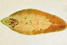
The helminth is a parasitic form of flukes that live in the organs of vertebrates (animals, humans) and invertebrates. Another name for the parasite is the cat fluke, since the cat is the most suitable host for the worm. An adult infects humans and cattle. The fluke has a leaf-shaped body and reaches a size of 3-5 cm. It can get into the organs of herbivores and fish, which act as a secondary host.
Main characteristics of the parasite:
- There is a special covering on the body that protects it from being digested by the host's juices.
- A variety of fastening devices: hooks, suction cups, etc.
- Simple structure of the digestive system.
- High fertility and asexual reproduction.
- Regressive development of the sense organs and nervous system.
The parasite is characterized by a complex life cycle with transformations and frequent changes of hosts. This leads to its dispersal and protects the main host from excessive overpopulation and death. Most often, human infection occurs when drinking unboiled or untreated water.
Structure liver fluke
The main difference between the flatworm and other parasites is its complex structure. The structure of the liver fluke is represented by the following organs and systems:
- Leaf-shaped, 3-5 cm, flattened in the dorsal-ventral direction.
- Developed attachment organs: oral and ventral suckers with a mouth opening.
- A branched digestive system and the absence of an anus.
- Protonephridial excretory system.
- Underdeveloped respiratory and circulatory systems.
- Asexual reproduction and development with a change of carriers.
- Developed nervous system (peripheral nerve ring, nerve cords along the body).
The development cycle of a helminth is characterized by constant transformations. Each stage of development has its own structure.
Internal structure of the liver fluke
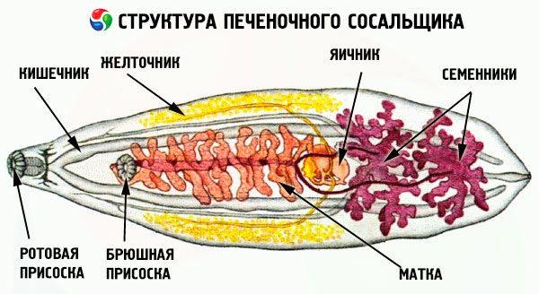
In humans, the causative agent of fascioliasis can be either the common or giant fasciola. Both have a specific and almost identical structure and functioning, which is due to their parasitic lifestyle. Let's consider the internal structure of the liver fluke:
- Oral sucker.
- Pharynx.
- Brain.
- Nerve ring.
- Esophagus.
- Abdominal sucker.
- The main branch of the intestine.
- Shell glands.
- Ventral nerve trunk.
- Uterus.
- Ovary.
- Testicle.
- Subpharyngeal commissure.
- Yolks.
The parasite has a leaf-shaped body, 2-7 cm in size and grayish-yellow in color. It lives in the bile ducts, liver and pancreas of vertebrates. With the help of oral and abdominal suckers it attaches and stays on the walls of the ducts.
The main life systems of a worm:
- Digestive - the mouth opening is connected to a muscular pharynx (sucking apparatus). Behind the pharynx there is a branched esophagus and blind-ending intestines.
- Nervous - is a peripharyngeal nerve ring, from which three pairs of nerve trunks depart (the lateral ones are the most developed). The nerve trunks are connected by means of bridges, which makes them look like a lattice.
- Excretory - developed protonephridia in the depth of the parenchyma. Thermal cells have channels with cilia that select tissue fluid and dissimilation products from the parenchyma. Cilia move the fluid through the channels and excretory pores, removing it from the body.
- Reproductive – the helminth is a hermaphrodite. The male reproductive system is represented by a pair of testicles, two vas deferens, which merge into the ejaculatory duct and cirrus. The female reproductive system is more complex: ovary, yolk glands, spermatheca, ootype (where fertilization and egg formation occur), uterus and genital opening. In some species, fertilization occurs in the spermatheca. In most cases, cross-insemination occurs, but there are cases of self-insemination.
The fluke is characterized by simplification and specialization in the structure of some organs. This is due to its parasitic lifestyle. The suckers, spines and other formations on the body of the worm, powerfully developed sexual organs and several complex life cycles act as specialization. Morphological simplification is expressed by the absence of sensory organs in sexually mature individuals, which act as endoparasites.
The digestive system of the liver fluke
The structure of the digestive system of the baked fluke is quite developed and consists of:
- Oral sucker.
- Pharynx.
- Abdominal sucker.
- Branching guts.
The digestive tract is branched and has two sections - the anterior and the middle. The anterior is the pharynx and esophagus, lined with ectoderm. The middle is the endodermal intestine, biramous, blind-closed. In some representatives of this class, the branches of the midgut have many blind processes. Parasites with a developed intestine have intraintestinal digestion of food, and helminths with rudimentary digestion absorb the digested food of the host through the tegument (body covering).
The parasite feeds on the tissues and blood of its host using sucking movements of the pharynx. Undigested food remains exit through the mouth. Flatworms that infect fish have an anus, which is represented by a separate intestinal trunk and an excretory bladder.
 [ 10 ], [ 11 ], [ 12 ], [ 13 ]
The nervous system of the liver fluke
The central nervous system of the liver fluke is represented by the following sections:
- Peripharyngeal ring.
- Nerve trunks: lateral, ventral.
- Jumpers.
The nervous system is located in the anterior third of the body at the level of the pharynx. It is a peripharyngeal ring, from which three nerve trunks extend. The end sections of the trunks are branched and enter the tegument. Two ventral, dorsal and lateral trunks extend from the brain ganglion, which reach the posterior end of the body and merge, forming an arch. The longitudinal nerve trunks are connected by bridges - transverse commissures. Due to this, the nervous system resembles a lattice that encircles the entire body.
The circulatory system of the liver fluke
Flatworms are parenchymatous, i.e., cavityless animals. The liver fluke has no circulatory system. The space between its internal organs consists of mesodermal connective tissue or parenchyma with many cells. The parenchyma fills all the spaces between the organs. It serves as a storage medium for nutrients and is responsible for metabolic processes.
The parasite also lacks a respiratory system. Special excretory organs, protonephridia, are located throughout the body. They are a system of branched canals that end in the parenchyma in the form of star cells with cilia. With the help of special excretory (excretory) openings, protonephridia contact the external environment.
External structure of the liver fluke
The causative agent of fascioliasis has a dense body adapted to life in the bile ducts of the host. The external structure of the liver fluke is a multilayer cuticle that protects against digestion, antitoxins and secretory fluid of the primary host. Gas exchange and the release of nitrogen-containing substances occur through the skin.
The outer part of the integument is an anuclear cytoplasmic plate with mitochondria and vacuoles. With the help of cytoplasmic strands, this layer is connected to the areas of the cytoplasm (immersed in the parenchyma), in which the nuclei are located.
The helminth has a leaf-shaped body and can reach sizes of 3-5 cm in length, up to 1.5 cm in width. The head end of the body is covered with spines, has an elongated proboscis, head and abdominal suckers. The skin is without cilia, but with a well-developed muscle layer. Due to its structure and parasitic lifestyle, the worm is able to survive in the absence of oxygen.
Fixation organs of the liver fluke
The adult helminth has a leaf-shaped, flattened form with a pointed rear end. The liver fluke's fixation organs are suckers and spines. With their help, the parasite attaches itself to the bile ducts, liver or pancreas of the host. This method of fixation protects against being washed away by secretory fluid.
At the front (wide) end of the body there is a narrow protrusion with a mouth sucker. In sexually mature individuals, the fixation organs, digestive and reproductive systems are well developed. Having attached to living tissues, the fluke does not change its location. It grows, feeds and lays eggs in the bile ducts. With the flow of bile, the eggs enter the intestines of the host and are excreted with excrement.
Sense organs of the liver fluke
The causative agent of fascioliasis has poorly developed sensory organs. The liver fluke, or rather its larvae floating in water, have several pairs of small eyes, arranged like turbellaria. In rare cases, appendages develop on the sides of the head end, resembling ears. Such growths are considered to be sensory organs (tactile and chemical).
Sensilla, or skin receptors, have a structure identical to turbellarians, and are an advantage in the larval stage of the parasite. The nervous system has a more complex structure. It consists of a peripharyngeal nerve ring, two ganglia and longitudinal nerve cords (innervating the sucker). Three pairs of powerful longitudinal nerve trunks with well-developed lateral nerves branch off from the nerve ring. They branch into numerous processes that run throughout the worm's body.
Organs of locomotion of the liver fluke
An important feature of the parasite's structure is its organs of movement. In the liver fluke, they are represented by a skin-muscle sac. It consists of an outer covering (tegument) fused with muscle threads. Actin spines are located in the cytoplasm of the connecting bridges.
The fluke has an archaic structure of muscle tissue. The muscle cell is represented by a myocyton, from which processes with contractile fibers extend. Each myocyton has from 2 to 10 processes.
Under the solid outer syncytial plate are the circular, diagonal and longitudinal muscles. The most pronounced muscle layers are in the locomotor section of the marita body. In the generative section, there are fewer muscle fibers and they are disordered.
Liver fluke egg
Among trematodes, the liver fluke egg is the largest. Its dimensions are 130-150x70-90 microns. The eggs are oval, and their color varies from yellowish to dark brown. They are covered with a smooth, dense, double-contour shell, on one side of which there is a small lid through which the miracidium emerges. At the opposite pole, the shell is thickened and is a tubercle. The contents of the nucleus are fine-grained.
- From the ovary, already formed eggs enter the ootype, where they are fertilized. The process of insemination occurs by inserting the copulatory organ into the uterus. Spermatozoa penetrate the seminal receptacle and the ootype.
- Yolk cells and cytoplasm with nutritious material penetrate into the ootype through the yolk glands and their ducts. Such an environment is necessary for the normal development of each fertilized egg.
- Each egg is surrounded by a nutrient membrane around which a dense shell is formed. The outer shell consists of yolk cell granules.
- The already formed egg enters the uterus and gradually moves towards the exit. The fertilized egg (marita) leaves the host's intestines and must enter water for further development. In the aquatic environment, it turns into a miracidium.
It is in this form that the helminth enters the body of a person or cattle. In order to become infected, it is enough to drink unpurified water or eat vegetables/fruits washed in liquid contaminated with parasites.
Miracidia of the liver fluke
The liver fluke larvae or miracidia develop from maritas, i.e. fertilized flatworm eggs that have fallen into the water. The larva appears after 2-3 weeks in the aquatic environment. They are very small in size – 0.02-0.34 millimeters. The lifespan out of water is 12-24 hours.
- Miracidia is an actively swimming form, the body of which is covered with cilia. Such skin covering provides fast movement.
- The behavioral adaptive reactions of the first stage larvae make it rise up to the light. Due to this, future parasites gather on the surface film of water, where the pond snails rise. Miracidia have a well-developed chemical sense, so they actively react to the mucus secreted by mollusks.
- The larva does not feed itself, but survives and develops thanks to the nutrients accumulated in the egg. It parasitizes freshwater pond snails. The host is a gastropod mollusk (snail). Its main task is to find the next host for further development.
Once the pond snail is found, the larva penetrates its body using special devices. At the front end of its body there is a large gland, the ducts of which open on a muscular proboscis. The parasite attaches itself to the body of the mollusk with its proboscis and secretes a secretion from the gland that dissolves the tissues of the host. This process is carried out with the help of rhythmic muscle contractions and takes about 30 minutes. After this, the miracidium sheds its cilia, turning into a sporocyst.
Cercaria of liver fluke
The larvae that emerge from the body of the first host to search for the next one are the cercariae of the liver fluke. Its body resembles an adult worm. The helminth has suckers, the digestive, excretory systems and the brain are already formed, but do not function. The worm has eyes, it perceives chemical and mechanical irritations.
The main difference between this stage of the fluke and the adult is the presence of a long tail with developed muscles at the rear end of the body. This structure ensures free swimming and mobility of the larva. Leaving the body of the mollusk, the cercariae gets back into the water. After a while, it crawls out onto the grass, sheds its tail and becomes covered with a cyst (a thick shell), inside which it maintains its viability.
Liver fluke cysts
Sporocyst is the developmental form of a flatworm in which reproduction occurs. Liver fluke or redia cysts are located in a large embryo sac. They gradually move away from the mother sporocyst, which leads to a large increase in the number of embryos. The larvae gradually migrate to the liver of the mollusk.
- The cyst has a well-developed skin-muscle sac.
- The nervous system, like the sense organs, is poorly developed.
- At the rear end of the body there are two motor processes, and in the front part there is a genital opening.
- The digestive system is a muscular pharynx and a sac-like sac. Redia feed on the liver tissue of the mollusk, absorbing nutrients with the entire surface of its body.
Cysts reproduce parthenogenetically (without fertilization). Germ cells in the worm cavity give rise to subsequent generations and cercariae.
[ 21 ]
Adolescaria of liver fluke
A motionless cyst attached to plants or objects in a body of water is an adolescaria of the liver fluke. It is formed in the external environment from a cercaria, i.e. an intermediate host. The process of transformation of a free cercaria into an adolescaria is cystogony.
- The outer shell of the larva has an uneven, layered surface.
- The lower shell is fibrous and thin. It separates the outer shell from the cyst.
- The inner shell lines the fluid-filled cavity of the worm.
Together with water or plant food, the adolescaria gets to the final host, turning into a sexually mature parasitic individual - marita.
[ 22 ]
Adaptations to parasitism in the liver fluke
The causative agent of fascioliasis has adaptations for parasitism. In the liver fluke, these are associated with its body shape, dense protective shell, the presence of suckers and hermaphroditism.
General adaptations to parasitism of the fluke:
- The cuticle (skin covering) protects against digestion by the host's juices.
- A variety of attachment organs to the carrier: suction cups, spikes, hooks.
- Regressive development of the sense organs and nervous system.
- Simple structure of the digestive system.
- High fertility.
- A complex development cycle with alternating reproduction methods and change of hosts.
The enormous fertility is due to the parasitic way of life, since the chance of getting into the body of the final host is minimal. To survive, the parasite lays many eggs using asexual reproduction (embryos divide many times).
Life cycle liver fluke
Fasciola is characterized by frequent transformations and host changes. The life cycle of the liver fluke is represented by the following chain:
- The final owner.
- Egg.
- Miracidium.
- Intermediate host (pond snail).
- Sporocyst.
- Mother redia.
- Daughter rediae (cysts).
- Cercaria.
- Adolescarius.
- Adolescarius in the external environment.
The liver fluke begins to develop from an egg, from which a miracidium emerges. The larva has a nerve ganglion, excretory organs, and a light-sensitive eye. The rear part contains germ cells. The front part of the body has a gland that produces an enzyme that dissolves living tissue and penetrates the intermediate host. The parasite is covered with cilia and actively moves in the aquatic environment. It feeds on substances accumulated in the egg.
In the next stage of its life cycle, the liver fluke becomes a sporocyst. This larva resembles a shapeless sac without organs, excretory system, or nervous system. At this stage, reproduction occurs without fertilization using germ cells. The sporocyst bursts and rediae emerge from it, which parasitize the same host.
Rediae have a number of formed organs: a mouth, a digestive tube and a pharynx, an opening for the release of eggs. Each cyst contains germ cells, from which the next larval generation is formed - cercariae. Cercariae have suckers, an intestine, an excretory and nervous system. The larva has a long muscular tail. Cercariae exit the mollusk and move in the water.
Free-floating cercariae attach to plant stems and objects in water, becoming covered with a shell. This stage is called adolescaria. The future fluke has a spherical shape. If the larva is swallowed by an animal from among the final hosts, the fasciola shell dissolves in the intestines of the carrier and the helminth enters the liver, where it develops to a sexually mature state. Animals become infected when they eat grass in flooded meadows and when they drink water from contaminated reservoirs. People become infected through contaminated vegetables.

Routes of infection with liver fluke
The causative agent of fascioliasis is indiscriminate in its choice of hosts: it can develop both in animals and humans. The ways of infection with liver fluke are related to its life cycle. The parasite is a hermaphrodite, that is, at any stage of development, the larva can produce its own kind and in large quantities. The helminth develops in the external environment, since its larvae get there after birth. As a rule, these are reservoirs or damp areas. The worms attach themselves to plants, getting into the body of a new victim.
There are risk groups that have an increased chance of becoming infected with fascioliasis:
- Peoples whose traditional cuisine includes dishes made from raw meat and fish.
- Hunters, fishermen and people working with the land or in nature.
- Children playing with dirt or sand while camping in nature.
- Salespeople in meat and fish shops.
The ways of infection of humans and animals are similar. The liver fluke enters the animal organism with contaminated grass or water. Humans become infected in the same way by eating dirty vegetables, fruits, and greens. Another source of infection is water with worm larvae. There are also known cases when fasciola penetrated the human liver when eating undercooked fish.
Fluke eggs are not dangerous for humans. They can enter the body with water or food, but their further development in the human intestine is impossible. The larvae are excreted from the body with excrement. But their life cycle does not end there. The future helminth gets into the sewer water and develops to the next stage, getting into water bodies, where they are eaten by animals. Therefore, it is very important to drink only purified water, thoroughly wash food before eating and heat-treat it.
[ 26 ]
Intermediate host of the liver fluke
The pond snail is an intermediate host of the liver fluke. The parasite larva penetrates the snail's body, where it lives and develops at its expense. The already grown individual leaves its host and attaches itself to the stems of aquatic and coastal plants with the help of suckers and spines on its body. At this stage, the helminth is covered with a protective shell - a carapace.
This stage is called aledoscaria. The worm can exist in an aquatic or humid environment for a long period of time, maintaining its viability. As soon as the larvae enter the body of the final host, which can be an animal or a human, they continue their development to sexually mature individuals. An acceptable environment for the survival of the parasite's offspring is animal and human excrement. With them, the worm's eggs enter water bodies, repeating their life cycle.
The primary host of the liver fluke
Herbivorous mammals (cattle, small cattle, pigs, horses, rabbits) and humans are the main hosts of the liver fluke. Infection occurs when consuming infected plants or water with eggs or larvae of the parasite.
Most often, helminths settle in the gallbladder or liver, but any other organs can be affected: kidneys, stomach, pancreas, bile ducts, spleen. Getting into the digestive system of the main host, the fluke loses its hard shell and moves into the blood through the intestinal walls. With the blood flow, the parasite "walks" throughout the body, settling in the liver or organs near it. At this stage, the transformation into a sexually mature individual occurs.
With the help of suckers, spines and hooks, the worm attaches itself to the living tissues of the host, lives and develops at their expense. After a while, the helminth begins to actively reproduce. Its eggs penetrate the host's intestines with the flow of bile, and from there with feces out.
Symptoms
Signs of fascioliasis manifest themselves in different ways. Symptoms of liver fluke at an early stage of infection are characterized by the following pathological conditions:
- Unexplained muscle pain.
- Gastrointestinal disorders.
- Dermatological reactions: itching, rash.
- A sharp increase in temperature, fever.
- Increased weakness and fatigue.
- Painful sensations in the liver area.
- Sudden weight loss.
- Deterioration of immunity.
In most cases, the above symptoms are not taken seriously. They are attributed to poor nutrition, failure to observe rest and work schedules, poor ecology, and much more. Since the symptoms are ignored, they become more pronounced and progress. Infected people begin to complain of:
- Problems with sleep.
- Stomach upset and biliary colic.
- Increased irritability and frequent mood swings.
- Frequent headaches, dizziness, migraines.
- Painful sensations in the right hypochondrium, radiating to the back.
In some cases, infection with the fascioliasis pathogen is asymptomatic. Helminths may not make themselves known for 3-5 months. Because of this, the pathological condition is detected at an advanced stage, which significantly complicates treatment.
Diagnostics
Liver fluke is diagnosed based on the presence of fluke eggs in feces. The parasite can be found in healthy people after consuming contaminated water or food. Eggs begin to be excreted with feces 3-4 months after infection. In the acute stage of fascioliasis, the diagnosis is based on painful symptoms.
The following methods are used in the diagnostic process:
- Collection of anamnesis, i.e. epidemiological data: bathing or drinking water from stagnant bodies of water, eating unwashed vegetables, fruits, as well as fish, meat or animal liver.
- Clinical signs of pathology: early symptoms and signs of the chronic form of fascioliasis.
- Laboratory tests depend on the stage of the disease. At an early stage, serological methods are used, i.e., blood tests for antibodies - ELISA, RNGA reactions. At advanced stages, copro-ovoscopy or duodeno-ovoscopy is performed.
Based on the results of the diagnostic procedures, the doctor makes a final diagnosis and prescribes a treatment regimen for the helminth.
[ 30 ]
Liver fluke test
Laboratory diagnostics of fascioliasis is carried out 1.5-3 months after the presumed infection. The liver fluke test is a highly effective immunological examination for the detection of specific antibodies in the blood serum.
Many patients have elevated eosinophil and leukocyte values in the general blood test. Chronic stages are characterized by normal leukocyte values and minor eosinophilia. If a bacterial infection is added to the background of fascioliasis, the erythrocyte sedimentation reaction increases.
A microscopic examination of fecal matter or duodenal contents is mandatory. In case of infection, yellowish-brown eggs measuring 135x80 µm are detected. If the results are questionable, a repeat examination of bile with microscopy is performed after 7-10 days.
[ 31 ], [ 32 ], [ 33 ], [ 34 ], [ 35 ]
Message about liver fluke
Routine laboratory tests may reveal the presence of Fasciola eggs. A message about the liver fluke obtained as a result of tests requires additional and more thorough diagnostics and, of course, treatment.
Particular attention is paid to the method of infection. If the cause of the invasion was the consumption of purchased meat or liver, then a sanitary and veterinary investigation is carried out. This is necessary to establish the source of infection, destroy it and carry out parasite prevention.
If fascioliasis appeared as a result of drinking contaminated water or swimming in a stagnant body of water, it is necessary to contact the sanitary and epidemiological service. This will significantly reduce the risk of disease and prevent a possible epidemic, both among people and animals.
Differential diagnosis
If infection with liver fluke is suspected, differential diagnosis is carried out with the following diseases:
- Allergic reactions.
- Hepatitis.
- Cholangitis.
- Cirrhosis.
- Gastroduodenitis.
- Cholecystitis.
- Leukemia.
- Helminthiasis (opisthorchiasis, clonorchiasis, trichinosis).
When eating the liver of an animal infected with the fluke, transit eggs that have passed through the human gastrointestinal tract are detected in the feces. Their detection does not carry diagnostic value. Therefore, during differentiation, a double examination of feces and duodenal contents (with an interval of 10-14 days) is carried out under a microscope for comparison with other helminthic lesions. Ultrasound and tomography of the abdominal organs are mandatory.
[ 36 ]
Differences between beef tapeworm and liver fluke
The fluke and tapeworm belong to the category of flatworms. The differences between the beef tapeworm and the liver fluke are that the former is a tapeworm, and the latter is from the class of flukes.
Let's consider the main characteristics of these types of flat parasitic worms:
View |
Liver fluke |
Beef tapeworm |
Primary (definitive) host |
Cattle, humans |
Human |
Intermediate host |
Pond snail |
Cattle |
Class |
Flukes |
Tape |
Size |
3-5 cm |
1-3 m |
Signs of parasitism |
Suckers, high fertility, simplified structure of organ systems. |
Suckers (located on the head), high fertility, no intestines. |
Habitat and nutrition |
The liver of a human or animal feeds on the tissues of the affected organ and blood. |
The small intestine of a person feeds on the contents of the intestine, absorbing food through the entire surface of the body. |
Eggs |
They come out with the feces of the final host, fall into the water and turn into a larva. It penetrates into an intermediate host, from which the next generation of the parasite emerges - a cyst. |
They come out with the feces of the final host, are eaten by pigs or cows. In the stomach of animals, larvae emerge from the eggs, they have hooks that help penetrate the blood vessels and spread throughout the body. Getting into the digestive organs, the egg passes into the Finna stage. |
Infection |
Unboiled water, swimming in stagnant bodies of water, eating dirty vegetables, fruits, herbs, meat or fish. |
Eating undercooked or raw meat. |
The above-described differences in parasites allow you to select the most informative methods for their detection, treatment and prevention.
Differences between white planaria and liver fluke
The main differences between the white planaria and the liver fluke are that the former is a parasitic predator and looks for its own victims. While the causative agent of fascioliasis waits until it is swallowed by a potential host.
Let's look at the main differences between parasites:
View |
Liver fluke |
White Planaria |
Class |
Flukes |
Ciliated worms |
Dimensions and features of body structure |
Leaf-shaped body from 3 to 5 cm. On the front end of the body and the peritoneum, there are oral and abdominal suckers. With their help, the worm is attached and held in the host's body. It moves due to the developed skin muscle sac. Ciliary epithelium is absent. |
The body length is about 1 cm, at the head end there are tentacles, which act as organs of smell and touch. There are also two eyes. The body is covered with ciliated epithelium. Movement is carried out by a developed skin-muscle sac. |
Habitat and feeding method |
The habitat depends on the stage of development. Adult worms live in the liver ducts of herbivores and humans. The intermediate studio is the pond snail, and the eggs are attached to plants in freshwater bodies of water. It feeds on blood and liver tissue. |
The worm lives in fresh water bodies. It feeds on sedentary animals, covers them with its body and captures them with its throat. |
Reproduction |
Hermaphrodite. Already fertilized eggs are excreted from the worm into the intestines of the host, and from there with feces into the external environment. Further development occurs in water. |
Hermaphrodite. Eggs are laid in dense cocoons that are attached to underwater objects. Sexually mature individuals emerge from the eggs. Has high regenerative properties |
The white planaria is not a threat to humans, while the fluke is the causative agent of fascioliasis.
Treatment
Fascioliasis has several stages of development, each of which is characterized by certain symptoms. Treatment of liver fluke depends on the stage of the pathological process and the individual characteristics of the patient's body. Therapy should be comprehensive, it is carried out with the help of special medications.
- Anthelmintic drugs for removing worms from the body:
- Chloksil
An anthelmintic drug used for liver helminthiasis. Especially for fascioliasis, clonorchiasis, opisthorchiasis. The powder is taken according to the doctor's prescribed regimen for two days. The first dose is taken one hour after breakfast - 0.1-0.15 g / kg of body weight, the daily dose is 6-10 g. If a five-day use of the drug is prescribed, then the powder is taken at 0.06 g / kg. The daily dose is calculated for 2-3 doses with an interval of 2 hours. It is recommended to wash down the drug with milk. If necessary, the course of treatment is repeated after 4-6 months.
The main contraindications are myocardial damage, liver disease, pregnancy. Possible side effects: painful sensations in the liver, allergic reactions of varying severity, drowsiness, general loss of strength.
- Praziquantel
A medicine for the treatment of diseases caused by trematodes and flatworms. Its mechanism of action is based on increasing the permeability of parasite membranes for calcium ions. This leads to spastic paralysis of the helminth. As a rule, patients undergo 1-2 days of treatment using a dosage of 0.03 g / kg 2 times a day.
Side effects include nausea, headaches, and dizziness. Abdominal pain, skin allergic reactions, and a sharp increase in temperature are possible. Overdose is manifested by more intense symptoms and is most often observed in patients with massive helminthic invasion.
Contraindications to the use of the drug are based on its mechanism of action. The drug is not prescribed in the early stages of pregnancy and during lactation, in case of hypersensitivity to the components of the drug. It is used with special caution for patients with ocular cysticercosis.
- Triclabendazole
Narrow-spectrum anthelmintic. It is used for fascioliasis, paragonimiasis and other parasitic infections. The drug is available in tablet form, each capsule contains 250 mg of the active substance. Its mechanism of action is associated with the suppression of the muscular system of worms, and affects both adult individuals and larval forms. In case of liver fluke infection, take 10 mg/kg of body weight at a time, in severe forms of invasion - twice with an interval of 12-24 hours. A repeated course of treatment is possible after 2-6 months.
Contraindications: patients with renal and hepatic insufficiency, pregnancy and breastfeeding, hypersensitivity to the components of the drug. The drug is not prescribed for children under 6 years of age. Side effects are rare and may manifest as nausea, dizziness, allergic reactions and indigestion, headaches.
- Choleretic drugs to accelerate the removal of helminths and restore the functioning of damaged organs:
- Hofitol
A medicinal product based on dry aqueous extract of field artichoke. The active components of the plant have a choleretic, diuretic and hepatoprotective effect. They reduce the level of urea in the blood, improve cholesterol metabolism and ketone body metabolism. Artichoke contains B vitamins, which normalize metabolic processes in the body and cleanse it of toxins, alkaloids and other harmful substances. If the medicine is used in combination with antibiotic therapy, it has a detoxifying effect.
Indications for use: hepatitis, fatty hepatosis, atherosclerosis, acetonemia, cholecystitis, cirrhosis and other liver lesions. The drug has several forms of release: tablets, injections and solution for oral administration. The dosage depends on the type of drug and the course of fascioliasis, so it is prescribed by a doctor, individually for each patient.
Side effects are possible when taking the drug for a long period of time or using high doses. In this case, patients experience various allergic reactions and gastrointestinal disorders. The main contraindication is obstruction of the bile ducts, acute liver and biliary tract diseases, renal failure, hypersensitivity to the components of the drug. In case of overdose, there is an increase in side effects.
- Allochol
A medication that increases bile formation. Its mechanism of action is based on the reflexes of the intestinal mucosa and the secretory function of the liver. The medication increases the volume of secreted bile, increases the motor and secretory functions of the gastrointestinal tract, and reduces the processes of putrefaction and fermentation in the intestine.
The tablets are prescribed for chronic hepatitis, cholecystitis, cholangitis, constipation caused by intestinal atony. The drug is taken 2 tablets 3 times a day after meals. The course of treatment is 1 month. Side effects are extremely rare and manifest themselves in the form of allergic reactions and diarrhea. Allochol is not prescribed for acute stages of hepatitis, liver dystrophy and obstructive jaundice. In case of overdose, there is an increase in side effects and an increase in the level of transaminases in the blood.
- Additional use of enzymes:
- Pancreatin
The medicine contains pancreatic enzymes, which are necessary for the normal functioning of the body. Pancreatin is used for insufficient secretory function of the pancreas, inflammatory-dystrophic diseases of the stomach, liver, intestines or gall bladder. It helps with digestion disorders, diarrhea and increased flatulence. The dosage and duration of treatment depend on the doctor's indications. As a rule, the medicine is taken 1-2 capsules 2-3 times a day for a month.
Contraindications: hypersensitivity to the components of the product, acute pancreatitis or its exacerbation. Side effects manifest themselves in the form of skin allergic rashes and gastrointestinal disorders.
- Mezim
An enzyme preparation with pancreatic protective activity. It is used for hyposecretion of pancreatic enzymes and functional disorders of the gastrointestinal tract, for pathologies and dysfunction of the digestive organs and to improve digestion. Tablets are taken during meals, 1-2 pcs. 2-3 times a day. The course of therapy varies from a single to three-day use.
Side effects are manifested in the form of allergic reactions, painful sensations in the epigastric region and nausea attacks. In case of overdose, an increase in the concentration of uric acid in the urine and blood is observed. Mezim is contraindicated in acute pancreatitis or its exacerbations, in case of individual intolerance to the components of the drug.
- Creon
A medicinal product in the form of gelatin capsules with pork pancreatin. It has a lipolytic and amylolytic effect, improves the absorption of food in the intestine. It is used for enzymatic deficiency caused by the following diseases: pancreatitis, condition after gastrectomy or pancreatectomy, cystic fibrosis, neoplasms in the pancreas and other diseases with a deficiency of pancreatic enzymes.
The dosage depends on the indications and the general condition of the patient's body, so it is prescribed by a doctor. Side effects are manifested in the form of allergic reactions and gastrointestinal disorders. Contraindications: intolerance to pancreatin of porcine origin, acute pancreatitis, hyperfunction of the pancreas. In case of overdose, hyperuricemia and hyperuricosuria may develop.
If the liver fluke has caused purulent complications, then patients are prescribed antibacterial drugs. In the case of a liver abscess, drainage is indicated, that is, surgical treatment of fascioliasis. In the acute phase of the disease, a diet is indicated, in which all products that put additional stress on the liver are excluded from the diet. If fascioliasis is accompanied by symptoms of hepatitis or myocarditis, then the patient is prescribed glucocorticosteroids.
To control the quality of the therapy, after six months a laboratory study of feces for helminthiasis, bile examination and blood analysis for antibodies are indicated. If the treatment was successful, then the titer of IgG antibodies is reduced, if the titer is increased, then repeated therapy is necessary.
Prevention liver fluke
To minimize the risk of infection with liver fluke, it is necessary to follow preventive recommendations. Prevention of fascioliasis consists of the following rules:
- Maintaining cleanliness in everything. Washing hands after using the toilet and before each meal. It is necessary to thoroughly wash salad herbs, vegetables and fruits, if possible, pour boiling water over them or blanch them before eating.
- Particular attention should be paid to the heat treatment of products. Fish caught from a pond should be boiled or stewed, even if it is intended for pets (cats are carriers of fascioliasis). Do not eat raw meat or liver.
- Avoid drinking unboiled or untreated water from stagnant bodies of water. Swimming in stagnant water is not recommended.
- Regularly carry out antihelminthic treatment of domestic animals. Observe sanitary and veterinary standards. It is also recommended to clean ponds and control mollusks (intermediate host of helminth) in water bodies.
The liver fluke is not the most terrible representative of flatworms, but since the ways of its infection are known, it is better to follow the rules of prevention. At the first symptoms or suspicion of invasion, it is necessary to contact an infectious disease specialist and gastroenterologist.
Forecast
With timely diagnosis and proper therapy, the prognosis for fascioliasis is favorable. But if the infection is detected at a late stage, it can lead to irreversible consequences.
The main complications of parasite infection are:
- Liver abscess.
- Cirrhosis.
- Subcutaneous abscesses.
- Purulent angiocholangitis.
- Chronic cholecystitis.
- Mechanical jaundice.
The helminth can severely injure the mucous membrane, causing blockage of the bile ducts. It can also lead to damage to the lungs and mammary glands. The prognosis for the above complications, massive invasion or secondary bacterial infection is not very favorable.
Liver fluke requires complex therapy. Self-medication is very dangerous. Since only a doctor can prescribe effective drugs to destroy the parasite and rehabilitation methods to restore organs after the invasion. Particularly severe and advanced cases can cause death.