
All iLive content is medically reviewed or fact checked to ensure as much factual accuracy as possible.
We have strict sourcing guidelines and only link to reputable media sites, academic research institutions and, whenever possible, medically peer reviewed studies. Note that the numbers in parentheses ([1], [2], etc.) are clickable links to these studies.
If you feel that any of our content is inaccurate, out-of-date, or otherwise questionable, please select it and press Ctrl + Enter.
Frontal sinus cyst
Medical expert of the article

Last reviewed: 12.07.2025
Today, medicine increasingly encounters various pathologies of the paranasal sinuses. These include various congenital, genetic anomalies, and the consequences of injuries, damage, and all sorts of complications of infectious and inflammatory processes. Hyperplastic processes and neoplasms are increasingly observed. Many patients are diagnosed with a frontal sinus cyst, which is a benign neoplasm.
Epidemiology
Most often observed in patients aged 11 to 20 years. This category accounts for approximately 54% of pathology. Least often, a cyst can be found in patients in the middle age group (up to 7%). In people aged 55 to 65 years, a cyst occurs in 30% of people, and in old age, in people over 65, such neoplasms do not occur. In all 100% of cases, it is localized strictly in the frontal sinus. In 47% of cases, the cyst is filled with mucous contents, in 50% - with purulent exudate. In 3% of cases, pneumocele is observed.
Causes frontal sinus cysts
Often the cause of cyst formation is mechanical damage to the frontal sinus, or an inflammatory process in the nasopharynx, sinuses, ear. Often the cause is long-term frontal sinusitis, which subsequently develops into a cyst. As a primary infection, a cyst develops extremely rarely. Long-term runny nose, frontal sinusitis, sinusitis, tonsillitis often end in inflammation of the frontal sinus, and then the formation of a cyst.
Risk factors
The risk group includes people who are prone to frequent and prolonged colds, inflammations in the upper respiratory tract, as well as those who have sinusitis and other inflammations of the sinuses. The risk increases significantly with head trauma, mechanical damage to the head or sinuses.
 [ 14 ]
Pathogenesis
Normally, the frontal sinus is covered with a layer of epithelial cells inside. It contains glands that produce a secretion. It is aimed at protecting the mucous membrane from drying out, bacterial infection, and moisturizing the nasal passages. This fluid also softens and moisturizes the incoming air. For various reasons, mucus can be produced in excessive quantities, or the excretory ducts through which the fluid should leave the sinuses become blocked. Despite the impossibility of removing mucus, its synthesis continues. As a result, a cavity filled with mucous contents is formed. Over time, an infection can join in, resulting in the formation of a neoplasm (cyst).
Symptoms frontal sinus cysts
With a cyst, as a rule, blood circulation is disrupted, lymph exchange is difficult. This leads to the development of edema, redness, and thickening of the mucous membrane. It has negative consequences for the entire respiratory system, since all the sinuses are connected to each other and to the nasal cavity through numerous ducts. When tapping and lowering the head, pain is often felt. The edema can increase and spread to other organs, the eyes. The most dangerous is the spread of edema or the ingress of the resulting exudate into the meninges and brain, since the frontal sinus has a direct connection with the brain through the eye socket.
A person with a cyst in the frontal sinus area develops pain in the sinus itself and nearby areas. Often the pain occurs in the area of the bridge of the nose, eyes, and radiates to the head. The pain can radiate to other parts of the body. If the cyst is not treated, the pain becomes more frequent, it becomes pulsating, and severe pain is felt in the temples. This condition is often accompanied by dizziness, weakness, and nausea. A common complication is frontal sinusitis - inflammation of the frontal sinus. When the infection is transmitted to the eye, conjunctivitis develops, vision is significantly reduced, and constant tearing from the eyes appears.
Inflammatory processes that occur against the background of a cyst are dangerous, since they can cause inflammation and lead to the formation of pus, which clogs the ducts between the sinuses. Clogging of the sinuses with pus can lead to the need for urgent surgery. The danger is that any benign cyst can always develop into a malignant, cancerous tumor.
They are often detected by chance during examination. If the cyst occurs with pronounced symptoms, it manifests itself in the form of headaches, impaired nasal breathing. Frequent sinusitis, maxillary sinusitis, frontal sinusitis and other inflammatory processes in the sinus area may indicate the development of a pathological process and the formation of a cyst. A person with a cyst may often get sick, recovery is slow, the disease is protracted. A person may be bothered by pain in the eye socket. When palpating, the neoplasm is quite well felt. In addition, any pressure, or even tilting, a sharp turn of the head, can cause severe pain. Also, palpation is often accompanied by a specific sound. If you press hard, the contents can come out through the resulting fistula.
Also, in severe forms, it acquires an abnormal location, which leads to visual impairment - diplopia may appear, in which the image doubles, and lacrimation appears.
First signs
It is important to remember that the cyst is often asymptomatic, so it is important to undergo preventive examinations, especially if a person falls into the risk group. Otherwise, it can be recognized by the following signs: nasal congestion, difficulty breathing, periodic or constant frontal sinusitis, which is an inflammatory process. Pain may be detected upon palpation. As the cyst increases in size, the pain increases.
Cyst of the right frontal sinus
A cyst of the right sinus can be recognized primarily by pain in the area of the right frontal lobe, nasal congestion, headache. It is necessary to treat it as quickly as possible. If the pathology is not treated in time, a fistula may develop, which is an opening through which pus and serous contents flow. The outpouring can occur in neighboring areas. The most dangerous are considered to be the outpouring into the brain, eye socket. Often, a cyst can be cured by conservative methods (only if treatment is started in a timely manner).
In other cases, surgical treatment is indicated.
Often, diagnostics are based on subjective sensations. It is also possible that the pathology is detected accidentally, during the diagnosis of another disease. The main diagnostic method is an X-ray. During treatment, you have to deal with ophthalmologists and neurologists. If the information obtained during the X-ray examination is insufficient, CT and MRI are performed. Additionally, various procedures are used to restore local immunity. The edema is removed and the frontal sinuses are washed. Often, the cystic contents are emptied spontaneously through the nose. It should be taken into account that clinical recovery does not occur in such a situation. Relapses are often observed, the cyst continues to fill with new contents. After spontaneous emptying, further treatment is imperative. The goal of such treatment should be to reduce the hypertrophied mucous membrane. This eliminates numerous symptoms of the disease. Previously, frontotomy was performed. Today, this method is practically not used, since it is highly traumatic. Bleeding and postoperative complications often occur. The recovery period lasts a very long time. Cases of postoperative stenosis occur.
[ 26 ]
Cyst in the left frontal sinus
A cyst is a small spherical cavity. It has elastic walls and is filled with liquid on the outside. The mucous membrane swells, forming a cavity that is filled with the resulting liquid. Under the pressure of the liquid, the cavity constantly expands. It is interesting that a cyst can be completely asymptomatic. Sometimes it manifests itself as pain, pressure in the left sinus, which increases when bending over or moving. During a routine examination by an otolaryngologist, pathology is not detected. Often, special instrumental diagnostics are required to detect it. X-ray examination is often used, which allows you to detect pathology by visualizing it on the image.
Treatment is most often conservative. Only if it is ineffective is surgical treatment used, during which the cyst is removed. Endoscopic methods are used more often. Open surgery is practically not used at present.
During conservative treatment, drainage of the cystic cavity is often used, aimed at gradual resorption of the cyst. Treatment takes place in several stages. At the first stage, the contents of the sinus are removed by washing with various drainage agents. Many specialists prefer to use herbal preparations, homeopathic remedies.
At the second stage, therapy is carried out aimed at consolidating the result, final removal of fluid from the cavity. At this stage, it is important to remove swelling and hypertrophy of the mucous membrane. This allows you to open the natural sinus ducts.
At the third stage, treatment is carried out aimed at resolving the cyst. In this case, special medicinal drops containing tanning agents are instilled into the nose. When they hit the cyst, a reaction occurs, during which the frontal sinus cyst gradually resolves.
Diagnostics frontal sinus cysts
In order to diagnose a cyst, you need to see an otolaryngologist. He will interview and examine the patient, after which he will prescribe the necessary additional studies. Sometimes the cyst can be felt using regular palpation. But a diagnosis is not made based on clinical studies alone, so it will be necessary to conduct several clarifying laboratory tests and instrumental studies.
[ 27 ], [ 28 ], [ 29 ], [ 30 ]
Tests
Of the tests, the first to be prescribed is a clinical blood test. If necessary, a biochemical blood test, a detailed immunogram, and rheumatic tests may be prescribed, which will make it possible to approximately determine the nature and severity of the neoplasm and the neglect of the pathological process.
When examining blood, the level of leukocytes has the greatest diagnostic value. It will allow you to immediately differentiate a cyst from a malignant neoplasm. In the presence of any malignant tumors in the body, organic leukopenia is detected, that is, a decrease in the number of leukocytes circulating in the blood. This may indicate a mild stage of bone marrow dysplasia, or already developed aplasia, as a result of which the bone marrow is replaced by fatty tissue.
An increase in leukocytes will indicate that an acute inflammatory or infectious process is occurring in the body, as well as hyperplastic processes, as a result of which a neoplasm is present in the body. It is benign, often a cyst or polyp. But such an analysis cannot be the basis for making a diagnosis. It only makes it possible to assume the direction of the main processes, since a similar picture can be observed in other diseases, for example, with prolonged bleeding, after recently suffered severe infections, against the background of bacteremia, under the influence of toxic substances, with necrotic processes, burns, endocrine disorders. As we can see from the presented, far from complete list, a number of additional studies will be required to make a final diagnosis.
[ 31 ], [ 32 ], [ 33 ], [ 34 ]
Instrumental diagnostics
Quite often, the method of microrhinoscopy is used, during which the nasal cavity is probed with rubber catheters. Sometimes special metal probes are used. The condition of various sinuses is assessed, a cyst can be detected and examined. Based on a visual examination, a preliminary conclusion can be made about the nature and severity of the tumor. Often, such a study is carried out using local anesthesia.
A modern and highly informative method of research is computed tomography, which makes it possible to comprehensively assess the condition of the nasal cavity and paranasal sinuses, to identify inflammatory and infectious processes, anomalies in it. The advantage of the method is that it allows for research in various projections, has no contraindications and side effects, and also allows for the detection of a tumor at the initial stages of its formation. It is possible to assess the condition of soft tissues and the skeletal system.
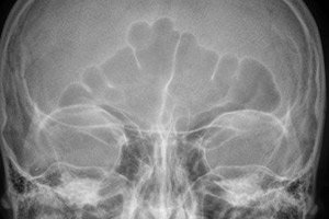
X-ray of a frontal sinus cyst
The main method of instrumental examination is X-ray. It allows visualizing the tumor, viewing the sinuses in various projections and identifying possible pathologies, as well as the nature of the tumor, its severity, size, tissue structure and localization features. Based on this method, a differential diagnosis can be made.
Differential diagnosis
First of all, the essence of differential diagnostics comes down to the need to separate the cyst from other tumors and neoplasms. It is determined whether it is malignant or benign, whether there is a risk of its transformation. For this, a biopsy is usually performed with the taking of a tissue sample for further histological examination. Thus, a piece of tissue is taken from the tumor, then it is placed in a sterile test tube or Petri dish.
After this, under sterile conditions, the culture is seeded on nutrient media intended for cultivating tissue culture. After primary cultivation under optimal conditions (usually in a thermostat or incubator), the culture is transferred to selective media for further identification. After this, a histological examination of the tissue is performed and its nature is determined. The direction and nature of growth can be used to determine the type of tumor and predict its further growth. This is the basis for the final diagnosis.
Who to contact?
Treatment frontal sinus cysts
Treatment of frontal sinus cysts includes measures to eliminate risk factors that contribute to the development of pathology, antibacterial therapy, restoration of normal blood circulation and respiration, restoration of the normal state of tissues (removal of swelling, hyperemia, redness), improvement of gas exchange, sanitation of chronic foci of infection, physiotherapy procedures, hardening of the body, climatotherapy. It is important to exclude smoking and alcohol consumption. If these methods are ineffective, surgical treatment is used.
Prevention
Prevention is based on timely detection of pathology and taking the necessary measures to eliminate it. To do this, it is necessary to undergo regular preventive examinations, conduct the necessary tests, and promptly treat identified concomitant diseases. Prevention also comes down to proper nutrition, maintaining the required level of immunity, normalizing microflora, and sanitizing foci of the infectious and inflammatory process. It is necessary for the body to receive the required amount of vitamins and microelements.
[ 35 ]
Forecast
If the pathology is detected in time and the necessary measures are taken, the prognosis can be favorable. If the cyst is detected at an early stage, it can be treated with conservative methods. If they are ineffective, surgical methods are used. Almost any cyst can be removed surgically, so if this is done in a timely manner, the prognosis can be favorable. If it is not removed in a timely manner, there is a high risk of complications. The most dangerous are inflammation, blockage of the sinus ducts with pus and liquid exudate, the spread of the infectious and inflammatory process to the membranes of the brain, and malignant degeneration.
Is it possible to live with a frontal sinus cyst?
People live with a cyst for quite a long time. The quality of life is significantly reduced. It is better to remove it, because living with a cyst is a constant risk. Complications can arise at any time, inflammation of the brain can develop, which will end in death or disability. It is also necessary to remember that a frontal sinus cyst can transform into a cancerous tumor at any time.