
All iLive content is medically reviewed or fact checked to ensure as much factual accuracy as possible.
We have strict sourcing guidelines and only link to reputable media sites, academic research institutions and, whenever possible, medically peer reviewed studies. Note that the numbers in parentheses ([1], [2], etc.) are clickable links to these studies.
If you feel that any of our content is inaccurate, out-of-date, or otherwise questionable, please select it and press Ctrl + Enter.
Fibrous polyp: what it is, types
Medical expert of the article

Last reviewed: 04.07.2025
Growths with a rounded top on legs, found on the mucous membrane of hollow organs - the digestive tract, genitourinary or respiratory system, as well as on the surface of the skin are called polyps, in Greek - polypus (many legs). Multiple growths of neoplasms are called polyposis, in this case their number should be close to two dozen or more. A fibrous polyp is a formation in the structure of which there are fibers of connective fibrous tissue (fibrous). Such tissue can be present to a greater or lesser extent in the structure of polyps of any localization. In addition to it, glandular tissue is found in the structure of this formation when polyps grow on the mucous membrane, skin epithelial tissue - on the surface of the skin (papillomas), its structure contains vessels that provide nutrition and development of the formation.
Polyps of different localizations represent a wide range of diseases, heterogeneous in origin and morphology, in general, not related to each other.
What does fibrous polyp mean?
This is a neoplasm of dense consistency, the basis of which is fibrous tissue covered with a thin layer of the cellular component of glandular epithelium or epidermis.
When the structure of a polyp is practically no different from the structure of the mucous membrane of the organ on which it is located, such a polyp is called glandular. The stroma in a glandular polyp has a loose structure and is penetrated by vessels. If the vascular pedicle of the polyp is not detected during histology, then the diagnosis itself is questionable.
When the neoplasm's structure is dominated by glandular epithelium and there is a certain amount of connective tissue fibers as a framework for the neoplasm, such a polyp is called glandular-fibrous.
These forms are also interpreted as stages of polyp development. First, a "young" polyp appears, having a soft, predominantly glandular structure; as it ages, with the development of connective tissue fibers, it strengthens, thickens, and acquires a fibrous form.
Causes fibrous polyp
The etiology of polyps has not been fully studied to date. Many factors can trigger the mechanism of polyposis development. As practice shows, the appearance of polyps of any localization was observed in people who already had such cases in their family history, that is, genetically predisposed to polyposis.
Risk factors for the development of these neoplasms also include:
- pathological disruptions in the functioning of the immune system;
- the presence of bad habits – overeating and, consequently, excess weight, the use of psychotropic substances (nicotine, alcohol, drugs);
- age – in adults over 35 years of age, such formations are found much more often;
- the presence of foci of chronic infection, allergic diseases - constant exacerbations create favorable conditions for the development of neoplasms, including polyps.
In addition, it has been noted that during periods of hormonal status changes, polyps are more likely to appear. Living in regions with increased radiation levels is also considered a risk factor for the appearance of tumor-like formations.
Polyps also differ somewhat in structure and age, which may affect the diagnostic conclusion. All polypous formations have connective tissue elements in their structure, which are the framework, and a vascular pedicle, which provides blood supply and growth development.
A fibrovascular polyp is diagnosed when it consists mainly of connective (fibrous) tissue and a whole network of vessels is found in its structure. This is a form of mature age polyp that did not appear yesterday. A synonym for this form is fibrous-angiomatous polyp.
The conclusion "polyp with fibrous stroma" may indicate a younger age of the formation. Apparently, this is a formation of unchanged epithelial cells attached to a framework of connective tissue. Such a polyp also contains vessels, perhaps not too many of them yet.
The conclusion glandular-fibrous polyp of the functional type indicates that in the structure of the formation, in addition to connective tissue cells, there are approximately the same number of cells of the functional layer of the endometrium, that is, hyperplastic changes occurred in this layer. Another type of polyp can also be diagnosed, in which the cells of the basal layer of the endometrium will predominate. In this case, the diagnosis will sound like glandular-fibrous polyp of the basal type.
The histological conclusion fibro-edematous polyp indicates that under the microscope, a predominance of cells of the edematous mucous membrane of the organ with signs of a chronic inflammatory process was detected. The presence of fibrous structures indicates a solid age of the polyp. This is one of the common forms of polyps found in the nasal cavity.
Researchers also identify specific factors that influence the appearance of polyps in a specific location. Pathogenesis and symptoms, as well as the consequences of formations in the digestive tract, in the nose or in the urethra, differ significantly from each other.
Symptoms fibrous polyp
These are completely separate diseases, so it makes sense to consider specific types of fibrous polyps by localization.
Fibrous nasal polyp
A tumor-like formation with a predominance of fibrous tissue cells has a solid age. Such polyps are usually located in the choanae of the nose. They are sometimes mistaken for benign tumors - fibromas.
In addition to the already listed reasons for the appearance of polyps, nasal localization is mainly associated with frequent runny noses that occur as a result of acute respiratory viral infections and become chronic, sinusitis, hay fever, aspirin triad, such an anatomical feature as narrow nasal passages, and other allergic and inflammatory diseases of the nasopharynx.
So, the main pathogenetic link in the appearance of a polyp in the nose is a respiratory infection. The penetration of an infectious agent into the mucous membrane of the nose and its increased reproduction cause changes in the epithelial cells, abundant mucus secretions in order to get rid of the pathogen. With a normal immune status and timely treatment, a complete recovery occurs. In addition, in the presence of a chronic infection, immunity suffers, a person may be prone to the formation of polyps, in short, general risk factors are added to the inflammatory process.
If the inflammation becomes chronic, a hyperplastic process begins in the mucous membrane - the mucous membrane of the nose tries to fight pathogens quantitatively, increasing its surface area. This is a kind of protective reaction of the body. When there is little space, polyps come out into the nasal passages.
The first signs of polyps are not very noticeable, the growth is small and does not cause significant discomfort. The patient is accustomed to a runny nose and perceives copious discharge from the nose as another rhinitis. However, already at the initial stage, the appearance of a polyp can cause complications in the form of inflammation of the adenoids, tonsils or otitis.
If the polyp is not detected, the hyperplastic process continues. Fibrous strands grow, the person's voice changes - it becomes nasal, the nasal passages become increasingly blocked - breathing becomes more difficult, the hearing organs are affected - deafness and speech distortion appear. At this stage, it is imperative to consult a doctor, otherwise the changes may become irreversible.
At the last stage, the air passage in the nose is completely blocked, the symptoms are pronounced - hearing loss, headaches, general weakness, constant nasal congestion and rhinorrhea. If an infection occurs, hyperthermia may occur.
The long-term growth of polyps in the nose can result in malocclusion in a child, poor appetite and underweight, in patients of any age - constant runny nose, sometimes with an admixture of purulent discharge, olfactory impairment, hearing impairment, taste perversion, and a strong pronunciation. The risk of inflammatory processes in the respiratory tract (tracheitis, bronchitis, pneumonia) increases, patients suffer from chronic sinusitis and tonsillitis with frequent exacerbations, inflammation of the Eustachian tube, otitis, and sometimes bronchial asthma develops. A serious complication of polyposis is sleep apnea, which can lead to the death of the patient. It is believed that the life expectancy of a person with nasal polyps is reduced by about six years, mainly due to the lack of normal nasal breathing and the need to breathe in an unnatural way - through the mouth, which leads to insufficient humidification, warming and purification of air that goes straight to the lower respiratory tract. And this is a direct path to the development of various complications.
A nasal polyp is the most common consequence of chronic rhinitis. Perhaps, polyps of this localization are most often found in children. Fibrous polyps of the maxillary sinus are typical for childhood, while in adults, the mucous membrane of the ethmoid labyrinth often grows. On average, nasal polyps are diagnosed in every fiftieth inhabitant of the planet, more often in adults than in children. Also, males are more susceptible to polyposis - such formations are found in them four times more often than in women.
 [ 10 ], [ 11 ], [ 12 ], [ 13 ], [ 14 ]
Fibrous polyp of the urethra
The main cause of tumor-like growths in this localization is considered to be long-term chronic urethritis - chlamydial, trichomonas, gonorrheal, herpetic, caused by opportunistic flora. In the pathogenesis of urethral polyps, intestinal inflammation, ischemia of the walls of the urethra, and its injuries are also considered. The likelihood of polyps appears increases during periods of decreased immunity and fluctuations in hormonal levels.
In a wide and short female urethra, polyps can be located along the entire length, although they are more often found at the exit on the back wall. In male patients - at the entrance to the prostate gland and at the exit to the urethra from the vas deferens.
At the initial stage, the polyp does not manifest itself in any way, only as the formation grows, discomfort appears during urination. Itching and burning, increasing during urination, a feeling of obstruction of the urine outflow, in men it often splashes to the sides, there may be bloody discharge and even significant urethrorrhagia. Large polyps can block the lumen of the urethra and the outflow of urine.
Women may complain of pain during sexual intercourse and bloody discharge after intercourse, men – of various kinds of dysfunctions in the sexual sphere: spontaneous release of sperm after urination, premature ejaculation, traces of blood in sperm, etc.
Fibrous polyps of the urethra are more common than other urethral neoplasms. Women are more prone to them, which is explained by the peculiarities of anatomy and morphology. Such formations are most often diagnosed in patients aged from fifty to seventy years.
[ 15 ], [ 16 ], [ 17 ], [ 18 ], [ 19 ], [ 20 ]
Fibrous polyp of the stomach
The current international classification by histological features divides gastric polyps into true (adenomatous) and pseudotumor formations. The second type includes hyperplastic and inflammatory fibrous polyps of the stomach. The reasons for their appearance have not been precisely established, however, as can be seen from the name, their appearance is associated with chronic inflammation of the gastric mucosa caused by infection with the bacterium Helicobacter pylori.
In addition to the infectious hypothesis, a chemical hypothesis is considered in the pathogenesis of gastric polyps. First of all, the mutagenic effect is blamed on nitric and nitrous acid salts (nitrates and nitrites) entering the stomach with food. These substances have a destructive effect on the epithelial cells of the stomach, which contributes to the growth of polyps.
Factors that increase the likelihood of developing gastric polyps are similar to other localizations.
Unlike adenomatous polyps, which consist of degenerated cells of the mucous epithelium, pseudotumoral formations consist of unchanged epithelial cells and fibrous stroma. They are usually located in the prepyloric and pyloric parts.
A fibrous polyp of the stomach does not carry the risk of malignant transformation, but it can cause profuse gastric bleeding.
Most gastric polyps do not manifest themselves in any way and are detected during gastroscopy, which is performed on patients with complaints of gastritis symptoms. Dyspepsia is the first sign of a fibrous gastric polyp, since it is a manifestation of inflammation, against the background of which the hyperplastic process has developed. Bloating, nausea, heartburn, constipation and diarrhea, rumbling in the stomach, distension after eating - such non-specific symptoms may be a reason for examination.
An increase in the size of the polyp leads to the appearance of ulcers on its surface and internal bleeding. Hidden bleeding leads to the development of anemia.
Polyps on a stalk are often damaged or twisted, which leads to rupture of the vascular membranes. This, in turn, is manifested by the appearance of traces of blood in the feces, brown vomiting, black feces. With massive gastric bleeding, the patient is pale, his blood pressure drops, his pulse quickens sharply, and sweat appears on the forehead.
Quite rarely, polyps manifest themselves as pain upon palpation or after eating.
Large polyps rarely block the pyloric canal and prevent food from moving from the stomach to the duodenum. It stagnates, initially periodically, after eating solid food, then more and more often and after eating pureed food. Symptoms include belching, persistent vomiting with a foul smell of the contents, the same smell from the mouth, and prolonged bursting pain after eating.
It is possible for a polyp on a stalk to penetrate into the duodenum. This is usually accompanied by vomiting, pain of varying intensity - epigastric, umbilical, under the right rib, constipation. In this case, there is a risk of pinching the polyp by the valve located between the stomach and the duodenum (pylorus). Symptoms of pinching are acute paroxysmal pains that cover the entire abdomen.
The most common localization of polyps in the stomach is the piloantral region. According to Russian researchers, polyp growth in this place occurs in 70-85% of cases. Americans also consider this localization to be the main one, however, their figure is lower - 58.5%.
Polyps are found in the body of the stomach in patients of Russian doctors in 17-25% of cases, American doctors cite approximately the same figure - 23.2%. The third most important localization is the cardiac section (researchers unanimously cite figures from 2 to 3%). In this place, polyps are localized mainly in children (cardioesophageal junction).
Solitary polyps occur with approximately the same frequency as multiple ones, among which the diffuse form accounts for about 10%.
There are differences in the gender composition of patients. Some authors claim that polyps are more common in men, but not everyone agrees with them. But regarding age, the authors are unanimous - in most cases, polyps were found in patients aged 40-50 years.
[ 21 ], [ 22 ], [ 23 ], [ 24 ], [ 25 ], [ 26 ]
Fibrous polyp of the intestine
These tumor-like formations are almost never found in the small intestine; their favorite place of localization is the large intestine. The exact reasons for the growth of the intestinal mucosa have not been established; however, the appearance of a fibrous polyp is considered to be the result of intestinal inflammation. Such polyps are most often found in the anal canal. The causes of their occurrence can be inflammation of the sinuses of the rectal canal, hemorrhoids, colitis, and incomplete internal fistula.
Some researchers believe that polyps form in places where the intestinal mucosa has been injured and the regeneration process has been disrupted.
A hollow hemorrhoidal node or hypertrophied anal papilla can transform into a fibrous polyp of the rectum.
In addition to the general risk factors for any neoplasms, people who consume little food containing fiber, rely heavily on carbohydrates and fats, suffer from constipation, dysbacteriosis, low or high acidity, diverticulosis, and lead a sedentary lifestyle are more susceptible to the development of intestinal polyps.
Polyps generally do not show any symptoms in the initial stages. When localized in the small intestine or duodenum, which happens extremely rarely, they grow asymptomatically to large sizes and block the lumen of the intestine. This is manifested by pain in the upper abdomen, a feeling of fullness in the stomach, rotten belching, heartburn, nausea and vomiting. Ignoring such symptoms can result in complete obstruction.
Polyps in the colon are manifested by pain in the area of the corresponding sections, bowel movements disorders - constipation alternates with diarrhea, tenesmus, moderate or significant discomfort during bowel movements, traces of blood or mucus in the feces, bloody or mucous discharge from the anus.
As the polyp grows, the patient begins to feel a foreign body, the polyp may begin to fall out of the anus, be injured by fecal matter, and become inflamed. Complications develop – burning, itching, pain, and the inflammation spreads to the skin surrounding the anus.
Polyps in the intestines can appear at any age, but after 50 years the likelihood of such formations increases, and they are found more often in men.
[ 27 ], [ 28 ], [ 29 ], [ 30 ]
Fibrous polyp in the uterus
Uterine formations have a hyperplastic origin, that is, they are a consequence of increased growth of cells of the inner layer lining the uterus. Polyps of the uterine body can be located anywhere in its inner layer, grow into the uterine cavity and are usually small in size, although sometimes they reach three centimeters. Vessels are present in polyps of all types, ensuring their growth and development.
The endometrium has a two-layer structure - the functional layer, which is rejected monthly, and the basal layer - its base. These layers differ in structure and polyps can have different shapes, corresponding to the cellular structure of the layers.
Functional polyps are formed with excess estrogen or progesterone, since this layer actively reacts to quantitative changes in sex hormones. In this layer, a glandular-fibrous polyp of the endometrium is formed, provided that the functional layer has not completely come out during menstruation. The glands that make up the majority of the polyp have the structure of the functional layer. Formations of this type are quite rare, mainly in women of childbearing age, after menopause - even twice as rare.
The basal layer does not react to hormonal fluctuations, polyps of this type have much more fibrous fibers, its structure is denser, and the glandular tissue is represented by cells of the basal layer. Such polyps are typical for mature patients, who still have high levels of estrogens.
A fibrous polyp of the endometrium can be formed only by connective fibers, with single inclusions of glands, and there are also not many vessels. In a fibrous polyp, the vascular pedicle has a thickened sclerotic membrane.
A fibrous polyp of the cervical canal grows on the mucous membrane of the cervix (synonyms - fibrous polyp of the endocervix, fibrous polyp of the cervix). Its structure is similar to an endometrial polyp - cells of glandular, connective and vascular tissue. Depending on the ratio of different types of cells, endocervical polyps can also be glandular-fibrous and fibrous.
The reasons for the appearance of polyps on the mucous membrane of the uterus and its cervix have not yet been fully clarified. There are several hypotheses and, perhaps, all of them have a right to exist. The process of development of any neoplasms is multifactorial.
The mechanism of proliferation of cells of the mucous membrane lining the uterus and cervical canal is triggered by inflammatory diseases. Almost all patients with polyps had other gynecological problems: endometritis, cervicitis, inflammation of the appendages, vaginitis, vaginal dysbacteriosis and other infectious and inflammatory processes in chronic form.
Damage to the cervix during childbirth (rupture), as a result of long-term contraception using an intrauterine device and destructive treatment methods also becomes the cause of polyps in this location.
The main cause of endometrial polyps is called hormonal disorders and, first of all, excess estrogen is blamed. Polyposis is considered a special case of endometrial hyperplasia, as a consequence of replacement therapy with estrogen-containing drugs in postmenopause.
It has already been established that endometrial polyps have not only estrogen but also progesterone receptors. It is believed that the development of polyposis is affected by a deficiency of the pregnancy hormone.
However, the hormonal theory, recognized as the main one in the development of intrauterine polyps, is not confirmed in relation to endocervical polyps. They are more common in the postpartum period and are practically not found in women who have crossed the half-century mark. In the pathogenesis of fibrous polyps of the cervix, the main role is given to injuries and inflammations.
Long-term corticosteroid therapy may also be associated with increased uterine polyp growth.
Polyposis is quite common in women who have taken the estrogen antagonist drug Tamoxifen as part of their breast cancer treatment regimen.
Also considered in the pathogenesis of pseudotumor growths of the endometrial mucosa are enzymatic hyperactivity of aromatase; ischemic processes in the tissues of the uterus associated with vascular occlusion, destructive processes (myoma, endometriosis, pseudo-erosions); surgical injuries (abortions, diagnostic curettage).
A hereditary factor (a gene, HNGIC-gene, responsible for the formation of polyps, was found in the endometrial cells) and other general reasons indicated above can also contribute to the initiation of the hyperplastic process.
The specifics of the symptoms also depend on many factors, about a fifth of cases (and maybe more), when polyps are up to 10 mm in size, are asymptomatic. And if a fibrous polyp of the cervix can sometimes be detected visually during a gynecological examination, then formations located inside the uterus - only on ultrasound or during diagnostic curettage prescribed for some other reason.
The main sign of the presence of polyps of both the endometrium and endocervix is considered to be discharge with streaks of blood or uterine bleeding in the intermenstrual or menopausal period, after coitus or a gynecological examination. Such symptoms are observed in a third of patients with endometrial polyps.
Long (up to seven days), heavy menstruation with multiple blood clots, dull aching pain in the lower abdomen should be a cause for concern. Sometimes painful sensations appear during or after sexual intercourse.
Large formations can cause profuse vaginal discharge, whitish or grayish in color.
In addition, infertility or habitual miscarriage may be symptoms that indirectly indicate the presence of a polyp. Experts believe that such consequences are caused not so much by the presence of a formation, but by a hormonal imbalance or an inflammatory (destructive) process that led to polyposis.
It is typical that fertile patients with glandular fibrous polyp of the endometrium usually have a stable menstrual cycle without disturbances.
Such formations are mostly found in women over 35 years old, and in the late reproductive period more often than after menopause. However, there are cases of polyposis in very young girls who are not yet sexually active.
The presence of a fibrous polyp of the endometrium and/or endocervix, even if it occurs without pronounced symptoms, reduces a woman’s quality of life – chronic inflammation, the possibility of bleeding, and pain after sex lead to a decrease in interest in intimate life; in addition, the risk of infection of the genitals increases, since the local immunity of their mucous membrane is reduced.
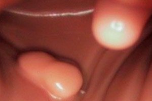
Fibrous polyp of the mucous membrane and skin
In addition to the polypous formations described above, which are quite common, such growths can form on the mucous membranes of any hollow organ - in the mouth, in the ear, on the vocal cords. Medicine is still studying the causes of their formation. Any infectious and inflammatory process that occurs on the mucous membrane for a long time increases the likelihood of the appearance of a fibrous polyp. While the polyp is small, it is usually discovered by chance, during an examination of the organ for some other problems, in particular, the same chronic inflammation. Later, some indirect symptoms appear, for example, a runny nose - with polyps in the nose, hoarseness - with polyps on the vocal cords, which can be interpreted as colds. Therefore, you should pay close attention to your health and get examined when any persistent symptoms of trouble appear.
Polyps, or more precisely papillomas, are fibroepithelial growths of a round or oval shape on legs that can also appear on the skin. Such formations appear in places that often suffer from friction against clothing or other areas of the skin. Their favorite places are in the armpits, on the inner upper surface of the thighs, on the eyelids, back and neck, under the bust in women.
They are also supplied with vessels, connective tissue fibers and cellular epithelium. Basically, they do not cause any discomfort, except for aesthetics. If a polyp is injured, slight bleeding may occur on the skin.
People at risk for developing skin polyps include those who are overweight – those who love sweets, flour, diabetics; pregnant women; and those with a corresponding hereditary predisposition. With age, the likelihood of developing such neoplasms increases. Women are more prone to developing papillomas, however, men, especially older and overweight ones, also have such formations.
Their pathogenesis involves the human papilloma virus, which can “sleep” in the body for a long time and manifest itself under the influence of one or a combination of several factors common to all polyps.
Skin polyps in the vast majority of cases are not dangerous, however, if they appear, you should see a doctor. After all, this is a neoplasm.
[ 33 ]
Fibrous polyp and pregnancy
One of the undesirable consequences of uterine polyposis can be infertility. A single large polyp or its unfortunate location can block the access of spermatozoa to the fallopian tubes, preventing implantation of the fertilized egg.
However, this is not always the case. In many cases, a woman can become pregnant with an endometrial or cervical polyp. Treatment of polyps during pregnancy is not carried out, except for cases when the polyps are large (exceeding 10 mm), bleed, manifest themselves as pronounced inflammation with elements of destruction or necrosis, have modified cells (not necessarily cancerous).
The pathology itself can lead to serious complications during pregnancy.
An intrauterine fibrous polyp located near the place of attachment of the placenta can provoke its partial detachment, which can result in premature termination of pregnancy or inadequate nutrition of the fetus.
A cervical polyp can lead to premature opening of the cervix (cervical insufficiency) and low placental location.
Fibrous polyps during pregnancy cause serious discomfort to most women: pain in the lower abdomen and in the lumbar region, bloody discharge from the vagina. Small polyps, as a rule, do not have a negative effect on the fetus. However, given the possible complications, it makes sense for a woman to be checked for polyps before pregnancy and get rid of them in advance.
Currently, hysteroscopy is used in most cases to remove polyps, which is a more gentle operation than the classic endometrial curettage, similar to a surgical abortion. The onset of pregnancy after surgical removal of polyps is quite possible in most cases.
Complications and consequences
A fibrous polyp of any localization is a benign tumor-like formation, a pseudotumor, which is formed from epithelial cells and connective tissue. What is the danger of a fibrous polyp? Why do specialists almost always insist on its removal?
The most serious consequence of having a fibrous polyp of the mucous membrane of any organ or skin is the risk of malignancy. Although such a process occurs with fibrous polyps in very rare cases, cellular degeneration is still considered possible. Experts estimate the frequency of malignancy at 0.5-1%, but such a probability exists.
In addition, it is impossible to determine the type of cells that make up a polyp only by the appearance of the formation. Such a prognosis is tentative. Even the smallest formation in the stomach, intestines, uterus, resembling a polyp in shape, may turn out to be a malignant tumor. And, naturally, the sooner this is established, the more favorable the prognosis. And conclusions about the cellular structure of the formation can only be made after a histological examination of the materials of the removed polyp.
A glandular-fibrous polyp with inflammation is considered even more dangerous in terms of malignancy than a simple fibrous one. Rapidly growing glandular components are more susceptible to transformations, the polyp first turns into an adenomatous one, and then, if left untreated, a neoplastic process may begin. The probability of malignancy of adenomatous polyps is estimated at 3-3.5%.
Even if we assume that the fibrous polyp remains benign, its presence and growth inside the organ leads to dystrophic changes, an ongoing inflammatory process. Large formations block natural openings, preventing breathing, the passage of food, sperm (depending on the location). And again, the risk of degeneration in large polyps increases several times.
Diagnostics fibrous polyp
The choice of diagnostic procedure depends on the location of the polyp. Sometimes they can be detected during a visual examination using gynecological mirrors (on the cervix, in the urethra), rhinoscopy (in the nose). To detect formations in the cavity of internal organs, instrumental diagnostics are used - ultrasound, contrast radiography, computed tomography or magnetic resonance imaging (uterine cavity, bladder, nasal sinuses), urethroscopy, hysteroscopy, endoscopic examination of the stomach and upper intestine, colonoscopy, rectoscopy. The choice of technique depends on the location of the polyp and the capabilities of the medical institution.
And if non-invasive studies (X-ray, ultrasound, CT, MRI) are purely diagnostic, then, for example, with the help of hysteroscopy, the polypous formation is immediately removed, after which separate diagnostic curettage of the cervix and uterine cavity is performed. If the formations are localized in the stomach or intestine, their endoscopic removal is performed. Often, diagnostic procedures are simultaneously therapeutic.
After polypectomy, histology of the fibrous polyp is mandatory. Only after a thorough examination of the materials from the removed tissues can a diagnostic conclusion be made with confidence - whether the polyp tissues are unchanged or whether a neoplastic process has already begun to occur in them.
In addition, inflammation that is almost always present requires tests to identify pathogenic flora - these can be PCR tests, cultures, microscopy, and others.
Differential diagnosis
Ultrasound diagnostic data are confirmed by histological studies in 80% of cases. This is a high level of accuracy, allowing to determine the necessity and extent of surgical intervention. In pregnant women, decidual pseudopolyp is differentiated from true polyp. Uterine polyps are distinguished with endometrial hyperplasia, small myomas that cannot be removed, early pregnancy, including missed pregnancy; endocervical polyps - with hyperplasia of the stromal wall of the cervical canal.
Based on the histological examination data, a malignant process is first excluded and the type of polyp is determined (glandular, adenomatous, fibrous, etc.).
The presence of infection or allergic reaction is excluded or confirmed.
In all cases, it is possible to accurately differentiate polyps of different localizations from other types of tumors of these organs (angiomas, lipomas, non-epithelial tumors and other formations) only by examining biopsies or tissues of the removed polyp.
This helps to determine the tactics for conducting the further course of therapy.
Treatment fibrous polyp
As practice shows, fibrous polyps of any localization do not resolve themselves, hormonal therapy is also ineffective in most cases. Polyps cannot be ignored due to possible malignancy. The presence of polyposis is interpreted as a precancerous condition. The only way to get rid of polyps is surgical. Treatment of fibrous polyps is usually prescribed after removal and a histological examination of the formation is mandatory. The main goal of postoperative therapy is to prevent relapses.
Prevention
To prevent the formation of polyps of any localization, it is necessary to promptly identify and treat various infectious and inflammatory diseases, preventing their chronicity, monitor hormonal and immune status, lead an active lifestyle, eat right, and give up bad habits.
If you do have to deal with this phenomenon, it is worth considering that polyps tend to recur, so after their removal you should not refuse the proposed course of therapy, you must strictly follow the doctor's recommendations and undergo regular examinations.
Forecast
The vast majority of fibrous polyps of any localization are benign formations. Their removal is possible using minimally invasive technologies, sometimes even on an outpatient basis.
With timely treatment and following the doctor's recommendations, the prognosis for life is favorable.