
All iLive content is medically reviewed or fact checked to ensure as much factual accuracy as possible.
We have strict sourcing guidelines and only link to reputable media sites, academic research institutions and, whenever possible, medically peer reviewed studies. Note that the numbers in parentheses ([1], [2], etc.) are clickable links to these studies.
If you feel that any of our content is inaccurate, out-of-date, or otherwise questionable, please select it and press Ctrl + Enter.
Escherichioses (genus Escherichia, E. coli)
Medical expert of the article

Last reviewed: 06.07.2025
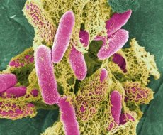
The main representative of the genus Escherichia - E. coli - was first discovered in 1885 by T. Escherich, after whom this genus of bacteria received its name. Key features of this genus: peritrichous (or non-motile), ferment lactose with the formation of acid and gas (or lactose-negative), do not grow on a starvation medium with citrate, the Voges-Proskauer reaction is negative, the MR test is positive, do not have phenylalanine deaminase, do not grow on a medium with KCN, the content of G + C in DNA is 50-51 mol%.
The genus Escherichia includes at least 7 species; of particular importance in medicine is the species E. coli, in particular those variants that cause human diseases. They are divided into 2 main groups: those that cause extraintestinal diseases and those that cause acute intestinal diseases (AID). Representatives of the first are divided into three pathological groups:
- meningeal (MENEC - meningitis E. coli);
- septicemic (SEPEC - septicemia E. coli) and
- uropathogenic (UPEC - uropathogenic E. coli).
In turn, the E. coli variants causing acute respiratory infections were initially divided into the following 4 categories: enterotoxigenic E. coli (ETEC); enteroinvasive E. coli (EIEC); enteropathogenic E. coli (EPEC) and enterohemorrhagic E. coli (EHEC). Subsequently, two more categories were identified: enteroaggregative E. coli (EAEC) and diffusely aggregative E. coli (DAEC).
In addition, E. coli is used in international standards as an indicator of the degree of faecal contamination of water, especially drinking water, and food products.
The standard strain of E. coli (E. coli K-12) is widely used in laboratories in many countries around the world to study the genetics of bacteria.
 [
[ Morphology
E. coli is a facultative anaerobe, grows well on conventional nutrient media - colonies on agar are round, convex, translucent. Growth on broth is in the form of diffuse turbidity. The temperature optimum for growth is 37 °C, grows in the range from 10 to 45 °C, the optimal pH is 7.2-7.5. On all differential diagnostic media, colonies of E. coli decomposing lactose are colored in the color of the indicator (on Endo medium - dark crimson with a metallic sheen).
Biochemical properties
In most cases, E. coli is capable of fermenting the following carbohydrates to form acid and gas: glucose, lactose, mannitol, arabinose, galactose, sometimes sucrose and some other carbohydrates; forms indole; usually does not form H2S ; reduces nitrates to nitrites, does not liquefy gelatin, does not grow on a starvation medium with citrate, gives a positive reaction with MR and a negative reaction with Voges-Proskauer. By these signs, it can be easily distinguished from pathogens of a number of diseases (dysentery, typhoid fever, salmonellosis, etc.). However, pathogenic E. coli very often do not differ from non-pathogenic ones either by cultural or biochemical properties.
Pathogenicity factors of E. coli
The ability of E. coli to cause various diseases is due to the presence of the following pathogenicity factors:
Adhesion and colonization factors. They are necessary for attachment to tissue cells and their colonization. Three variants of the colonization factor have been discovered: a) CFA/I-CFA/VI (colonization factor) - they have a fimbrial structure; b) EAF (enteropathogenic E. coli adherence factor) - intimin - an outer membrane protein, encoded by the eaeA gene. Found in 4 and EHEC, it is detected by the ability of bacteria to attach to Hep-2 cells; c) Adhesion Henle-407 - fimbrial structures, detected by the ability of bacteria to attach to Henle-407 cells. All of them are encoded by plasmid genes. In addition to them, other colonization factors have been described, which can also include bacterial lipopolysaccharides.
Invasion factors. With their help, EIEC and EHEC, for example, penetrate into intestinal epithelial cells, multiply in them and cause their destruction. The role of invasion factors is performed by proteins of the outer membrane.
Exotoxins. Pathogenic E. coli have been found to contain exotoxins that damage membranes (hemolysin), inhibit protein synthesis (Shiga toxin), and activate secondary messengers (messenger - communication) - toxins CNF, ST, CT, CLTD, EAST.
Hemolysins are produced by various pathogens, including E. coli. Hemolysin is a pore-forming toxin. It first binds to the target cell membrane and then forms a pore in it, through which small molecules and ions enter and exit, leading to cell death and erythrocyte lysis.
Shiga toxin (STX) was first discovered in Shigella dysenteriae, and then a similar toxin (Shiga-like toxin) was found in EHEC. The toxin (N-glycosidase) blocks protein synthesis by interacting with 28S rRNA, resulting in cell death (cytotoxin). There are two types of Shiga-like toxin: STX-1 and STX-2. STX-1 is almost identical to Shiga toxin in its antigenic properties, while STX-2 differs from Shiga toxin in its antigenic properties and, unlike STX-1, is not neutralized by antiserum to it. Synthesis of STX-1 and STX-2 cytotoxins is controlled in E. coli by the genes of the moderate converting prophages 9331 (STX-1) and 933W (STX-2).
- Toxin L (heat-labile toxin) is an ADP-ribosyltransferase; by binding to a G protein, it causes diarrhea.
- ST toxin (thermostable toxin), interacting with the guanylate cyclase receptor, stimulates its activity and causes diarrhea.
- CNF (cytotoxic necrotic factor) is a deamidase protein that damages so-called RhoG proteins. This toxin is found in UPEC, which causes urinary tract infections.
- CLTD toxin is a cytolethal disintegrating toxin. The mechanism of action is poorly understood.
- EAST toxin is a heat-stable toxin of enteroaggregative E. coli (EAEC), probably similar to heat-stable toxin (ST).
Endotoxins are lipopolysaccharides. They determine the antigenic specificity of bacteria (which is determined by the repeating side chain of sugars) and the shape of the colonies (loss of side chains leads to the transformation of S-colonies into R-colonies).
Thus, the pathogenicity factors of E. coli are controlled not only by the chromosomal genes of the host cell, but also by genes introduced by plasmids or temperate converting phages. All this indicates the possibility of the emergence of pathogenic variants of E. coli as a result of the spread of plasmids and temperate phages among them. Below is a brief description of 4 categories of E. coli causing acute respiratory infections; information on the recently identified categories DAEC and EAEC was not found in the sources available to us.
ETEC includes 17 serogroups. The adhesion and colonization factors of the fimbrial structure of the CFA type and enterotoxins (LT or ST, or both) are encoded by the same plasmid(s). They colonize the villi without damaging them. Enterotoxins cause a violation of water-salt metabolism. The localization of the process is the small intestine. The infective dose is 108-1010 cells. The disease proceeds as cholera-like diarrhea. The type of epidemics is waterborne, less often foodborne. Children aged 1 to 3 years and adults are affected.
EIEC includes 9 serogroups, pathogenicity is associated with the ability to penetrate into the epithelial cells of the intestinal mucosa and multiply inside them, causing their destruction. These properties are encoded, in addition to chromosomal genes, by plasmid genes (140 MD). The plasmid encodes the synthesis of proteins of the outer membrane, which determine the invasion. Both the plasmid itself and the proteins it encodes are related to those of dysentery pathogens, which explains the similarity of EIEC with shigella. The infective dose is 10s cells. Localization of the process is the lower ileum and large intestine. The disease proceeds as dysentery: initially watery diarrhea, then colitic syndrome. Children 1.5-2 years old, adolescents and adults are ill. Type of outbreaks - food, water.
Epidemiology
E. coli is a representative of the normal microflora of the intestinal tract of all mammals, birds, reptiles and fish. Therefore, to clarify the question of which variants of E. coli and why cause escherichiosis, it was necessary to study the antigen structure, develop a serological classification necessary for identifying pathogenic serovariants, and find out what pathogenicity factors they possess, i.e. why they are capable of causing various forms of escherichiosis.
E. coli has 171 O-antigen variants (01-0171), 57 H-antigen variants (H1-H57) and 90 surface (capsular) K-antigen variants. However, in reality there are 164 O-antigen groups and 55 H-antigen serovariants, since some of the previous 0:H serogroups were excluded from the E. coli species, but the ordinal numbers of the O- and H-antigens remained unchanged. The antigenic characteristics of diarrheagenic E. coli include the O- and H-antigen numbers, for example, 055:116; 0157:H7; O-antigen indicates belonging to a certain serogroup, and H-antigen is its serovariant. In addition, a more in-depth study of O- and H-antigens revealed the so-called factor O- and H-antigens, i.e. their antigenic subvariants, such as H2a, H2b, H2c or O20, O20a, O20ab, etc. In total, the list of diarrheagenic E. coli includes 43 O-serogroups and 57 OH-serovariants. This list is constantly being supplemented with new serovariants.
Symptoms
The group includes 9 class 1 serogroups and four class 2 serogroups. Class 1 serogroups have a plasmid (60 MDa) that controls the synthesis of the adhesion and colonization factor of the EAF type. It is represented by a protein localized in the outer membrane and is detected by the ability of bacteria to attach to HEp-2 cells. The protein has an m. w. of 94 kDa. Class 2 serogroups do not have this plasmid; their pathogenicity is due to some other factors. Some strains of both classes have been found to be able to synthesize STX. 4 colonize the plasma membrane of enterocytes, causing damage to the epithelial surface with the formation of erosions and moderate inflammation. The infectious dose is 105-10 12 cells. The process is localized in the small intestine. The disease is characterized by watery diarrhea and severe dehydration. Mostly children in the first year of life are sick. The method of infection is contact-household, less often food-borne.
Serogroups EIEC and 4 are the most common causes of nosocomial outbreaks.
EHECs produce cytotoxins STX-1 and STX-2. They cause hemorrhagic colitis in humans with severe complications such as hemolytic uremia and thrombotic thrombocytopenic purpura. The toxins destroy the endothelial cells of small blood vessels. The formation of blood clots and fibrin deposition lead to impaired blood flow, bleeding, ischemia, and necrosis in the cell wall. Uremic hemolytic syndrome can be fatal. EHECs are represented by many serotypes (-150), but the main epidemiological role is played by E. coli 0157-H7 and its flagellate mutant E. coli 0157:NM, since only they produce STX. These strains of bacteria can secrete only one of the cytotoxins or both simultaneously. It is believed that the natural reservoir of EHEC serovars, including E. coli 0157:H7, are cattle and sheep. The most common route of infection is food (meat, especially minced meat; milk). E. coli 0157:H7 is unusually resistant to unfavorable factors. This contributes to its survival and reproduction in various products. Infection is possible through contact with household items. The onset of the disease is acute: intestinal spasms occur, then diarrhea, initially watery, then bloody. Children and adults are ill. A sick person is contagious.
Laboratory diagnostics
It is based on the isolation of a pure culture of the pathogen and its identification, as well as on testing toxins using PCR. The causative agent of escherichiosis is identified using a set of polyvalent OK-sera and a set of adsorbed sera containing antibodies only to certain antigens. A keratoconjunctival test can be used to identify EIEC. Some representatives of EIEC are immobile and do not ferment lactose and salicin. Identification of E. coli 0157:H7 is facilitated by its inability to ferment sorbitol (Endo medium with sorbitol is used instead of lactose). But it is best to use PCR test systems for identifying and differentiating OKZ pathogens (all categories). If necessary, the sensitivity of the isolated pathogens to antibiotics is determined.