
All iLive content is medically reviewed or fact checked to ensure as much factual accuracy as possible.
We have strict sourcing guidelines and only link to reputable media sites, academic research institutions and, whenever possible, medically peer reviewed studies. Note that the numbers in parentheses ([1], [2], etc.) are clickable links to these studies.
If you feel that any of our content is inaccurate, out-of-date, or otherwise questionable, please select it and press Ctrl + Enter.
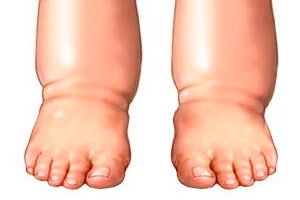
Elephant feet
Medical expert of the article

Last reviewed: 04.07.2025
Elephantiasis is a rare disease caused by a disruption of lymph flow. Let's consider the features of this pathology, types, stages, diagnostic and treatment methods.
According to the international classification of diseases ICD-10, elephantiasis of the legs falls under category IX Diseases of the circulatory system (I00-I99):
I95-I99 Other and unspecified diseases of the circulatory system.
- I97 Post-procedural disorders of the circulatory system, not elsewhere classified.
- I97.2 Postmastectomy lymphedema syndrome (elephantiasis, obliteration of lymphatic vessels, mastectomy).
Elephantiasis of the lower extremities occurs due to lymph stagnation. Lymphatic fluid performs important functions in the body. It cleanses tissues and cells from accumulated toxins and normalizes water balance. The colorless liquid supplies blood to all organs and systems and participates in the formation of immunity.
With persistent edema, metabolic products are not removed from the tissues, protein compounds disintegrate and provoke the formation of fibrin. This leads to the appearance of coarse connective tissue between the muscles. The limbs increase in size, acquiring a cylindrical shape, which outwardly resembles the legs of an elephant. The skin on the affected tissues ulcerates, becomes covered with cracks, rashes and warts.
The disease can occur due to congenital weakness of the lymphatic system, injuries, parasitic and bacterial infections. In 70% of cases, the disorder appears only on one leg; cases of bilateral lymphedema are extremely rare. But the problem is not only in the deformation of the limbs. The pathology affects internal organs and systems, disrupting the functioning of the entire body. In particularly severe cases, elephantiasis affects not only the legs, but also the arms, face, mammary glands, and genitals.
Epidemiology
More than 300 million people – 13% of the world’s population – face the problem of lymphatic edema. All of them belong to the high-risk group for elephantiasis. At the same time, medical statistics indicate that filaria infection alone causes the disease in 100 million people in the tropics.
In European countries and on continents with a temperate climate, the disease has a low prevalence. Here, elephantiasis occurs due to a number of other factors, both congenital and acquired.
According to statistics, lymphedema most often affects the lower extremities - about 95% of all cases of the disease. Less often, persistent swelling of the hands, mammary glands, face, genitals is diagnosed. In 70% of cases, the problem is one-sided.
Causes elephantiasis
Elephantiasis is associated with pathological changes in the lymphatic system. Fluid accumulation occurs due to blockage or narrowing of the lymphatic ducts. The causes of elephantiasis of the legs depend on the type of disease.
Secondary elephantiasis, that is, acquired, is directly related to a disorder of the lymphatic system of various etiologies and can occur at any age.
- Tumor lesions and removal of lymph nodes, chemotherapy. The affected lymph node passes lymph with certain disorders. The fluid accumulates in the vessels, stretches them and even gets into the tissues. Long-term stagnation provokes severe edema and proliferation of connective tissue.
- Erysipelas and phlegmon caused by streptococcal infection. Microorganisms multiply in the lymphatic capillaries, and the toxins they secrete provoke allergic reactions. The immune system fights the problem by provoking increased cell division and tissue enlargement.
- Damage to lymphatic vessels occurs with frostbite, extensive injuries, burns. A large amount of lymph stagnates in the tissues, which causes persistent swelling.
- Varicose veins and post-thrombophlebitis syndrome. Damage to deep veins disrupts the functioning and nutrition of soft tissues. Gradually, changes affect the lymphatic vessels, disrupting their patency, which leads to lymph stagnation. Pathogenic microorganisms multiply in the altered cells, causing intoxication of the body. This leads to tissue proliferation and skin rashes.
- Parasitic infestations from insect bites are another cause of elephantiasis. Mosquitoes and midges can infect a person with filariae, worms that parasitize the lymphatic vessels. Helminths intertwine into balls, clogging and stretching the lumen of the vessels. The toxic-allergic reaction of the body is accompanied by edema and proliferation of connective tissues.
Primary lymphostasis (congenital) may be associated with the following causes:
- Increased lymph production.
- Nonne-Milroy-Meige syndrome (tissue trophic disorder).
- Shershevsky-Turner syndrome (chromosomal pathology).
- Anomalies in the development of lymphatic vessels (aplasia, dysplasia, hypoplasia, hyperplasia).
- Lesions of the central nervous system.
- Endocrine disorders.
- Valve insufficiency.
In 3-5% of cases, it is difficult to establish the primary causes, so this form of the disease is called idiopathic. Congenital pathology very often leads to damage to both limbs.
Risk factors
Elephantiasis develops due to many reasons associated with both congenital and acquired factors.
The occurrence of the disease can be influenced by risk factors such as:
- Oncological pathologies with damage to the lymphatic vessels.
- Chemotherapy or radiation.
- Venereal diseases.
- Circulatory disorders.
- Diseases of the hematopoietic system.
- Varicose veins.
- Surgical interventions with removal of lymph nodes.
- Systemic lupus erythematosus.
- Autoimmune pathologies.
- Parasitic infestations.
- Severe frostbite.
- Injuries to soft tissues of the lower extremities.
- Overweight, obesity.
- Chronic eczema.
The above-mentioned diseases are dangerous not only because of the high risk of developing elephantiasis, but also because of the significant disruption of the normal functioning of the body.
Pathogenesis
The lymphatic system is involved in metabolic processes and the cleansing of cells from toxins. It consists of vessels, nodes, trunks and capillaries. Free passage of fluid through the vascular bed ensures normal lymph flow.
The mechanism of elephantiasis development is associated with the insufficiency of the lymphatic system functions and the disruption of fluid outflow. Normally, the tissues of the lower extremities synthesize about 2 liters of lymph daily, but with vascular blockages, congestion occurs, which manifests itself as persistent edema.
The pathogenesis of elephantiasis is based on the sequential development of the following pathological changes:
- Violation of lymph drainage.
- Fluid retention in tissues.
- Lymphatic edema due to tissue impregnation with proteins.
- Pathological restructuring of the lymphatic system.
- Fibrous processes affecting the dermis, subcutaneous tissue, and fascia.
Disruption of the fluid flow leads to increased intralymphatic pressure and decreased resorption (absorption). Fluid and protein accumulate in the tissues. Protein compounds disintegrate and transform into fibrin fibers. Fibroblasts penetrate the altered tissues and form collagen fibers. Against this background, serious disruptions occur in the cells of the connective tissue.
Fibrous changes in elephantiasis affect the skin, subcutaneous fat layer, muscles, fascia, walls of arterial, venous and lymphatic vessels. Growing edema worsens hemo and lymphodynamics. Metabolic products accumulate in the tissues, hypoxia occurs. This leads to a weakening of the protective properties of the immune system. Soft pasty edema appears. When it intensifies, the dermis becomes easily injured. Against this background, soft tissue pathologies and trophic disorders develop. Rapid progression of lymphostasis leads to deformation of the damaged limb.
Symptoms elephantiasis
The signs of elephantiasis depend entirely on the causes, type and stage of the pathological process. Symptoms of elephantiasis of the legs arise as the disease progresses, let's consider the main ones:
- Edema most often appears on only one limb. Unilateral lesions are typical for the acquired form of the disease. In congenital pathologies, lymphostasis of both legs is possible at the same time.
- As the swelling increases, a feeling of distension appears in the limb. Discomfort is accompanied by increased fatigue and deterioration of general well-being.
- The swelling appears on the foot or hand, i.e. below the affected area of the lymphatic vessels. It gradually moves to the ankle and then to the thigh. The swelling is soft, so when pressing on the tissue, pits appear.
- The disruption of lymph drainage leads to the proliferation of pathogenic microorganisms in the lymphatic system and the thickness of the skin. The lymph nodes that filter the lymph in the affected area increase in size and become inflamed.
- The fluid in the intermuscular space and subcutaneous fat are gradually replaced by connective tissue. Because of this, the leg becomes hard to the touch, that is, the swelling hardens. The skin is almost impossible to gather into a fold, and when pressed, no pits remain.
- Persistent progression of edema leads to deformation of the limb. All bulges on the ankle are smoothed out, the leg acquires a cylindrical shape. The limb increases in volume several times.
- Impaired blood circulation leads to atrophy of the sebaceous and sweat glands (their secretion protects the dermis from bacteria and viruses). Due to the disruption of the protective layer, various rashes, papillomas, warts, abscesses, ulcerative lesions, and cracks appear on the skin. The presence of bacterial flora is dangerous due to the development of allergic reactions.
- Excess lymph fluid begins to be excreted through the skin. Fistulas form on the tissues, through which yellowish fluid flows out. Most often, the holes are localized in places with thin skin, that is, in the interdigital folds.
- Due to impaired blood circulation, the tissues begin to produce a lot of melanin. Brown spots appear on the extremities. Increased cell division leads to the growth of shapeless bumps and other growths, separated by transverse folds.
The above-described symptom complex may be accompanied by disturbances in other organ systems, significantly worsening well-being.
Swollen legs in elephantiasis
Lymphedema is a pathological condition with progressive swelling of the soft tissues of the affected area. Swelling of the legs in elephantiasis develops as a result of a disruption in the flow of lymph through the lymphatic vessels. This pathology can be associated with both congenital and acquired factors.
There are several types of lymphatic edema, let's look at them:
- Mechanical – appear after tissue trauma.
- Cachectic - associated with cardiovascular pathologies and exhaustion of the body.
- Congestive – increased capillary pressure, pronounced vascular permeability and decreased albumin levels.
- Neuropathic – endocrine pathologies, alcoholism.
- Hydremic - accumulation of lymph caused by kidney diseases.
In mild elephantiasis, swelling disappears after adequate rest and wearing compression garments. In moderate severity, persistent, non-disappearing swelling with growth of connective tissues is observed. The skin becomes taut and dense. The patient complains of painful sensations and distension of the legs, and general well-being worsens. Temporary cramps and paresthesia are possible.
Severe edema, i.e. the last stage of elephantiasis, leads to irreversible damage to the lymph flow, fibrocystic changes in the tissues. The limb is severely deformed and cannot function normally. Because of this, contractures, deforming osteoarthrosis, eczema, erysipelas, trophic ulcers develop. Another danger of persistent edema is the increased risk of lymphosarcoma.
 [ 24 ], [ 25 ], [ 26 ], [ 27 ]
First signs
A feature of lymphedema is that at first its symptoms are so blurred that the patient does not attach much importance to them. In the evening, a slight swelling appears on the feet and ankles, which is often attributed to fatigue during the day. The swelling is especially noticeable in hot weather, after prolonged physical exertion and during the menstrual cycle. At the same time, the joint retains normal mobility and there is no pain in the leg.
The first signs of elephantiasis:
- Periodic swelling of one or both limbs.
- The swelling is especially noticeable at the end of the day, but disappears completely after a night's rest.
- Swelling increases with vertical positioning of the limbs, after increased physical exertion and with limited mobility.
- At the early stage, irreversible tissue growth and other pathological changes do not occur.
Moreover, the above-described symptoms of the disease can persist for many years, accompanied by a deterioration in general well-being and weakness.
Stages
The symptom complex of elephantiasis of the lower extremities has the following stages:
- At first, small swellings appear. They are associated with the growth of fibrous tissue and disruption of tissue metabolism. The swelling begins with the foot and gradually moves above the knee to the thigh.
- Asymmetrical swelling of the extremities.
- Soft swelling (after pressing on the skin, a pit remains).
- The tissues appear very pale, are easily displaced, but are difficult to gather into folds.
- Rashes and itchy skin appear.
The duration of the first stage is about 6-8 months.
- The symptoms become more pronounced. The swollen area thickens and steadily increases in size.
- Enlarged lymphatic vessels can be felt.
- The swelling affects not only the feet, but also the shins and thighs.
- Joint movement is limited.
- There is no pain, but there is slight discomfort.
- The skin of the affected limb is very tight and immobile.
- The tissues are very sensitive, even light pressure causes discomfort.
- The affected leg is significantly enlarged in size.
The second stage begins 2-7 years after the onset of lymphedema.
- This stage is considered the most severe and incurable. The skin becomes very rough, various neoplasms appear on it (warts, papillomas, blisters, ulcers). The affected limbs are deformed, folds form on them, making movement difficult.
- Thickening of the stratum corneum of the epidermis.
- Various neoplasms and cracks in tissues.
- Rupture of lymphatic vessels, leakage of lymph through fistulas.
- The lymph nodes are enlarged, inflamed and very painful.
- The leg has a cylindrical shape and is 2-3 times larger than a healthy one.
- Blood poisoning.
- Muscle tissue atrophy and cell death.
The third stage develops 7-15 years after the first signs of the disease appear.
If elephantiasis of the legs is detected at an early stage, then drug treatment in combination with physiotherapy allows to restore the patient's condition. The last stage cannot be corrected. In this case, treatment is aimed at alleviating the patient's painful condition.
Forms
Elephantiasis of the legs occurs due to many different factors. The types of the disease depend on its etiology, so the following forms of lymphostasis are distinguished:
- Primary (idiopathic) – associated with congenital functional disorders of the lymphatic system. Pathology occurs when:
- Hypoplasia of lymph nodes and vessels.
- Hyperplasia of the lymphatic ducts.
- Valve insufficiency.
- Lymphangiectasia.
The first signs appear in childhood, but become worse as people grow older.
- Secondary – associated with traumatic injuries to the limbs, disruptions in the lymphatic system and other pathological processes in the body. Can be of inflammatory and non-inflammatory origin.
Elephantiasis of the legs has several types, based on the deformation of the limbs:
- Grade I – swelling and slight deformation of the foot.
- Stage II – the pathological process spreads to the foot and lower leg.
- Stage III – persistent swelling of the foot, lower leg, and thigh occurs.
- IV degree – damage to the foot, lower leg, thigh in combination with trophic disorders (cracks, papillomatosis, lymphorrhea).
The disease is also divided by age criterion. Juvenile lymphostasis is distinguished at 15-30 years of age and late - after 30 years. According to the clinical course, there is stable, slowly and rapidly progressing. According to duration: acute, latent, transitional and chronic elephantiasis.
Complications and consequences
The human lymphatic system consists of nodes and vessels. Lymphatic vessels run parallel to blood vessels and drain into lymph nodes, filtering out viruses, dying cells, bacteria and other pathogens. With lymphedema, fluid does not move through the vessels, but accumulates in the tissues, causing persistent swelling.
- The consequences and complications of elephantiasis at the first stage are associated with secondary skin infections. Against this background, deep vein thrombosis very often develops.
- At the second stage, due to the growth of connective tissue, the swelling hardens, the tissues are very stretched and painful sensations arise. If treatment is not started at this stage, elephantiasis will progress, worsening the patient's quality of life.
- The affected limb is severely deformed, so its functioning is impaired. In addition to mobility problems, cosmetic defects are also observed. Due to impaired blood supply, reddish areas form in the area of edema, which gradually turn into trophic ulcers.
Patients with chronic elephantiasis that lasts more than 10 years are at risk of developing lymphangiosarcoma (cancer of the lymphatic vessels). The prognosis for this complication is very poor, since even with amputation of the affected limb, the risk of death is quite high. Infectious processes trigger another complication - sepsis, that is, blood poisoning.
[ 38 ], [ 39 ], [ 40 ], [ 41 ], [ 42 ], [ 43 ], [ 44 ], [ 45 ]
Diagnostics elephantiasis
The diagnostic tests for elephantiasis largely depend on the causes of the disease. If elephantiasis is caused by erysipelas, then an infectious disease specialist is responsible for diagnostics and treatment. If infected with filaria, you should consult a parasitologist. All other cases are handled by a surgeon.
Diagnosis begins with collecting anamnesis and questioning the patient:
- When swelling started to appear.
- Do tissues recover after prolonged rest?
- Does the swelling go away if the limb is in an elevated position?
- Are there any venous diseases or erysipelas?
- Recent visits to tropical countries (risk of filaria infection).
- Does swelling cause joint pain or loss of mobility?
- Presence of cardiovascular, renal or hepatic diseases.
The next stage involves laboratory diagnostics: clinical and biochemical analysis of blood and urine. Instrumental examinations include ultrasound examination of the veins of the lower extremities, abdominal organs and the pelvis.
Magnetic resonance imaging, duplex scanning of the vessels of the extremities, and X-ray examinations are also performed. Differential diagnostics with diseases with similar symptoms is mandatory.
[ 46 ], [ 47 ], [ 48 ], [ 49 ], [ 50 ], [ 51 ]
Tests
Laboratory diagnostics of lower limb lymphostasis begins with a general blood test. The study is conducted to count all types of blood cells and their characteristics. The analysis is aimed at determining the level of eosinophils, albumin and the degree of blood clotting. Based on the results obtained, the doctor can draw conclusions about the presence of inflammatory processes in the body.
Serological testing of blood serum is also indicated. This test is prescribed if elephantiasis caused by filaria infection is suspected. In this case, specific antibodies to parasites can be detected. Tests are performed at all stages of treatment to monitor the patient's condition and the effectiveness of the prescribed therapy.
Instrumental diagnostics
To confirm lymphedema of the lower extremities, determine its type and stage, the patient is prescribed a set of instrumental studies. Diagnostics consists of:
- X-ray (angiography) – is performed to visualize changes in soft tissues. The image may show signs of osteoporosis, thickening of the bone (the last stage of the disease), layers on the surface of the bone, potassium deposits in parasitic invasions.
- Ultrasound examination – reveals areas of narrowing and blockage in the lymphatic vessels, the presence of blood clots, and damage to the valves in large vessels. Also, dilated varicose areas with impaired flow of lymphatic fluid can be revealed.
- Magnetic resonance imaging is a layer-by-layer visualization of a section of the affected limb. Elephantiasis is characterized by the presence of the following symptoms:
- Narrowing or blockage of blood/lymphatic vessels.
- Varicose veins of lymphatic capillaries and their rupture.
- Strong proliferation of coarse connective tissue fibers.
- Balls of filariae in the lumen of blood vessels and calcium deposits due to their death.
- Reduced density of subcutaneous fat (early stages).
- Invasion of tissues by fibrous fibers (last stages).
- Doppler ultrasound of the extremities reveals enlarged lymph nodes and their swelling, the presence of thrombophlebitis and varicose veins.
- Thermography - examination of the affected limb is carried out with infrared radiation. The presence of lymphostasis is indicated by a decrease in the temperature of the diseased area in comparison with healthy tissues by 1.5 degrees, circulatory disorders. Local increase in temperature in the foci of inflammation may also occur.
- Lymphoscintigraphy – a special drug is injected into the lymphatic vessels, which confirms pathological changes. The disease is characterized by a slowdown in the rate of drug distribution and its slow absorption in tissues.
- McClure-Aldrich blister test - a saline solution is injected into diseased and healthy tissues to form a small blister on the skin. In elephantiasis, the defect disappears within 5-10 minutes, since the affected tissues have an increased ability to absorb liquid. While on a healthy leg, the drug is absorbed within an hour.
Based on the results of instrumental diagnostics, the doctor draws up a treatment plan or prescribes additional examinations.
Differential diagnosis
Despite the fact that the main symptom of elephantiasis of the legs is an increase in the volume of the affected limbs, differential diagnostics of the disease can be significantly complicated. This is due to the fact that there are many other pathologies with a similar course.
Elephantiasis is differentiated from the lymphatic form of arteriovenous fistulas. This pathology is characterized by elongation and thickening of the limb, elevated temperature and spots on the skin, increased oxygenation of venous blood.
In widespread hemangiomatosis, the limbs have multiple swellings with a soft-elastic consistency. The swellings are painful to palpation and cause discomfort during physical exertion. The skin is very thin and pigmented, its temperature is elevated.
Lymphedema is necessarily compared with the following diseases:
- Edema-pain form of post-thrombophlebitic syndrome.
- Parkes-Weber-Rubashov syndrome.
- Klippel-Trenaunay syndrome.
- Hemangioma.
- Obesity.
- Tumor lesions of the extremities.
- Metastatic and traumatic lesions of the lymphatic tract.
- Hysterical edema.
- Neurofibromatosis.
- Diseases of the cardiovascular system and kidneys.
- Erythromelalgia.
In obesity, diffuse lipomatosis of the legs is characterized by the appearance of symmetrical edema of soft consistency. The skin is not changed and easily gathers into a fold. The fingers and foot are of normal size, but there is pain when pressing on the edema. The mechanism of obesity development is associated with disorders of the central nervous system and endocrine glands.
Post-thrombotic disease is characterized by a soft, painful swelling, which produces a pit when pressed. The tissues are cyanotic, and a network of dilated subcutaneous veins is visible. Sharp pain occurs when palpating the calf muscles.
Elephantiasis of the legs is differentiated from myxedema. This disorder is a specific edema with the deposition of mucinous substance in tissues due to damage to the thyroid gland. Protein deposits disrupt the structure and elasticity of the skin. In thyrotoxicosis, the pathological process occurs locally, affecting the pretibial region.
Treatment elephantiasis
Restoring normal lymph flow is the main goal of lymphostasis treatment. This can be achieved using a comprehensive approach that aims to:
- Strengthening the immune system's protective properties.
- Strengthening of vascular walls.
- Acceleration of biological and metabolic processes.
Prevention
There is a set of preventive measures to prevent lymphostasis of the lower extremities. Particular attention should be paid to people at risk for developing the disease: patients with extensive injuries, burns and frostbite of the legs, those who have had lymph nodes removed, erysipelas or thrombophlebitis. People with obesity, severe sunburn and skin fungal infections.
Preventive recommendations:
- A comprehensive examination of the body to identify pathological factors causing persistent edema.
- Maintain personal hygiene. It is necessary to thoroughly wash the lower extremities 2 times a day, wipe them well, wear socks, stockings or tights made of natural materials.
- Treat any tissue damage with antiseptics, for example, brilliant green solution.
- Regular physical activity – gymnastics, swimming.
- Giving up bad habits: alcoholism, abuse of sleeping pills or antidepressants, smoking.
- Rational balanced diet with a minimum amount of salt. Compliance with the drinking regime.
If swelling in the legs often occurs, you should consult a surgeon. Timely diagnosis and treatment will help eliminate the disease at an early stage and avoid the development of serious complications.
Forecast
With timely diagnosis and treatment, elephantiasis has a favorable prognosis. Conservative therapy at an early stage of the disease prevents its further progression. A good prognosis is given by surgical treatment in combination with medication and physiotherapy methods.
If elephantiasis of the legs is detected at late, rapidly progressing stages, its prognosis worsens significantly. This is due to the risk of complications, the most dangerous of which are oncological lesions of the lymph nodes and sepsis.
[ 58 ]