
All iLive content is medically reviewed or fact checked to ensure as much factual accuracy as possible.
We have strict sourcing guidelines and only link to reputable media sites, academic research institutions and, whenever possible, medically peer reviewed studies. Note that the numbers in parentheses ([1], [2], etc.) are clickable links to these studies.
If you feel that any of our content is inaccurate, out-of-date, or otherwise questionable, please select it and press Ctrl + Enter.
Dysenteric amoeba: characterization, signs, diagnosis and prevention
Medical expert of the article

Last reviewed: 06.07.2025
The dysenteric amoeba is a protozoan parasite that, when it gets inside a person, causes severe pathologies: amoebic dysentery and amoebic colitis. Like other amoebas, they have adapted to a parasitic existence inside a person in the large intestine, but under certain conditions they can cause a severe disease - amoebiasis. Described for the first time in 1875 by the scientist Lesch, they are widespread throughout the globe, but the inhabitants of tropical and subtropical countries are most susceptible to the disease. In other climatic zones, people are more often carriers of the dysenteric amoeba, and outbreaks of amoebiasis are quite rare.
 [ 1 ]
[ 1 ]
Structure dysenteric amoeba
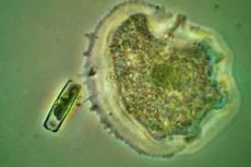
The structure of the dysenteric amoeba is as follows: it is an individual that constantly changes its contours, 20-30 microns in size, has a spherical core inside the endoplasm - internal contents, covered with ectoplasm - the outer layer of cellular cytoplasm, no skeleton, very mobile, moves with the help of peculiar processes called pseudopodia or pseudopods. Its movement resembles flowing from one outline to another. There are luminal, tissue, large vegetative forms of amoeba and in the form of cysts. The size of the luminal is about 20 microns, it is located in the lumen of the upper section of the large intestine, feeds on its bacteria and does not cause any harm to the carrier.
Life cycle dysenteric amoeba
The life cycle of the dysenteric amoeba begins when it enters the gastrointestinal tract. The routes of infection with dysenteric amoeba are fecal-oral and household. Together with feces, cysts enter the environment, more than 300 million of them are excreted per day. They are highly resistant to temperature changes and other adverse effects. Thus, cysts can survive for a month at a temperature of 20ºC, a week in a humid and darkened environment, up to a week in chilled food, several months at sub-zero temperatures. They enter a person with dirty hands, unwashed food, contaminated water, through tactile contact with the hands of a sick person. They are carried by flies and cockroaches. Factors that contribute to the development of pathology are pregnancy, protein deficiency, dysbacteriosis, worms - everything that reduces immunity.
Cyst of dysenteric amoeba
Cysts of dysenteric amoeba appear from the vegetative phase after the acute phase of the disease has subsided. Some of them turn into lumen, others, finding themselves in the environment of thickened feces, become smaller, become covered with a membrane and encyst. They have 4 nuclei and are arranged in the same way as the nuclei of the vegetative form. Immature cysts can have from one to three nuclei. This is the most viable form of dysenteric amoeba, capable of surviving in an unfavorable external environment and, having entered a person, renewing the life cycle.
Invasive stage of dysenteric amoeba
The invasive stage of the dysenteric amoeba is characterized by an incubation period that lasts up to two weeks. During this time, the cysts move through the intestinal sections. Along the way, they penetrate its mucous membrane. In this case, the transverse and descending sections of the large intestine are most susceptible to damage. At this stage, moving, the cysts turn into a vegetative form, containing enzymes that are destructive to the intestinal walls - pepsin and trypsin. This helps the parasite penetrate its layers, right down to the muscular ones, which becomes noticeable to humans.
Tissue form of dysenteric amoebas
The tissue form of dysenteric amoeba is formed when the luminal form penetrates the intestinal walls. Scientists have not yet figured out why this happens. But at this stage, the amoeba causes damage to the mucous membrane of the colon. It is this form of its existence that is found in patients with amebiasis. Reproducing, it provokes the formation of ulcers on the intestinal walls, which lead to the accumulation of pus, blood, and mucus. Conditions are created for the transformation of the luminal and tissue forms into a large vegetative form. They increase to 30 microns and are able to absorb erythrocytes. Coming out, the vegetative form dies.

Symptoms
From the moment the walls are damaged, clinical symptoms of dysenteric amoeba appear. Signs of acute amoebiasis increase gradually with obvious dynamics. At first, stool frequency increases to 4-6 times a day, feces are of liquid consistency with mucus, having a sharp and unpleasant odor. Gradually, visits to the toilet increase and can reach 20 times, false urges to defecate appear, blood clots are found in the glassy mucus. Body temperature rises to 38ºС, which lasts for several days, the abdomen is swollen and painful. Treatment of the disease can last up to one and a half months, if it is not carried out, then remission occurs and the pathology becomes chronic. Its symptoms are expressed in a white coating on the tongue, bad breath, poor appetite, weight loss, signs of vitamin deficiency (hair loss, brittle nails, pale skin), abdominal pain. Over time, problems with the heart and liver may develop.
Diagnostics
Diagnostics are carried out using a method from simple to more complex and are initially based on the patient's story about the symptoms: frequency and nature of stool, pain, dynamics of the disease, and temperature is measured. Then material is taken for laboratory tests. If it is not possible to obtain feces, biopsies are taken using endoscopy, and the intestinal walls are also examined for damage, the presence of ulcers. As additional methods, ultrasound of the kidneys and abdominal organs is used to assess their condition.
Laboratory diagnostics
Laboratory diagnostics include microscopic examination of feces and biopsies taken from damaged areas. In case of complications, nasopharyngeal scrapings are taken. The presence of cysts and vegetative forms of amoeba (trophozoites) in the material being examined confirms the diagnosis. Smears are stained for better detection. In biopsies for amoebiasis, trophozoites with erythrocytes inside are detected. Express diagnostics using the Coons method for determining antibodies is also used. It consists of staining the smear with luminescent serum; bacteria against this background have a green rim around the perimeter. Another similar method, the enzyme immunoassay, based on the antigen-antibody reaction, is also used in laboratory diagnostics.
When a case of dysentery is detected, it is necessary to determine the source-carrier in order to prevent the spread of mass infection. To do this, the dysentery amoeba is reported to the sanitary service, which carries out disinfection of public catering points, if the infection occurred there, or other places. Also, persons in contact with the patient or working in the catering industry are examined for carriage of cysts.
Differential diagnosis
The task of differential diagnostics is to distinguish the dysenteric amoeba from the intestinal amoeba. They differ in structure: the dysenteric amoeba has a double-contour shell that refracts light, it has 4 nuclei (the intestinal amoeba has 8) located eccentrically, it includes blood cells, which is not the case with the intestinal amoeba. The dysenteric amoeba is more energetic in its movements.
In many ways, the symptoms are similar to malaria. Its causative agent is the malarial plasmodium. Plasmodium is carried by mosquitoes, and humans are the intermediate host. With an insect bite, unlike the dysenteric amoeba, the plasmodium enters the blood and then the liver, where asexual reproduction occurs, the so-called tissue schizogony. As a result of multiple division, which occurs during the incubation period, many daughter individuals appear, absorbing hemoglobin and destroying liver cells. Malaria is accompanied by severe attacks of fever, chills and signs of intoxication of the body.
Treatment
Several groups of drugs are aimed at treating dysenteric amoeba. Some of them kill the luminal form of the amoeba, are used at the stage of remission in the chronic course of the disease and for the prevention of the disease. Such drugs are called "direct amoebicides", they include: diiodoquine, quiniofon. In the acute course of dysentery, drugs are used that are aimed at tissue and luminal forms: quinamine, emetine, ambilgar, dihydroemitin. There are universal drugs such as furamide, trichopolum. Antibiotics, enzymes and drugs that restore intestinal microflora are also used. In combination with drug treatment, a special protein sparing diet is mandatory, excluding coarse spicy food. Meals should be frequent, but in small portions, at first - food in a mashed form. In the presence of serious complications, even surgery is possible.
Prevention dysenteric amoeba
There are no special preventive measures. The best prevention is to follow sanitary and hygienic rules: frequent hand washing, washing vegetables and fruits, boiling drinking water, preventing feces from toilets from getting into the beds, fighting cockroaches. When detecting outbreaks of diseases, it is important to identify carriers of the dysenteric amoeba.
[ 22 ]
Forecast
The prognosis for amebiasis is favorable if treatment is started in a timely manner. If the patient's condition is complicated by abscess rupture, intestinal bleeding, narrowing of intestinal areas, or penetration of dysenteric amoebas into other organs: liver, brain, lungs, then a fatal outcome is possible.
Interesting facts
It is known that 50 million people on Earth are infected with dysenteric amoeba. And if we take into account that in many African countries no records of the disease are kept, and the environment for the spread of amoebiasis is the most suitable, then it is not difficult to imagine the scale of the spread. Statistics claim that about 100 thousand people die from this disease every year. It is interesting that scientists still cannot understand why in some organisms individuals peacefully coexist with the host, while in others they aggressively penetrate the tissues of its entrails, eating living cells and causing significant harm to the body.
The amoeba was discovered in 1757 by the German entomologist (a science that studies insects) Roesel von Rosenhof, thanks to water accidentally spilled on a microscope. After 200 years, it turned out that the single-celled organism he observed was completely different. The amoeba received its name in 1822 and it means "variability" due to its ability to constantly change shape. When moving, the amoeba stretches out in length, pseudopodia appear in its front part. For a long time, scientists could not figure out this mechanism, and when they did, they were surprised by such a complex movement device that could only have arisen as a result of long-term evolution. Geneticists also discovered a genome that is too long for a single-celled organism. Having observed this form of life for several centuries, scientists are only convinced that not everything is so simple with this individual. Surely, new discoveries related to the amoeba await us.