
All iLive content is medically reviewed or fact checked to ensure as much factual accuracy as possible.
We have strict sourcing guidelines and only link to reputable media sites, academic research institutions and, whenever possible, medically peer reviewed studies. Note that the numbers in parentheses ([1], [2], etc.) are clickable links to these studies.
If you feel that any of our content is inaccurate, out-of-date, or otherwise questionable, please select it and press Ctrl + Enter.
Duodenum
Medical expert of the article

Last reviewed: 07.07.2025
The duodenum is the initial section of the small intestine, located on the back wall of the abdominal cavity. The duodenum begins at the pylorus of the stomach and ends at the duodenojejunal flexure, located at the left edge of the second lumbar vertebra. In typical cases, the duodenum has the shape of a horseshoe, enveloping the head of the pancreas. The duodenum is divided into the upper, descending, horizontal and ascending parts.
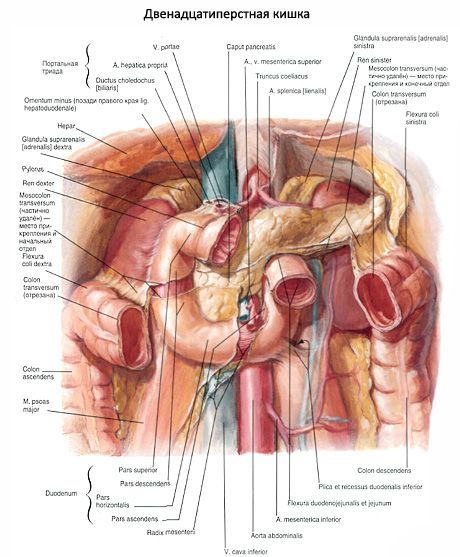
The superior part (pars superior), or bulb, is the shortest (3-6 cm) and widest (up to 4 cm), runs from the pylorus to the right and back and forms the superior flexure of the duodenum. Almost 3/4 of the circumference of this part of the intestine is covered by the peritoneum. When the stomach is moderately or heavily filled, the superior part is located almost sagittally, when empty - more transversely. Its upper surface borders on the posterior part of the square lobe of the liver, then crosses the right part of the proper hepatic artery and the common hepatic duct. Below, the superior part of the duodenum comes into contact with the upper part of the head of the pancreas and the transverse colon. Behind the superior part, in the thickness of the hepatoduodenal ligament, are the common hepatic duct (on the right), the proper hepatic artery (on the left), and the portal vein (behind and between them).
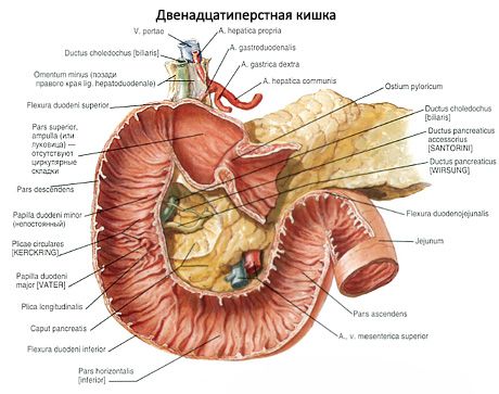
The descending part (pars descendens) begins at the superior flexure of the duodenum at the level of the 1st lumbar vertebra and descends along the right edge of the spine. The descending part ends at the level of the 3rd lumbar vertebra with a sharp turn to the left, forming the inferior flexure of the duodenum. The length of the descending part is 8-10 cm. The gate of the right kidney and the upper part of the ureter are located behind it. Medially, the posterior surface of the descending part borders on the inferior vena cava, and in the area of the transition of the upper part to the descending part of the intestine, on the right adrenal gland. In front, the descending part is covered by the peritoneum and intersects with the root of the mesentery of the transverse colon. On the left, the descending part borders on the head of the pancreas and is closely fused with its capsule. Between the descending part and the head of the pancreas are the terminal part of the common bile duct and the anastomosing superior and inferior pancreaticoduodenal arteries.
The horizontal part (pars horizontalis) begins at the lower flexure of the duodenum, runs horizontally to the left at the level of the third lumbar vertebra, then turns upward and passes into the ascending part at the level of the intersection with the superior mesenteric artery and vein. Behind the horizontal part are the inferior vena cava (on the right) and the aorta (on the left). The anterior surface of the horizontal part is covered with peritoneum, and loops of the small intestine are adjacent to it.
The ascending part (pars ascendens) begins at the point where the superior mesenteric artery and vein emerge from under the lower edge of the pancreas onto the anterior surface of the duodenum. The ascending part ends at the upper edge of the body of the second lumbar vertebra with a sharp bend of the intestine downwards, forwards and to the left - the duodenojejunal flexure (flexura duodenojejunalis). The bend is fixed to the diaphragm by the muscle and ligament that suspend the duodenum (m. et lig.suspensorii duodeni). Behind the ascending part is the aorta, and in front is the parietal peritoneum.
Innervation: The duodenum receives parasympathetic nerve fibers from the vagus nerves, and sympathetic fibers from the gastric, hepatic, and superior mesenteric plexuses. The jejunum and ileum are innervated by fibers of the vagus nerves, as well as the superior mesenteric plexus.
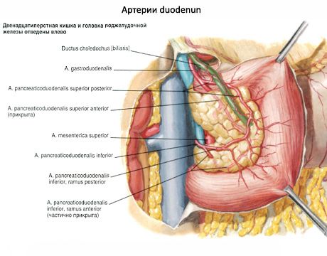
Blood supply: the duodenum is supplied with blood by the anterior and posterior superior pancreaticoduodenal arteries (from the gastroduodenal artery), the inferior pancreaticoduodenal artery (from the superior mesenteric artery); the jejunum and ileum are supplied by the jejunal and ileocolic arteries (from the superior mesenteric artery). Venous outflow occurs through the veins of the same name into the portal vein.
Lymph drainage: from the duodenum - to the pancreaticoduodenal, superior mesenteric, celiac, lumbar lymph nodes, from the jejunum and ileum - to the mesenteric and ileocolic (from the final part of the ileum) lymph nodes.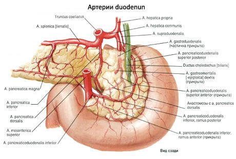
 [
[ Where does it hurt?
What do need to examine?
What tests are needed?