All iLive content is medically reviewed or fact checked to ensure as much factual accuracy as possible.
We have strict sourcing guidelines and only link to reputable media sites, academic research institutions and, whenever possible, medically peer reviewed studies. Note that the numbers in parentheses ([1], [2], etc.) are clickable links to these studies.
If you feel that any of our content is inaccurate, out-of-date, or otherwise questionable, please select it and press Ctrl + Enter.
Coral nephrolithiasis (coral kidney stones)
Medical expert of the article
Last reviewed: 12.07.2025
Coral-shaped kidney stones (coral-shaped nephrolithiasis) are an independent disease that differs from all other forms of urolithiasis in its pathogenesis features and has its own clinical picture.
What causes coral kidney stones?
Staghorn kidney stones develop against the background of impaired hemo- and urodynamics and are complicated by pyelonephritis, which leads to a progressive decrease in kidney function. The development of staghorn nephrolithiasis is most often promoted by various congenital and acquired tubulo- and glomerulopathies, which are based on enzymopathies. The most common enzymopathy in staghorn nephrolithiasis leads to oxaluria (85.2%); tubulopathies leading to fructosuria, galactosuria, tubular acidosis, and cystinuria are much less common. If these factors are decisive in the development of the disease, then all other exogenous and endogenous factors act only as contributors to the development of the disease, i.e. are less significant. Of great importance are climate conditions, especially for people who have changed their place of residence to hot countries, water, food products, air pollution. Stone formation is promoted by diseases of the gastrointestinal tract, liver, hyperfunction of the parathyroid glands, bone fractures that require long-term bed rest. In some cases, the formation of coral stones during pregnancy is noted, which is caused by a violation of water-electrolyte balance, urodynamics, hormonal shifts. A number of researchers draw attention to the role of hereditary factors in the development of the disease, which make up about 19%.
Many authors consider hyperparathyroidism to be the etiologic factor of nephrolithiasis, acting in 38% of cases. Despite the obvious changes in the patient's body with primary hyperparathyroidism, it is not possible to prove the leading role of changes in the function of the parathyroid glands in the occurrence of kidney stones. The triad of symptoms of primary hyperparathyroidism (hypercalcemia, hypophosphatemia and hypercalciuria) is not characteristic of all patients with coral nephrolithiasis, and not all patients with hyperparathyroidism have a coral stone.
To diagnose parathyroid gland adenoma, ultrasound and radioisotope scintigraphy are most often used.
At the same time, the cause of kidney stones in general and coral stones in particular remains an unresolved issue, which creates difficulties in developing treatment tactics for patients with coral nephrolithiasis, effective prevention of stone formation and its recurrence.
How do coral kidney stones develop?
The core of most stones is formed by an organic substance. However, when studying the chemical composition of stones, it was found that their formation can also begin on an inorganic basis. In any case, for stone formation, even with oversaturation of urine with salts, a binding component is necessary, which is an organic substance. Such an organic matrix of stones is colloidal bodies with a diameter of 10-15 microns, found in the lumens of the tubules and lymphatic capillaries of the stroma. Glycosaminoglycans and glycoproteins are found in the composition of colloidal bodies. In addition to the usual components (cystine, phosphate, calcium, urates, etc.), the stone contains mucoproteins and plasma proteins of various molecular weights. Most often, it is possible to detect uromucoid, albumin and immunoglobulins IgG and IgA.

The most interesting data were obtained from immunochemical analysis of the protein composition of urine, which revealed the excretion of small plasma proteins into the urine, such as alpha-acid glycoprotein, albumin, transferrin and IgG, which is a sign of the tubular type of proteinuria, but sometimes proteins of higher molecular weight are also detected, such as IgA and a2-macroglobulin.
These proteins penetrate into the secondary urine due to a disruption of the structural integrity of the glomeruli, namely the glomerular basement membranes. This confirms the data that coral stones in the kidneys are accompanied not only by tubular disorders, but also by glomerulopathy.
Electron microscopic examination of renal tissue revealed abnormalities in the plasma membrane region that provides obligatory and optional reabsorption processes. Changes in the microvilli of the brush border were found in the nephrocytes of the renal tubules of the proximal and distal sections. Electron-loose flocculent material was found in the lumen of the loop of Henle and collecting tubules.
The nuclei of the cells lining the loop of Henle are always deformed, and the greatest changes are found in the basement membrane.
Studies have shown that in coral nephrolithiasis, the renal parenchyma is altered in all areas.
A study of the immune status of patients based on the results of blood and urine tests showed no significant deviations from the norm.
Symptoms of Coral Kidney Stones
Symptoms of coral nephrolithiasis are non-specific, as are complaints that are characteristic only of patients with this disease.
Upon detailed analysis, it can be noted that the clinical picture is expressed by symptoms of impaired urodynamics and renal function.
Based on the clinical picture, four stages of coral nephrolithiasis are distinguished:
- I - latent period;
- II - the onset of the disease;
- III - stage of clinical manifestations;
- IV - hyperazotemic stage.
Stage I is called the latent period, since at this time there are no obvious clinical manifestations of kidney disease. Patients complain of weakness, increased fatigue, headache, dry mouth and chills.
The onset of the disease (stage II) is characterized by weak dull pain in the lumbar region and sometimes intermittent changes in the urine.
In the stage of clinical manifestations (stage III), dull pain in the lumbar region is constant, subfebrile temperature appears, increased fatigue, weakness and malaise progress. Hematuria and passage of small stones, accompanied by renal colic, often occur. Signs of chronic renal failure appear - latent or compensated stage.
In stage IV - hyperazotemic - patients complain of thirst, dry mouth, general weakness, increased fatigue, pain in the lumbar region, dysuria and symptoms of exacerbation of pyelonephritis. This stage is characterized by intermittent or even terminal stage of chronic renal failure.
Where does it hurt?
Classification of coral kidney stones
Depending on the size and location of the coral stone in the renal pelvis and its configuration, four stages of coral nephrolithiasis are distinguished:
- Coral-shaped nephrolithiasis-1 - the calculus fills the renal pelvis and one of the calyces;
- Coral-shaped nephrolithiasis-2 - located in the extrarenal pelvis with processes in two or more calyces;
- Coral-shaped nephrolithiasis-3 - located in the renal pelvis of the intrarenal type with processes in all cups;
- Coral-shaped nephrolithiasis-4 - has processes and fills the entire deformed renal pelvis-calyceal system.
Retention changes in coral nephrolithiasis are varied: from moderate pyelectasis to total expansion of not only the renal pelvis, but also all the calyces.
The main factor in choosing a treatment method is the degree of renal dysfunction. Four phases of renal dysfunction reflect the deficiency of their secretory capacity:
- Phase I - tubular secretion deficit 0-20%;
- Phase II - 21-50%;
- Phase III - 51-70%:
- Phase IV - over 70%.
Thus, with the help of this classification, which allows for a comprehensive assessment of the size and configuration of the stone, ectasia of the renal pelvis-calyceal system, the degree of renal dysfunction and the stage of the inflammatory process, indications for one or another treatment method are developed.
Diagnosis of coral kidney stones
Staghorn stones are usually discovered by chance during an ultrasound or on a plain X-ray of the urinary tract.
Diagnosis of coral nephrolithiasis is based on general clinical signs and additional research data.
Patients with coral kidney stones often have elevated blood pressure. The cause of arterial hypertension is a violation of hemodynamic balance.
Chronic pyelonephritis accompanying coral nephrolithiasis can be diagnosed at any stage of the clinical course.
A detailed study of the patients' lifestyle, anamnesis and clinical picture of the disease, X-ray and laboratory data, indicators of radioisotope and immunological studies made it possible to identify signs of various stages of chronic renal failure (latent, compensated, intermittent and terminal). It should be noted that due to technical progress and improvement of diagnostic methods over the past decade, patients with coral stones in the terminal stage of chronic renal failure are extremely rare.
In the latent stage of chronic renal failure, the SCF is 80-120 ml/min with a tendency to gradual decrease. In the compensated stage, the SCF decreases to 50-30 ml/min, in the intermittent stage - 30-25 ml/min, in the terminal stage - 15 ml/min. A marked weakening of glomerular filtration always leads to an increase in the content of urea and creatinine in the blood serum. The sodium content in the plasma fluctuates within the normal range, excretion is reduced to 2.0-2.3 g/day. Hypokalemia (3.8-3.9 meq/l) and hypercalcemia (5.1-6.4 meq/l) are often observed. In the compensated stage of chronic renal failure, polyuria occurs, which is always accompanied by a decrease in the relative density of urine. Changes in protein metabolism lead to proteinuria, dysproteinemia, and hyperlipemia. A relative increase in aspartate aminotransferase activity and a decrease in alanine aminotransferase activity in the blood serum were noted.
In chronic renal failure in patients with coral stones, plasma proteins were found among the uroproteins: acid glycoprotein, albumin, transferrin. In severe cases, proteins with a higher molecular weight enter the urine: immunoglobulins, a2-macroglobulins, beta-lipoproteins. This confirms the assumption of a violation of the integrity of the glomerular basement membranes, which normally do not allow the said plasma proteins to pass into the urine.
Changes in the functional activity of the kidneys are always accompanied by a disruption in carbohydrate metabolism, which is caused by increased insulin levels in the blood.
Dull pain in the lumbar region, weakness, and increased fatigue can serve as clinical symptoms of many kidney diseases, such as chronic pyelonephritis, other clinical forms of urolithiasis, polycystic kidney disease, hydronephrotic transformation, kidney tumor, etc.
Based on the complaints presented by patients, one can only suspect kidney disease. The leading place in diagnostics is occupied by ultrasound and X-ray examination. Ultrasound in 100% of cases determines the size and contours of the kidney, the shadow in its projection, the size and configuration of the coral stone, establishes the presence of expansion of the calyceal-pelvic system.
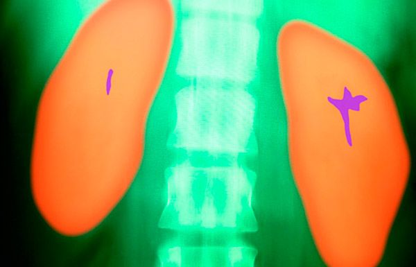
On the plain radiograph in the projection of the kidney, the shadow of a coral stone is visible.
Excretory urography allows for a more accurate assessment of the functional activity of the kidneys and confirmation of the presence of dilation of the renal pelvis.
Clinical diagnostics of coral kidney stones
Patients complain of dull pain in the lumbar region, often intensifying before an attack of renal colic, the passage of small stones, fever, dysuria, and changes in the color of urine. In addition to the listed symptoms, patients experience thirst, dry mouth, weakness, increased fatigue, and itching of the skin. The skin is pale, with a yellowish tint in the most severe group of patients.
Laboratory diagnostics of coral kidney stones
Laboratory tests help to assess the severity of the inflammatory process, establish the functional state of the kidneys, other organs and systems. In all patients at the stage of clinical development of the disease, an increase in ESR, leukocytosis and pyuria can be detected.
With a sharp disruption of the filtration process, creatinine clearance is reduced to 15 ml/min. An increase in the concentration of amino acids in the blood plasma is associated with a disruption of liver function.
Instrumental diagnostics of coral stones in the kidneys
Instrumental methods of examination, in particular cystoscopy, allow to identify the source of bleeding in case of macrohematuria. Ultrasound of the kidneys helps not only to detect a coral stone, but also to study its configuration, changes in the renal parenchyma and the presence of dilation of the calyceal-pelvic system. The main place in the diagnosis of coral kidney stones is given to X-ray methods of examination. A coral stone is visible on a general image of the urinary tract, its shape and size can be assessed.
Excretory urography allows us to determine the size of the kidney, its contours, segmental changes on nephrograms, slowing of the release of contrast agent, its accumulation in dilated calyces, and the absence of renal function.
Retrograde pyelography is performed extremely rarely, immediately before surgery if there is a suspicion of a violation of urodynamics.
Renal angiography allows to establish the place of origin of the renal artery from the aorta, the diameter of the renal artery and the number of segmental branches. Renal angiography is indicated in cases when it is planned to perform a nephrotomy with intermittent clamping of the renal artery.
The method of isotope renography with assessment of blood clearance allows determining the level of functional activity of the kidneys.
Dynamic nephroscintigraphy helps to assess the functional state of not only the affected but also the contralateral kidney.
Indirect renal angiography is a valuable study that allows us to establish qualitative and quantitative hemodynamic disturbances in individual segments of the kidneys.
To diagnose parathyroid gland adenoma, ultrasound and radioisotope scintigraphy are most often used.
Who to contact?
Treatment of coral kidney stones
A patient with coral nephrolithiasis in stage KN-1, if the disease proceeds without pain, exacerbations of pyelonephritis and renal dysfunction, can be observed by a urologist and receive conservative treatment. Antibacterial drugs are prescribed taking into account the bacteriological analysis of urine. Litholytic drugs, diet and diuretics are widely used.
Drug treatment of coral kidney stones
To reduce the formation of uric acid, patients can be prescribed uriuretics. If necessary, nitrate mixtures (blemaren) are recommended at the same time to maintain urine pH in the range of 6.2-6.8. To increase urine pH, baking soda can also be used in a dose of 5-15 g/day.
In oxaluria, good results were achieved by treatment with a combination of pyridoxine or magnesium oxide with marelin. In hypercalciuria, dairy products are excluded, hydrochlorothiazide is recommended at a dose of 0.015-0.025 g 2 times a day. The potassium level in the blood is well maintained by introducing dried apricots, raisins, baked potatoes or 2.0 g of potassium chloride per day into the diet. The use of calcitonin in patients with primary hyperparathyroidism leads to a decrease in hypercalcemia.
To prevent purulent-inflammatory complications, antibiotic prophylaxis is necessary.
Surgical treatment of coral kidney stones
In cases where the disease occurs with frequent attacks of acute pyelonephritis, complicated by hematuria or pyonephrosis, surgical treatment is indicated.
The introduction of new technologies - PNL and DLT - have reduced the indications for open surgical interventions and have greatly improved the treatment of severe patients with coral nephrolithiasis. Open surgical interventions themselves, aimed at preserving the renal parenchyma, have also been improved.
The optimal and most gentle method of removing coral stones at stages KN-1 and KN-2 is PNL. At these stages, this type of treatment is considered as a method of choice, and at stage KN-3 as an alternative to open surgery.
DLT is used mainly at stage KN-1. Its high efficiency in children has been noted. DLT is effective for intrarenal type stones in the renal pelvis, a decrease in kidney function by no more than 25% and normal urodynamics against the background of remission of chronic pyelonephritis.
Many authors prefer combined treatment. The combination of open surgery and EBRT or PNL and EBRT most fully meets the principles of treatment for this category of patients.
Advances in medicine in recent years have expanded the indications for open surgical treatment of patients with coral nephrolithiasis. The most gentle open surgery for coral kidney stones is lower, posterior subcortical pyelolithotomy or with transition to the calyces (pyelocalicotomy). However, pyelolithotomy does not always succeed in removing stones located in the calyces. The main method of treatment for coral stones at stages KN-3 and KN- remains pyelonephrolithotomy. Performing one or more nephrotomy incisions with intermittent clamping of the renal artery (ischemia period is usually 20-25 minutes) does not significantly affect the functional state of the kidney. The operation ends with the installation of a nephrostomy.
The introduction of new technologies in the treatment of coral nephrolithiasis (PNL and DLT) reduced the number of complications to 1-2%. Open surgical interventions with appropriate preoperative preparation, improvement of anesthesiology and methods of pyelonephrolithotomy with clamping of the renal artery made it possible to perform organ-preserving operations. Nephrectomy for coral stones is performed in 3-5% of cases.
Further management
Coral kidney stones can be prevented by dynamic monitoring by a urologist at the place of residence. In case of metabolic disorders (hyperuricosuria, hyperuricemia, decreased or increased urine pH, hyperoxaluria, hypo- or hypercalcemia, hypo- or hyperphosphatemia), it is necessary to prescribe corrective therapy. It is necessary to reduce the amount of food consumed, including fats and table salt, exclude chocolate, coffee, cocoa, offal, broths, fried and spicy foods. The amount of fluid consumed should be at least 1.5-2.0 liters per day with normal glomerular filtration. Since the xanthine oxidase inhibitor allopurinol reduces the level of uricemia, they are indicated for purine metabolism disorders.


 [
[