
All iLive content is medically reviewed or fact checked to ensure as much factual accuracy as possible.
We have strict sourcing guidelines and only link to reputable media sites, academic research institutions and, whenever possible, medically peer reviewed studies. Note that the numbers in parentheses ([1], [2], etc.) are clickable links to these studies.
If you feel that any of our content is inaccurate, out-of-date, or otherwise questionable, please select it and press Ctrl + Enter.
Coccidia
Medical expert of the article

Last reviewed: 06.07.2025
Order Coccidia
The order Coccidia is a fairly broad group of protozoan parasites, which has approximately 400 varieties. They take root inside various living organisms: inside worms, arthropods, etc. Coccidia penetrate into the cells of tissues and organs, and some species can parasitize inside the body of pets, birds and certain species of fish.
Only one type of coccidia is capable of infecting the human body.
Coccidia are able to reproduce sexually and asexually, thus a change of generations occurs, which is sometimes accompanied by a change of the carrier (host). Most often, the parasite stops and develops in the intestinal epithelium, bile ducts, liver tissue, blood cells and endothelial cells.
The order Coccidia are considered to be highly specific parasites. This is due to the fact that almost all of their varieties stick to one host, but are not capable of parasitizing even in close and similar host species. For example, parasites that infect a rabbit are not capable of infecting a hare, and vice versa. Inside the host, coccidia do not parasitize throughout the entire body, but stick to certain "favorite" areas. For example, coccidia does not infect the entire intestine, but only certain parts.
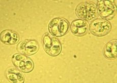
Structure of coccidia
The adult generation of coccidia has a round or oval shape. Their structure is quite complex, especially at the mobile stages of development.
On the outside, the coccidia is covered by a multilayered pellicle, under which is a tubular-fibrillar system, or so-called subpellicular microtubes. Together with the membranes, they form the outer skeleton of the zoite.
The outer shell of the pellicle is integral, and the underlying layers have interruptions in front and behind, at the location of the posterior and anterior support rings. Along the inner circle of the anterior ring there is a specific cone-shaped dense structure - a conoid, with walls in the form of spirally bent fibrils. The function of the conoid is to support the moment the zoite enters the host cell.
The anterior 1/3 of the zoite are tubular structures with an internal expansion – rhoptries. Their edges pass through the openings of the conoid. Presumably, the function of the rhoptries is to secrete a substance that facilitates the entry of the zoite into the host cell.
The anterior end of the merozoite contains dense, curling strands called micronemes. Their function remains unclear.
In addition to the structures listed above, the cytoplasmic layer of zoites also contains components that are common to all cells. These are mitochondria and endoplasm with ribosomes, the Golgi complex. There are also other components: carbohydrate, protein and fat particles, which are a reserve of energy resources.
Life cycle of coccidia
The life cycle of coccidia begins when it enters the host's intestinal cavity. The first stage of this cycle is the formation of a sporozoite, which comes out of the swallowed (eaten) oocyst. The sporozoite is a small spindle-shaped cell formation with one nucleus. The sporozoite immediately penetrates the epithelial cells of the intestine, where it immediately acquires a rounded shape and becomes like a ball. Then the parasite begins to actively develop: it increases in size in a short time. Coccidia feeds osmotically. At this stage, coccidia is called "schizont", which characterizes its method of reproduction.
The coccidia development cycle goes through a period of nuclear division: the schizont receives several nuclei instead of one. Their number can vary from eight to sixty. The developing schizont outgrows the epithelial cell and gradually passes into the subepithelial connective tissue layer. The schizont development cycle ends with asexual reproduction. A cytoplasmic zone is determined around the circumference of each individual nucleus, as a result of which the schizont disintegrates into mononuclear spindle-shaped cell structures. Here, the asexual reproduction of the schizont comes to an end: it is also called multiple division or schizogony. The described cycle lasts approximately 90 hours. The resulting spindle-shaped cells are called "merozoites".
The merozoites again appear in the intestinal epithelial cells and continue to reproduce: this is how the next generation of schizonts is born. This process takes a little longer – about 120 hours. The second generation, in turn, gives rise to the third. Those merozoites that are not capable of forming schizonts contribute to the birth of gametes (reproductive cell structures). Such cells have a clear division into male and female macrogametes.
Fertilization is the most important stage of coccidia development. The interaction of micro- and macrogametes occurs with the formation of an internal membrane, and the zygote ends up in the intestinal lumen. Such a zygote with a two-layer membrane is called an oocyst.
Then the internal stage of the parasite's development ends, since the oocyst requires oxygen for its vital functions. To do this, the coccidia oocyst must leave the host's intestines.
Coccidia in humans
Coccidia are very rarely found in humans: isolated cases of infection have been recorded in Uzbekistan, the Caucasus, and Crimea.
Only coccidia Isospora belli or Isospora hominis can affect humans. Domestic animals can play a major role in human infection, if personal hygiene rules are not observed. The patient becomes the host of the parasite when ingesting oocysts with food or liquids, which subsequently begin to be excreted with the feces of the patient with coccidiosis. Over the course of several days, the coccidia matures in soil conditions.
In the intestinal cavity of the human body, sporozoites are released from oocysts. They then penetrate the epithelial tissue with its subsequent destruction. An inflammatory process develops, in some cases ulcerative surfaces are formed. Fever with an increase in temperature to 39 ° C, general weakness, lack of appetite, bowel disorder, apathy, drowsiness are observed.
The disease (coccidiosis) can last for several weeks or up to 1 month. A person who has recovered from coccidiosis can still excrete coccidia oocysts with feces for another month.
A patient who has had a certain type of coccidiosis cannot get sick again.
Coccidia in cats
Coccidia are more common in cats than in humans. Kittens are more susceptible to infection, although adults also suffer from coccidiosis. Cats are mainly affected by such types of coccidia as Isospora felis or rivolta.
Under what conditions can a cat become infected:
- through the feces of individuals carrying parasites (oocysts in the feces of other cats);
- as a result of eating contaminated food, such as raw fish;
- when eating caught rodents or birds infected with coccidia.
Coccidia settle in the intestines of the animal, where they develop and reproduce. Externally, the disease manifests itself as enterocolitis, and in young individuals and kittens the disease is more severe.
The main symptoms of infection are: diarrhea (feces with mucus, in some cases even with blood), anemia. The animal becomes lethargic, refuses food, and loses weight.
If you suspect coccidiosis, it is essential to contact a veterinary clinic.
Coccidia in dogs
Coccidia that affect dogs are Isospora canis or I. ohioensis. Coccidia infection is primarily characterized by digestive disorders, emaciation, which can subsequently lead to the death of the dog.
The parasites settle mainly in the posterior third of the small intestine after the dog has swallowed the pathogen.
In puppyhood, the disease manifests itself especially acutely, sometimes with damage not only to the intestines, but also to the liver of the animal. Vomiting, diarrhea, increased temperature, and abdominal distension are observed.
Unsanitary conditions and numerous carriers of parasites, such as flies, rodents, and birds, play a major role in the infection of animals with coccidia.
In most dogs, when examining feces, attention is paid to changes in the color of feces (with a green tint, dark, grayish, yellow, etc.) and odor, which indicates active reproduction of microflora in the intestines.
In some cases, coccidiosis is combined with helminthic invasion.
 [ 10 ]
Treatment of coccidia
A treatment regimen for coccidia in humans has not been developed due to the fact that this disease is extremely rare. Cases of the disease have been isolated for many decades.
Sick animals should be isolated. For their treatment use:
- sulfonamides;
- nitrofurans (furazolidone);
- antifungal (nystatin);
- antiprotozoal (osarsol);
- silver preparations (albargin);
- tetracyclines;
- chloramphenicol (syntomycin), etc.
The most commonly used are:
- sulfadimethoxine at 5 mg/kg, mixed into feed, for 4 days;
- norsulfazole (phthalazole) 3-5 g/kg with liquid, 2 times a day for five days;
- iodine solutions with drinking water, iodinol, etc.;
- furazolidone and furatsilin 2 g each;
- coccidin 0.05 g/kg for 4 days.
Treatment is aimed at disrupting proteolytic processes in cellular structures and inhibiting the consumption of para-aminobenzoic acid, which leads to disruption of the growth and reproduction of coccidia, as well as damage to the ability of parasites to secrete toxins.
Separately, drugs can be used to prevent anemia and restore the body's immune defenses.
Prevention of coccidia
Prevention of coccidia consists of following the following rules:
- compliance with sanitary and hygienic measures;
- maintaining cleanliness in areas where animals are kept or bred;
- complete feeding of young animals, puppies, kittens.
If a sick animal is found, it must be isolated and treated, after the incubation period.
For some species of animals, preventive vaccination against coccidia is provided, which can protect the pet from several types of the parasite at once. You can find out about the availability of such vaccines and the possibility of using vaccination at your nearest veterinary clinic.
A person can protect themselves from coccidiosis by simply following sanitary and hygienic standards. This includes frequent hand washing, eating only washed or heat-treated vegetables and fruits, and maintaining cleanliness in living and utility rooms. Much attention is paid to nutrition: food should be fresh and balanced, and drinking water should be clean and taken from well-known, proven sources.
It has been proven that coccidia cannot be transmitted from domestic animals to humans, however, compliance with basic hygiene rules when keeping an animal is mandatory.