
All iLive content is medically reviewed or fact checked to ensure as much factual accuracy as possible.
We have strict sourcing guidelines and only link to reputable media sites, academic research institutions and, whenever possible, medically peer reviewed studies. Note that the numbers in parentheses ([1], [2], etc.) are clickable links to these studies.
If you feel that any of our content is inaccurate, out-of-date, or otherwise questionable, please select it and press Ctrl + Enter.
Chlamydiae
Medical expert of the article

Last reviewed: 06.07.2025
Chlamydia are small gram-negative coccoid parasitic bacteria belonging to the order Chlamydiales, family Chlamydiaceae. Currently, this family includes two genera that differ in antigenic structure, intracellular inclusions and sensitivity to sulfonamides: Chlamydia ( Chlamydia trachomatis ): Chlamydophila (Chlamydia pneumonia, Chlamydia psittaci ).
The name "chlamydia" (from the Greek chtamys - mantle) reflects the presence of a membrane around the microbial particles.
All types of chlamydia have common morphological features, a common group antigen, and a separate reproduction cycle. Chlamydia are considered gram-negative bacteria that have lost the ability to synthesize ATP. Therefore, they are obligate intracellular energy parasites.
Chlamydia trachomatis and Chlamydia pneumoniae are classified as microorganisms that are definitely pathogenic for humans and are the causative agents of anthropogenic chlamydia. Depending on the type of pathogen and the entry point (respiratory tract, genitourinary system), respiratory and urogenital chlamydia are distinguished.
More than 20 nosological forms caused by Chlamydia trachomatis have been described, including trachoma, conjunctivitis, inguinal lymphogranulomatosis, Reiter's syndrome, urogenital chlamydia, infections caused by Chlamydia trachomatis, according to WHO estimates, rank second among sexually transmitted diseases after trichomonas infections. About 50 million cases are registered worldwide every year.
Chlamydophila pneumonia causes severe pneumonia, a disease of the upper respiratory tract. There are suggestions that Chlamydophila pneumonia is involved in the development of atherosclerosis and bronchial asthma.
Chlamydophila psittaci is the cause of ornithosis (psittacosis), a zoonotic disease.
Morphological and tinctorial properties of chlamydia
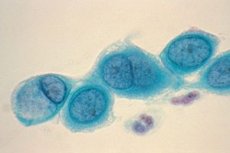
Chlamydia are small gram-negative bacteria of spherical or ovoid shape. They do not have flagella or capsules. The main method for detecting chlamydia is Romanovsky-Giemsa staining. The color of the stain depends on the stage of the life cycle: elementary rings are stained purple against the background of blue cell cytoplasm, reticular bodies are stained blue.
The structure of the cell wall resembles that of gram-negative bacteria, although there are differences. It does not contain typical peptide glycan: N-acetylmuramic acid is completely absent from its composition. The cell wall includes an outer membrane, which includes LPS and proteins. Despite the absence of peptide glycan, the cell wall of chlamydia is rigid. The cell cytoplasm is limited by an internal cytoplasmic membrane.
Analysis of the outer membrane (OM) of chlamydia showed that it contains LPS, the main protein of the outer membrane (MOMP), and cysteine-rich proteins Ompl and Omp3, associated with the inner surface of the OM. LPS and MOMP of Chlamydia psittaci and Chlamydia trachomatis, in contrast to MOMP of Chlamydia pneumoniae, are localized on the outer surface of the cell. The Omp proteins of Chlamydia psittaci and Chlamydia pneumoniae with a molecular weight of 90-100 kDa are also located here.
Chlamydia are polymorphic, which is related to the peculiarities of their reproduction. The unique (two-phase) development cycle of chlamydia is characterized by the alternation of two different forms of existence - the infectious form (elementary bodies - EB) and the vegetative form (reticular, or initial, bodies - RT).
Microorganisms contain RNA and DNA. In RT, RNA is 4 times more than DNA. In these, the content is equivalent.
Reticular bodies can be oval, crescent-shaped, in the form of bipolar rods and coccobacilli, 300-1000 nm in size. Reticular bodies do not have infectious properties and, undergoing division, ensure the reproduction of chlamydia.
Elementary bodies of oval shape, 250-500 nm in size, have infectious properties, are able to penetrate into a sensitive cell, where the development cycle occurs. They have a dense outer membrane, which makes them resistant in the extracellular environment.
Cultivation of chlamydia
Chlamydia, being obligate parasites, do not reproduce on artificial nutrient media, they can be cultivated only in living cells. They are energy parasites, as they are not able to independently accumulate energy and use the ATP of the host cell. Chlamydia is cultivated in HeLa, McCoy cell culture, in yolk sacs of chicken embryos, and in the body of sensitive animals at a temperature of 35 °C.
 [ 3 ], [ 4 ], [ 5 ], [ 6 ], [ 7 ], [ 8 ], [ 9 ]
Antigenic structure of chlamydia
Chlamydia has three types of antigens: specific antigen (common to all types of chlamydia) - LPS; info-specific antigen (different for all types of chlamydia) - protein in nature, located in the outer membrane; type-specific (different for serovars of Chlamydia trachomatis) - LPS, multiplying in the cell wall of the microorganism; variant-specific antigen of protein nature.
Serovars A, B, and C are called ocular, as they cause trachoma, serovars D, E, K, O, H, I, J, K (genial) are the causative agents of urogenital chlamydia and its complications, serovar L is the causative agent of venereal lymphogranulomatosis. The causative agent of respiratory chlamydia Chlamydia pneumoniae has 4 serovars: TWAR, AR, RF, CWL. Chlamydia psittaci has 13 serovars.
[ 10 ], [ 11 ], [ 12 ], [ 13 ], [ 14 ], [ 15 ], [ 16 ], [ 17 ], [ 18 ]
Cellular tropism of chlamydia
Chlamydia trachomatis has a tropism for the mucous membrane of the urogenital tract epithelium, and can remain locally on it or spread over the entire surface of the tissue. The causative agent of venereal lymphogranuloma has a tropism for lymphoid tissue.
Chlamydia pneumoniae replicates in alveolar macrophages, monocytes, and vascular endothelial cells; systemic dissemination of infection is also possible.
Chlamydia psittaci cause infection in a variety of cell types, including mononuclear phagocytes.
Chlamydia life cycle
The development cycle of chlamydia lasts 40-72 hours and includes two forms of existence that differ in morphological and biological properties.
At the first stage of the infectious process, the adsorption of elementary bodies of chlamydia on the plasma membrane of the sensitive host cell occurs with the participation of electrostatic forces. The introduction of chlamydia into the cell occurs by endocytosis. The sections of the plasma membrane with EBs adsorbed on them invaginate into the cytoplasm with the formation of phagocytic vacuoles. This stage lasts 7-10 hours.
Then, within 6-8 hours, the infectious elementary bodies are reorganized into metabolically active non-infectious, vegetative, intracellular forms - RT, which divide repeatedly. These intracellular forms, which are microcolonies, are called chlamydial inclusions. Within 18-24 hours of development, they are localized in a cytoplasmic vesicle formed from the host cell membrane. The inclusion may contain from 100 to 500 reticular bodies of chlamydia.
At the next stage, during 36-42 hours, maturation (formation of intermediate bodies) and transformation of reticular bodies by division in elementary bodies occur. Destroying the infected cell. Elementary bodies leave it. Being extracellular, elementary bodies penetrate new host cells after 40-72 hours, and a new cycle of chlamydia development begins.
In addition to such a reproductive cycle, other mechanisms of interaction between chlamydia and the host cell are realized under unfavorable conditions. These are the destruction of chlamydia in phagosomes, L-like transformation and persistence.
Transformed and persistent forms of chlamydia are capable of reverting to the original (reticular) forms with subsequent transformation into elementary bodies.
Outside host cells, metabolic functions are reduced to a minimum.
Pathogenicity factors of chlamydia
The adhesive properties of chlamydia are due to proteins of the outer membrane of cells, which also have antiphagocytic properties. In addition, microbial cells have endotoxins and produce exotoxins. Endotoxins are represented by LPS, in many ways similar to LPS of gram-negative bacteria. The thermolability of the substance is made up of exotoxins, they are present in all and cause death of mice after intravenous administration.
Chlamydia has been found to have a type III secretory system, through which chlamydial proteins are injected into the host cell cytoplasm as an integral part of the infectious process.
Heat shock protein (HSP) has the ability to induce autoimmune reactions.
[ 29 ], [ 30 ], [ 31 ], [ 32 ], [ 33 ], [ 34 ]
Ecology and resistance of chlamydia
Chlamydia are very common microorganisms. They have been found in more than 200 species of animals, fish, amphibians, mollusks, and arthropods. Microorganisms similar in morphology have also been found in higher plants. The main hosts of chlamydia are humans, birds, and mammals.
The causative agent of chlamydia is unstable in the external environment, is very sensitive to high temperatures and quickly dies when dried. Its inactivation at 50 °C occurs after 30 minutes, at 90 °C - after 1 minute. At room temperature (18-20 °C), the infectious activity of the pathogen decreases after 5-7 days. At 37 °C, a drop in virulence by 80% is observed after 6 hours in a thermostat. Low temperature (-20 °C) contributes to the long-term preservation of the infectious properties of the pathogen. Chlamydia quickly dies under the influence of UV radiation, from contact with ethyl ether and 70% ethanol, under the influence of 2% lysol for 10 minutes, 2% chloramine.