
All iLive content is medically reviewed or fact checked to ensure as much factual accuracy as possible.
We have strict sourcing guidelines and only link to reputable media sites, academic research institutions and, whenever possible, medically peer reviewed studies. Note that the numbers in parentheses ([1], [2], etc.) are clickable links to these studies.
If you feel that any of our content is inaccurate, out-of-date, or otherwise questionable, please select it and press Ctrl + Enter.
The causative agent of adiaspiromycosis
Medical expert of the article

Last reviewed: 06.07.2025
Adiaspiromycosis (synonym: haplomycosis) is a chronic mycosis with predominant damage to the lungs.
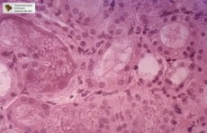
Morphology
Emmorisia crescens and E. parva are dimorphic fungi. The mycelial form of these fungi is identical. The mycelium is rarely septate. Microconidia of 2-4 µm, sometimes 5-6 µm, are formed on conidiophores singly or in short chains. Attachment of aleuriae or their clusters to the mycelium without conidiophores is possible; in the anamnesis, a tissue non-dividing form of the fungus, adiaspore, develops. Adiaspores of E. crescens are multinuclear, 700 µm in diameter, mononuclear, 40 µm in diameter.
Cultural properties
Undemanding to the nutrient substrate. Grows well on simple nutrient media. Grows in a wide temperature range - from 4 to 30 °C in a wide range of pH of the medium.
Ecological niche - soil. E. parva predominates in arid ranks.
The resistance in the environment is high. The ability to grow at low temperatures ensures the elimination of the competitive action of normal soil microflora.
Sensitivity to antiseptics and disinfectants. Sensitive to the action of commonly used antiseptics and disinfectants.
 [
[ Pathogenesis of adiaspiromycosis
In natural conditions, infection is caused by aleuria, which, due to their small size, are able to penetrate the respiratory system right up to the alveoli. Inhaled aleuria settle in small bronchi and alveoli, causing a minimal tissue reaction to a foreign body. Aleuria transform into adiaspores, which, increasing in size, cause proliferation of connective tissue. The severity of the disease depends on the massiveness of lung seeding; the severity of fibrosis determines the degree of cardiopulmonary insufficiency. In addition to the lungs, the pathogen can penetrate damaged tissues when wounds are contaminated with soil. Immunity is cellular. Its intensity and duration have not been studied. Clinical picture. With the formation of single adiaspores (solitary type), the infection is asymptomatic; massive penetration of aleuria leads to disseminated lesions. In such cases, the disease may proceed as bronchopneumonia of unknown etiology, tuberculosis, allergic alveolitis, hemosiderosis, reticulosis, sarcoidosis with pulmonary insufficiency and subfebrile condition. Pathognomonic symptoms are absent.
Epidemiology of adiaspiroamicosis
Adiaspiromycosis - sapronosis. The source of the infectious agent is the soil. A sick person is not dangerous to others, the death of infected animals can lead to the formation of additional foci of fungal reproduction in the soil. The mechanism of transmission is aerogenic, the route of transmission is airborne dust. The susceptibility of the population is universal.