
All iLive content is medically reviewed or fact checked to ensure as much factual accuracy as possible.
We have strict sourcing guidelines and only link to reputable media sites, academic research institutions and, whenever possible, medically peer reviewed studies. Note that the numbers in parentheses ([1], [2], etc.) are clickable links to these studies.
If you feel that any of our content is inaccurate, out-of-date, or otherwise questionable, please select it and press Ctrl + Enter.
Pancoast cancer
Medical expert of the article

Last reviewed: 12.07.2025
Oncologists diagnose Pancoast cancer when a malignant tumor – primary lung carcinoma – arises at the top of the lung (apex pulmonis), invading or putting pressure on any of the nearby structures.
Professor of radiology at the University of Pennsylvania (USA) Henry Pancoast, who described this neoplasm in the first third of the last century, defined it as an apical (top) tumor of the lung.
Another name for this pathology is known as lung apex cancer with Pancoast syndrome.
Epidemiology
Among all oncological lung diseases, Pancoast cancer accounts for no more than 5%. It is rarely found in young people, and the majority of patients are in the 40+ age category. And, basically, these are smoking men and women.
For example, in the UK, lung cancer is diagnosed in approximately 44,500 people each year, while in the US (according to the National Institutes of Health) it is diagnosed in more than 200,000. The most common type of tumor is non-small cell, which accounts for more than 80% of cases.
 [
[ Causes of Pancoast cancer
Although lung cancer can also occur in those who have never smoked, oncologists associate the key causes of its development – in at least 85% of cases – with smoking. And they explain this by the fact that smokers regularly inhale almost two hundred toxic and more than four dozen carcinogenic substances with smoke. The lungs suffer no less from so-called passive smoking, that is, someone smokes, and those around them inhale cigarette smoke containing carcinogens – polycyclic aromatic hydrocarbons.
Risk factors
Pulmonologists take into account such risk factors for the development of malignant pulmonary tumors as the aggressive effect on lung tissue of formaldehyde, radon, asbestos dust in the air, industrial gaseous emissions, automobile exhaust gases, etc.
Also, malignant neoplasms in the lungs, including cancer of the apex of the lung with Pancoast syndrome, can develop in the presence of a genetic predisposition (chromosomal abnormalities).
Pathogenesis
The pathogenesis of Pancoast tumor is determined by both its localization and the adjacent structures affected.
Arising at the apex of the lung – at the superior thoracic aperture and in the region of the superior pulmonary groove (superior sulcus) formed by the subclavian artery, these bronchogenic tumors gradually spread to the upper ribs, periosteum, vertebral bodies of the thoracic spine; causing compression of the sympathetic thoracic nerves, the stellate ganglion of the neck, trunks and nerve roots of the brachial plexus (plexus brachialis).
Under the pressure of the neoplasm, the lumens of the subclavian blood vessels and lymphatic vessels narrow.
According to their histology, Pancoast tumors are squamous cell carcinomas (up to 45-50% of cases), adenocarcinomas (36-38%), undifferentiated large cell carcinomas (11-13%), and small cell carcinomas (2-8%).
Symptoms of Pancoast cancer
Such symptoms typical of common lung cancer as cough with bloody sputum and difficulty breathing (dyspnea) are usually not observed in Pancoast cancer at the initial stages of the disease due to the peripheral location of these tumors. And the sequence of symptoms in Pancoast cancer often leads to diagnostic errors.
The fact is that the first symptoms of Pancoast tumor, which spreads to the chest wall and brachial plexus, are manifested by pain in the shoulder and elbow, radiating to the forearm, neck, sternum, armpit and shoulder blade on the side of the tumor. And the soon occurring paresthesia of half of the fourth and fifth fingers of the hand, muscle weakness (atrophy) on the inside of the hand indicate compression of the nerves by the tumor. In fact, this set of clinical signs is Pancoast syndrome in lung cancer or Pancoast-Tobias syndrome.
As the disease progresses and tumor alteration of the sympathetic trunks of the thoracic nerves and the stellate ganglion of the neck occurs, Bernard-Horner syndrome appears - with partial drooping of the upper eyelid (ptosis), narrowing of the pupil of the same eye (miosis), deepening of the eyeball into the orbit (enophthalmos) and almost complete cessation of sweating (anhidrosis) on the ipsilateral side of the face.
By the way, this syndrome is observed in 25% of patients with a pulmonary tumor localized in the mediastinum, which is diagnosed as a mediastinal form of lung cancer. But an X-ray examination clearly shows that this tumor is located in the tracheobronchial tree, which is usually the first to be involved in the pathological process.
Complications and consequences
Due to the fact that Pancoast cancer is often diagnosed too late, and the proliferative activity of such a tumor is high, it is simply impossible to prevent its consequences and complications – metastasis.
As experts note, such tumors are detected at stages T3 – IIIa or IIIb (according to the TNM Classification of Malignant Tumors), and if the vertebral bodies, nerve trunks or blood vessels are involved in the pathological process, the tumor rises to stage T4.
First of all, metastases affect adjacent structures, regional lymph nodes (supraclavicular, thoracic and mediastinal), bones and the brain. According to some data, cerebral metastases develop in 24-55% of cases; in 36% - distant.
When the tumor grows into the vertebral bodies (which occurs in 10-15% of patients), it can lead to compression of the spinal cord and paraplegia – paralysis of the lower body and both legs.
Diagnostics of Pancoast cancer
At an early stage, diagnosis of Pancoast cancer is extremely difficult: the clinical picture and patient complaints are not typical for malignant lung diseases.
Instrumental diagnostics help, including:
- chest x-ray;
- chest computed tomography (CT);
- magnetic resonance imaging (MRI):
- whole body positron emission computed tomography (PET-CT).
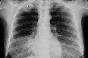
In the early stages, Pancoast cancer is difficult to detect on X-ray due to the lack of clear visualization and the large number of shadows of nearby structures. Chest X-ray may reveal asymmetry of the apex of the lungs (a small area of thickening of the pleura in the apical zone of one lung); an increase in tissue mass; damage to 1-3 ribs or part of the vertebrae.
Pancoast carcinoma is defined on chest CT by the presence of a pathological tissue formation in the area of the superior thoracic orifice and the superior pulmonary groove and its penetration into the chest wall, spine, blood vessels, nerves, or space between the lungs.
But MRI is recommended to determine a more complete picture of the local spread of tumor cells and the amount of involvement of nerve endings.
To establish an accurate diagnosis, endoscopic mediastinoscopy is also used to examine the lymph nodes. And if there is palpable adenopathy of the supraclavicular lymph nodes, their fine-needle transdermal aspiration is performed.
The tests to confirm the diagnosis of the tumor and to accurately assess its stage are a biopsy (tumor cells) obtained by transthoracic fine-needle aspiration biopsy. In some situations, an endoscopic or open thoracotomy may be required for histological examination of the neoplasm.
Differential diagnosis
Differential diagnosis should distinguish Pancoast tumor from: Hodgkin's lymphoma and lymphoma, pleural mesothelioma, echinococcal cyst of the lung, thyroid carcinoma and adenoid cystic carcinoma, desmoid tumors of the mediastinum, breast cancer, as well as scalene muscle and cervical rib syndromes.
Who to contact?
Treatment of Pancoast cancer
A positive result – cessation of tumor growth and its regression, reduction of local and distant relapses and increased survival – is ensured by treatment using induction chemoradiation therapy and subsequent surgical treatment – performing an operation to resect the affected structures.
Types of chemotherapy for Pancoast cancer:
- before surgery – a combination of certain chemotherapy drugs with radiation for 5-6 weeks;
- after removal of part or all of the lung, affected adjacent tissues or upper ribs (which is carried out after a course of chemoradiation therapy) – final postoperative chemotherapy.
Chemotherapy regimens use the cytostatic drug Cisplatin (Platinotin) in combination with other antitumor drugs, in particular, Etoposide and Vindesine (Eldisine).
For example, intravenously administered Cisplatin is a platinum derivative; the drug is effective, but like all anticancer cytostatics, it causes many side effects and negative consequences. The most common consequences of chemotherapy for Pancoast cancer are described in detail in the publications:
Radiation therapy regimens for Pancoast cancer usually include fractions of 45 Gy/27 over 5-6 weeks, followed by surgery (4-6 weeks later).
In this case, contraindications to surgical treatment are metastases, damage to the supraclavicular and mediastinal lymph nodes, more than half
Bodies of the vertebrae, trachea and esophagus.
Patients with inoperable Pancoast tumor are given palliative treatment.
Forecast
As with other malignancies, the prognosis for patients with Pancoast cancer directly depends on the stage of the disease. The presence of symptoms of Bernard-Horner syndrome is considered a poor prognostic factor.
Over the past few decades, survival rates for patients with apical lung cancer with Pancoast syndrome have improved significantly.
After induction chemoradiotherapy and subsequent surgery, in 33-40% of cases (according to other data, 54-72%) the average survival time is five years, and the incidence of complications varies in the range of 10-28%.
Almost 75% of patients continue to live for two years.