
All iLive content is medically reviewed or fact checked to ensure as much factual accuracy as possible.
We have strict sourcing guidelines and only link to reputable media sites, academic research institutions and, whenever possible, medically peer reviewed studies. Note that the numbers in parentheses ([1], [2], etc.) are clickable links to these studies.
If you feel that any of our content is inaccurate, out-of-date, or otherwise questionable, please select it and press Ctrl + Enter.
Caliciviruses
Medical expert of the article

Last reviewed: 08.07.2025
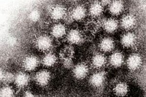 ">
">Caliciviruses were first isolated from animals in 1932 and were found in the feces of children suffering from acute gastroenteritis in 1976. They are now classified as a separate family, Caliciviridae.
The virions are spherical and 37 nm in diameter, with no supercapsid. The genome is represented by positive single-stranded RNA with a molecular weight of about 2.6-2.8 MD. Negative-contrast microscopy reveals 32 deep (about 10 nm) cup-shaped depressions on the surface of the virions, which is why they were named caliciviruses (from the Greek calyx - cup). Caliciviruses do not reproduce in cell cultures, which makes them difficult to detect. Immune electron microscopy is mainly used for diagnostics.
 [ 1 ], [ 2 ], [ 3 ], [ 4 ], [ 5 ], [ 6 ], [ 7 ], [ 8 ], [ 9 ]