
All iLive content is medically reviewed or fact checked to ensure as much factual accuracy as possible.
We have strict sourcing guidelines and only link to reputable media sites, academic research institutions and, whenever possible, medically peer reviewed studies. Note that the numbers in parentheses ([1], [2], etc.) are clickable links to these studies.
If you feel that any of our content is inaccurate, out-of-date, or otherwise questionable, please select it and press Ctrl + Enter.
Osteoma of bone: causes, surgical removal
Medical expert of the article

Last reviewed: 04.07.2025
A benign tumor process that develops in bone tissue is called osteoma of the bone. This tumor grows slowly, during its growth, adjacent tissues move apart, and no growth occurs in them. Osteoma is not capable of metastasizing, can grow to large sizes, and often has a peculiar capsule.
As a rule, osteoma of the bone responds well to treatment, the outcome of which can be classified as favorable.
Epidemiology
Osteoma of the bone is most often detected in childhood and adolescence, as well as in young people aged 20-25. Mostly men are affected, however, damage to the facial bones is more often diagnosed in women.
Osteomas account for about 10% of all bone tumors.
Most often, the disease affects the flat cranial bones, paranasal sinuses, tibia, femur, humerus, and less often the vertebrae and ribs.
Causes bone osteomas
The exact causes of the appearance and growth of bone osteoma are not fully defined. Presumably, the pathological process may be associated with mechanical damage to the bone area, or with a hereditary predisposition. Such pathologies as gout, rheumatism, syphilis also contribute to the development of the disease. But in such situations, exostoses are formed in the bone tissue - bone growths that are not tumors as such.
Inflammatory processes and injuries play a significant role in the development of osteoma. For example, when the bones of the nasal sinuses are affected, the provoking factors can be both inflammatory ENT diseases and directly the puncture of the sinus during the treatment of chronic sinusitis.
Experts also do not rule out a certain role of the peculiarities of intrauterine development, calcium metabolism disorders, and negative environmental background.
Risk factors
The onset of the pathological process associated with osteoma of the bone can be provoked by the following factors:
- processes of metaplasia with the replacement of healthy cells with pathological structures;
- unfavorable heredity;
- pathologies of embryonic development;
- inflammatory processes, infectious diseases;
- chronic systemic pathologies;
- gout;
- violation of calcium metabolism;
- post-inflammatory complications.
Pathogenesis
Until relatively recently, osteoma was considered one of the signs of chronic sclerosing osteomyelitis and the tumor was not considered as a separate pathology. The first bone formation that was considered an independent disease was osteoid osteoma of the bone. This tumor develops in tubular structures and looks like a small area with sparse bone tissue, up to 20 mm in diameter. With more detailed visualization, one can pay attention to the obvious sclerotic reaction along the edge of the tumor focus. Such osteomas can be cortical or spongy. Histology reveals many osteoblasts and osteoclasts.
Examination of the pathology with a microscope allows one to notice clear contours separating the rarefied tissue permeated with vessels. In the central part of the osteoma there are osteoid trabeculae and strands, as if entangled with each other. In the altered tissue there are large osteoblasts with a large nucleus.
The osteoma structure does not contain hemocytoblasts and lipid tissue. In some areas, osteoclasts can be identified, with a single or group arrangement. If the integrity of the bone is compromised at the site of the osteoma, then cartilaginous tissue can be seen inside it, which is also present in formations developing below the articular cartilage. This is the structure of the central part of the tumor. Along the perimeter, there is fibrous connective tissue, which has the appearance of stripes reaching two millimeters in width. Further, a layer of sparse cortical plate can be seen - but this does not always happen.
Symptoms bone osteomas
Osteoma most often develops slowly, without any specific signs or manifestations. The predominant location of osteoma is the outer surface of the bone. The tumor can occur in any part of the skeletal system (the exception is the sternum bone). The most common localization is the bone surfaces of the paranasal sinuses, the bones of the skull, shoulder and hip.
Osteoma most often looks like a hard and smooth elevation on the outer part of the bone, which is immobile and painless. When the formation develops on the inner surface of the skull, the first signs are especially clear, in the form of headaches, increased intracranial pressure, memory impairment, convulsions. If osteoma appears in the "Turkish saddle" area, it can manifest itself in hormonal disruptions.
Osteoma of the paranasal sinuses is often accompanied by the following symptoms:
- protrusion of the eye ( exophthalmos type );
- deterioration of vision;
- double vision;
- drooping eyelid;
- differences in pupil size.
If the osteoma is localized in the vertebral area, the patient will complain of pain. The diagnostic method determines compression of the spinal cord, deformation of the spine.
Forms
The pathogenetic division of osteomas is as follows:
- hard osteomas, which are distinguished by their particular strength and density;
- spongy osteomas with a corresponding spongy structure;
- medullary osteomas, consisting of relatively large cavities with a bone marrow component inside.
Hard formations include osteophytes - these are specific bone deposits located around the circumference (hyperostoses), on one convex section of the bone (exostoses) or inside the bone tissue (endostoses).
Hard formations are often found in the area of the skull, on the pelvic bones.
According to the etiological factor, the following types of osteomas are distinguished:
- hyperplastic, which arise directly from bone tissue (osteoid osteomas, simple bone osteomas);
- heteroplastic, which arise from connective tissue (osteophytes).
Osteomas are always solitary. Multiple formations are typical for Gardner syndrome, a disease in which adenomatous polyps are combined with osteomas of the cranial bones and skin neoplasms. The syndrome belongs to the group of familial polyposis with an autosomal dominant type of inheritance.
- Osteoid osteoma of bone occurs in the diaphyseal zone of long tubular bones. The tibia is most often affected, less often - flat bones, vertebrae. If the pathology is localized near the growth zone, then bone growth can be stimulated, which in childhood can cause asymmetry of the supporting apparatus. In addition, symptoms associated with compression of peripheral nerves often appear.
- Spongy osteoma of bone is characterized by a porous structure resembling a sponge. The neoplasm is penetrated by a network of vessels and contains a lot of lipid and connective tissue. The predominant localization of spongy osteoma is tubular bones. A distinctive feature of such pathology is the ability to separate from the bone element with strong growth.
- Osteoma of the cranial bone in many cases develops in the area of the lower jaw - on the back surface, or on the jaw branch, below the molars. Such a tumor can be round or oval, with a smooth surface and clear crust-like contours. The size of the formation can be different: in advanced cases, osteoma displaces nearby tissues, causing asymmetry and disruption of muscle function.
- Osteoma of the frontal bone is the most common. With a significant increase in the tumor, the face swells (without pain), breathing may be difficult. Patients are often bothered by headaches and visual impairment. The tumor usually ranges in size from 2 to 30 mm, sometimes larger. The affected bone tissue may become inflamed, which becomes a direct indication for surgical intervention.
- Osteoma of the occipital bone is considered a rare pathology. The disease is not accompanied by painful symptoms and is detected mainly by chance - using an X-ray. In some patients, the tumor manifests itself as increased sensitivity to external irritants, dizziness and general discomfort associated with the creation of pressure on the inner ear. Occipital osteoma does not disrupt the structure of bone tissue, developing from the cranial vault.
- Osteoma of the parietal bone may be represented by osteoid osteoma or osteoblastoma. Osteoblastoma is characterized by large sizes and is prone to further enlargement. The parietal bone is most often affected in children, without any specific symptoms. However, tumors with such a location are subject to mandatory removal, due to the danger of their localization.
- Osteoma of the temporal bone in most cases is of concern only because of the existing aesthetic defect, since other signs of pathology are usually not manifested. With large sizes of the formation, patients may complain of constant headaches.
- Ethmoid osteoma is a benign disease of the cranial bones. It is located in the center between the facial bones and is in contact with many of them. The ethmoid bone itself is involved in the formation of the nasal cavity and eye sockets, so when the formation reaches large sizes, it can cause problems not only with nasal breathing, but also with visual function.
- Osteoma of the femur is most often an osteoid tumor consisting of osteoblasts, vascular network, and bone tissue itself. Such a tumor has a central zone of mineralization or vascular-fibrous borders and can appear on any part of the femur.
- Osteoma of the tibia may have a hard, spongy or combined structure, but most often this tumor is dense, like ivory. There are no bone marrow cells in its structure. Among all neoplasms affecting long tubular bones, the most common is a tumor of the femur. The second most common is osteoma of the tibia, and the third is osteoma of the fibula. The listed pathologies often manifest themselves as lameness, painful sensations at rest (for example, during a night's rest), muscle atrophy. Some patients experience repeated fractures of the limbs.
- Osteoma of the ilium is diagnosed relatively rarely, since at small sizes it does not reveal itself with clinical symptoms. Pelvic bone tumors in women can significantly complicate the course of labor.
- Osteoma of the calcaneus can develop at almost any age. This is one of the types of osteomas that, due to their specific localization, almost immediately reveal themselves with pronounced symptoms. Patients complain of severe pain when walking and standing, which often significantly worsens the quality of life. The formation on the heel includes cartilaginous cells and grows on the bone surface.
- In most patients, osteoma of the metatarsal bone is asymptomatic, and only with pronounced sizes of the pathological focus can pain be felt after or during exercise. There is also a deformation of the metatarsal bone, which can create discomfort to the patient to varying degrees.
- Osteoma of the pubic bone is a pelvic formation and is relatively rare. The pathology does not have clear symptoms and is detected by chance - during X-ray or computed tomography.
- Osteoma of the ischium is a focus of round configuration with smooth, clear sclerotic borders. Along the lower edge, a compacted zone of round shape is found, as well as thin striped periosteal layers. Such a bone defect is a rare benign pathology.
- Osteoma of the humerus is common, but has some difficulties with identification. Thus, on an X-ray, the formation resembles a healthy normal bone, or is manifested by a slight thickening. The accuracy of diagnosis depends on the qualifications of the medical specialist.
- Osteoma of the humeral head, if relatively large, may be accompanied by pain in the upper shoulder area, for example, during passive movements. During examination, a disturbed configuration of the shoulder joint may be detected. To clarify the diagnosis, radiography is prescribed in two projections: in the anteroposterior direction, as well as in the axial direction, in which the rays pass from top to bottom through the axillary fossa.
- Osteoma of the radius can be located on any part of the bone tissue, but most often this pathology is represented by osteoid osteoma. In most cases, the disease does not have bright symptoms and does not bother the patient with pain or other uncomfortable sensations.
Complications and consequences
The most unfavorable complication of osteoma of the inner bone surface of the skull is visual impairment in the form of loss of the ability to separately perceive two points that are at a distance from each other. If the osteoma continues to increase in size, the following problems may occur:
- severe and frequent migraine attacks;
- convulsive attacks, sometimes with loss of consciousness;
- uncontrolled muscle contractions;
- disruption of nervous activity, changes in the body's response to the influence of external or internal factors;
- disruption of bioelectrical activity and, as a consequence, disruption of breathing and cardiac activity.
The listed negative consequences can only occur with tumor damage to the bones of the head. With damage to the spinal column, paresis, innervation disorders, and deterioration of the motor ability of the limbs can be observed.
 [ 33 ]
Diagnostics bone osteomas
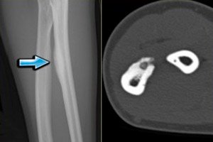
Osteoma is determined by examining an X-ray image. Since the symptoms of the disease have much in common with osteogenic sarcoma and chronic osteomyelitis, an X-ray is mandatory, as it allows for accurate differentiation of the disease.
Additional instrumental diagnostics may include computed tomography. Histologically, a discrepancy with the typical composition of the bone marrow is determined. The channels are located chaotically, there are relatively few of them. Spongy osteoma is devoid of channels, chaotically located bone beams are visualized. The layers of fibrous tissue are expanded against the background of an increase in brain spaces.
Less often, diagnostics are supplemented by ultrasound scanning, thermography, angiography, and radioisotope examination. The listed diagnostic procedures can help with the detection of compact or spongy osteoma of the bone, which occur with almost the same frequency.
A compact tumor grows within a bone formation and does not manifest itself as a protrusion. The formation has a hemispherical or spherical configuration, and an X-ray image reveals an unstructured darkening. This pathology is discovered accidentally in most patients.
In spongy osteoma, the lesion is large: a convex swelling of the bone tissue layer on the outer side of the bone is observed. The cortical layer remains intact.
The tests are an addition to a comprehensive diagnostic examination:
- blood test to assess the level of the enzyme alkaline phosphatase;
- General blood test to assess the general condition of the body.
Differential diagnosis
Differential diagnostics are carried out:
- with sclerosing processes in the bone (pay attention to the lack of a contour between the affected and normal tissue);
- with exostases (practically do not cause pain or functional impairment);
- with osteoid osteoma (typically aching pain that intensifies at night).
Who to contact?
Treatment bone osteomas
If during diagnostic procedures an osteoma of the bone is detected without indications for surgical treatment, then conservative therapy may be prescribed. In case of large sizes of the formation, the only method of treatment is considered to be surgical intervention, which is also indicated in case of disruption of the functioning of nearby organs, or in case of visible changes in the configuration of the bone.
Medicines are prescribed mainly for symptomatic effects - for example, to relieve pain, improve overall well-being and strengthen the immune system.
Method of administration and dosage |
Side effects |
Cautions |
|
Ortofen |
Take 100-150 mg per day. |
Hypersensitivity, drowsiness, tinnitus, abdominal pain, irritability. |
The drug should not be taken for a long time. Optimally, 3-4 days in a row. |
Ibuprofen |
Take at a rate of 20-30 mg per kg of weight per day. |
Nausea, dizziness, hypersensitivity reactions. |
Not prescribed to children under six years of age. |
Calcemin |
Take one tablet 1-2 times a day, but no more than 4 tablets per day. |
Rarely – allergy, nausea. |
Not prescribed to children under 5 years of age. |
Calcium D 3 Nycomed |
Take one tablet twice daily. |
Rarely – dyspepsia, allergic reaction. |
Not prescribed to patients with phenylketonuria, sarcoidosis, or children under 5 years of age. |
Chondroitin Complex |
Take one capsule twice a day, half an hour before meals. |
Rarely – allergies, dizziness, nausea. |
The recommended duration of treatment is at least six months. |
Surgical treatment
The method of surgical intervention is chosen by the doctor, taking into account the symptoms of the osteoma, the patient's complaints, the degree of tumor growth and its localization. As a rule, the removal of the bone osteoma is carried out after the results of the histological analysis are received.
The location of the pathological formation is the main point that influences the choice of the type of surgery. For example, when osteoma is localized on the bones of the skull, the intervention is most often entrusted to neurosurgeons, and if the tumor affects the bones of the limbs, then to a trauma surgeon.
The technical features of the operation are discussed by doctors in advance and depend on the presence of symptoms, the stage of development of the pathology and the presence of complications from neighboring organs. In recent years, laser has been widely used to remove osteoma.
The use of a laser is especially in demand when flat cranial bones are affected. The operation is performed under general anesthesia. The doctor makes an incision in the skin. If necessary, he trepans the skull and performs a thorough resection of the tumor tissue. Damaged blood vessels are also subject to removal.
However, laser removal is not the most modern surgical method. A more effective intervention is considered to be excision of the tumor focus using radiofrequency irradiation with computed tomography guidance. This procedure helps to avoid possible recurrences of the disease, bleeding and infectious complications. Treatment can even be carried out using local anesthesia. To detect the tumor focus, thin computed tomography sections are used, after which a radiofrequency transmitting device is inserted into the affected tissue. The formation is heated to 90 ° C - at this temperature, the tumor is destroyed, and nearby normal tissues are not affected. The operation is performed on an outpatient basis. The rehabilitation period is short: after a week, the patient can return to work.
Prevention
Experts do not have specific recommendations for the prevention of osteoma of the bone - primarily because the exact causes of the disease are unknown. Among the general recommendations, the following can be highlighted:
- avoid injuries and damage to the musculoskeletal system;
- For any inflammatory diseases or injuries, consult a doctor;
- If the doctor has prescribed treatment, follow all instructions exactly and complete the course of therapy;
- Eat a balanced diet and ensure that your body is constantly receiving important minerals and vitamins.
Timely medical attention and thorough diagnostics will help avoid the development of unpleasant consequences of osteoma.
Forecast
The prognostic data on the disease are favorable. The tumor develops gradually, without intensive aggressive growth. To date, there have been no cases of its transformation into a malignant tumor: osteoma of the bone does not metastasize and is not prone to growing into nearby tissues
You should not treat osteoma on your own: the only possible solution to this problem is surgery. In no case should you apply heat to the tumor, put compresses, or act on it in any other physical way - this can only increase the growth of the tumor. It is necessary to take into account that in the vast majority of cases, osteoma of the bone can be successfully treated and does not pose a danger to human life.
[ 40 ]