
All iLive content is medically reviewed or fact checked to ensure as much factual accuracy as possible.
We have strict sourcing guidelines and only link to reputable media sites, academic research institutions and, whenever possible, medically peer reviewed studies. Note that the numbers in parentheses ([1], [2], etc.) are clickable links to these studies.
If you feel that any of our content is inaccurate, out-of-date, or otherwise questionable, please select it and press Ctrl + Enter.
Blepharochalasis
Medical expert of the article

Last reviewed: 04.07.2025
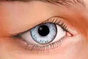
A condition in which the skin of the upper eyelids hangs down like a bag over the edge of the eyelid is blepharochalasis. Let's look at the main causes of this pathology and treatment methods.
Bilateral atrophy of the skin of the upper eyelids is a pathological process caused by damage to the elastic fibers of the connective tissue and characterized by increased flabbiness of the epidermis. According to the International Classification of Diseases of the 10th revision ICD-10, blepharochalasis is included in the following category VII Diseases of the eye and its adnexa (H00-H59):
H00-H06 Diseases of eyelids, lacrimal ducts and orbit.
- H02 Other diseases of eyelids.
- H02.3 Blepharochalasis (dermatolysis).
Hypertrophy of the upper eyelid tissues is usually formed under the influence of individual features of the skin structure, with age-related changes or prolonged swelling of the eyelids. As a rule, this disease is diagnosed in elderly people.
Many doctors claim that eyelid atrophy is associated with endocrine, vascular or neurotrophic disorders. In some cases, the disorder manifests itself in hereditary pathology - premature skin aging syndrome "cutis laxa". To treat inflammatory eyelid edema, you need to see an ophthalmologist and a plastic surgeon.
Epidemiology
Blepharochalasis has an age dependence. Statistics indicate that eyelid pathologies account for about 10% of the overall structure of ophthalmological diseases. This is due to the fact that the eyelids consist of different tissues that react differently to the same irritation. That is why eyelid diseases are classified by anatomical features, and not by etiopathogenetic ones.
According to medical statistics, an increase in the amount of tissue in the upper eyelid is most often diagnosed in patients with a hereditary predisposition and advanced endocrine diseases.
Causes blepharochalasis
To date, reliable causes of blepharochalasis have not been established. The disease has an idiopathic origin. Possible causes include the following:
- Endocrine disorders and diseases.
- Pathologies of the vascular system.
- Hereditary predisposition.
- Neurotrophic disorders.
- Inflammatory diseases of the eyelids.
- Allergic reactions.
- Vasomotor disorders.
- Usher syndrome.
- Progressive atrophy of the skin.
Regardless of the cause, treatment of the disorder is carried out only by surgery.
Risk factors
Since eyelid skin atrophy is idiopathic in origin, a number of risk factors are identified that can provoke it. First of all, these are endocrine system disorders and genetic factors. Sporadic variants of the development of pathology are also identified:
- Inflammatory lesions of the eyelids.
- Allergic reactions.
- Thyroid dysfunction.
- Vasomotor disorders.
Dermatolysis has been suggested to be associated with progressive skin atrophy. In some cases, atrophy occurs simultaneously with goiter and double lip, suggesting Usher syndrome.
The peculiarity of the disorder is that it begins unnoticed, sometimes after chronic, frequently recurring inflammation. Gradually, the skin of the eyelids loses elasticity, becomes flabby, thin with translucent vessels and small folds, hanging over the upper part of the eye and limiting the field of vision.
Pathogenesis
The mechanism of origin of blepharochalasis depends entirely on the cause that provoked it. Pathogenesis in most cases is associated with periodic inflammation of the upper eyelid. The inflammatory process is accompanied by swelling. Frequent relapses of swelling lead to atrophy of the fibrous structures of the eyelid, which provide its elasticity. As the disease progresses, the function of the muscles responsible for lifting the eyelid is impaired.
Symptoms blepharochalasis
Excess tissue of the upper eyelid has a number of characteristic signs. Symptoms of blepharochalasis occur with equal frequency among both men and women. Moreover, most often in elderly patients. There are also isolated cases of the disease developing during puberty.
The main symptoms of the pathology:
- Excessive growth of tissue in the upper eyelid.
- Overhanging skin fold on the upper eyelashes.
- Loss of skin elasticity and its increased stretchiness.
- Dilated blood vessels are visible through the skin of the eyelid.
The sagging tissue is especially noticeable when viewed from the side. With each passing year, the atrophy becomes more pronounced. Blepharochalasis can progress so much that the skin begins to hang over the upper eyelashes, covering the pupil and impairing vision.
First signs
According to the conducted research, it has been established that the disease can have an unnoticeable onset, or it can manifest itself as a chronic, frequently recurring inflammatory process on the eyelids. Due to constant swelling, the skin becomes thinner and a bag is formed that hangs over the eyes.
Let's consider the first signs of hypertrophy of the tissues of the upper eyelid and their progression:
- Tissue swelling/edema.
- Formation of a skin fold that is particularly noticeable when viewing the head from the side and when moving the facial muscles.
- Significant expansion of the blood supply in the eyelid area.
- Loss of elasticity, flabbiness and thinning of the skin.
As the disease progresses, the skin stretches more and more, hanging over the eye and partially obscuring vision.
 [ 18 ]
Blepharochalasis of the lower eyelids
Such pathology as blepharochalasis of the lower eyelids is much less common than atrophy of the upper eyelid tissues. Enlargement and drooping of the skin fold of the lower eyelid creates the impression of a bag under the eyes. Very often, this type of pathology is confused with eversion of the eyelid, when the eyelash edge does not adhere to the eyeball, as it is turned outward. Because of this, the skin sags strongly, revealing the eyeball.
The disease may develop with inflammatory processes of the conjunctiva and eyelid. The disorder occurs with facial nerve neuropathy. The origin of blepharochalasis may be associated with insufficiently strong connection of the skin with the levator tendon, defects of the tarso-orbital fascia, or thinning/overstretching of the skin due to recurrent angioedema. The pathology causes cosmetic inconvenience, so treatment is based on surgical excision of the excess skin fold.
[ 19 ]
Stages
Blepharochalasis goes through several stages in its development. At the initial stage, the skin loses its elasticity and gradually becomes thinner. Most often, this is associated with a recurring inflammatory process. As the disease progresses, small blood vessels appear, which quickly become pronounced. At the last stage, the eyelid takes on a baggy shape, covering part of the eye and impairing vision.
Forms
There are no specific types of the disorder. There are unilateral and bilateral blepharochalasis. Eyelid edema is also distinguished by the location of the skin fold: upper or lower eyelid.
The following forms of dermatolysis are distinguished:
- Autosomal recessive - signs of the disease are already evident at birth or develop rapidly as the child grows older. The loose skin hangs in large folds, but it is not atrophic or hyperelastic. Bag-like folds give the face a tearful expression.
- Autosomal dominant (limited) – hypertrophy of the eyelids appears simultaneously with Usher syndrome. Progressive enlargement of the lips (usually the upper one) occurs due to swelling of the mucous membrane and the formation of transverse grooves.
Atrophy may be associated with genetic and acquired factors.
Complications and consequences
The increase in the amount of tissue in the upper eyelid tends to progress. The consequences and complications manifest themselves in the form of a pronounced cosmetic defect. Surgical intervention is used to treat the pathological condition. Blepharoplasty can lead to the following problems:
- Retrobulbar hematoma – occurs due to bleeding in the postoperative period and accumulation of blood in the orbital cavity. It is manifested by significant edema, increasing exophthalmos, a sharp decrease in vision and limitation of eyeball mobility. To eliminate this complication, a wound revision, drainage and removal of blood clots are performed.
- Suppuration of the postoperative wound - manifests itself as infiltration of the skin of the upper eyelids, severe swelling and pain. Treatment involves washing with antiseptics and drainage of the wound area.
- Hypertrophic scars – are formed due to the disruption of the development of a normal scar. They look like dense red strands that pull together thin tissues, forming skin folds. To eliminate such a consequence of surgery, myogymnastics is prescribed.
- Conjunctivitis (bacterial, viral, allergic) - occurs against the background of reduced local immunity after surgery, due to intolerance to medications or failure to follow eyelid care rules. Manifested by a sensation of a foreign body in the eye, photophobia, itching, increased lacrimation. Treatment is local.
- Ptosis of the upper eyelid most often occurs due to a violation of the surgical technique, that is, injury to the levator aponeurosis and its subsequent scarring.
- Ectropion – this complication is associated with a violation of the supporting structures of the lower eyelid. Most often, this occurs due to injury to the pre-pelvic part of the orbicularis oculi muscle.
- Enophthalmos is a posterior displacement of the eyeball. It occurs due to the excision of most of the orbital fat. The size of the palpebral fissure decreases.
- Dark circles under the eyes – occur due to hyperinsolation in the postoperative period and the formation of a hematoma. Moderate hyperpigmentation disappears within 4 weeks after treatment. In other cases, bleaching preparations with hydroquinone, glycolic acid and hydrocortisone are used.
Without proper treatment, hypertrophy causes folds of skin to hang over the upper eyelashes, obscuring the field of vision from both the sides and above. This leads to visual impairment.
Diagnostics blepharochalasis
Atrophy and enlargement of the skin of the upper eyelids refers to ophthalmological diseases. Diagnosis of blepharochalasis begins with checking visual acuity and external examination of the eye. The ophthalmologist evaluates the condition of the eyelids and conjunctiva, and if necessary, conducts biomicroscopy.
In most cases, diagnosis is not difficult, as it is based on the clinical manifestations of the disease. Blepharochalasis is characterized by a saccular hanging of the skin fold above the eye, which can partially cover the pupil. Edema in most cases occurs as a recurrent inflammatory process.
[ 28 ], [ 29 ], [ 30 ], [ 31 ], [ 32 ]
Tests
A set of laboratory tests for diagnosing blepharochalasis is carried out at all stages of the disease development and its treatment. The tests consist of:
- A general blood test is the first thing you need to take. It provides information about the general condition of the body: the content of cellular elements of the blood (erythrocytes, leukocytes, lymphocytes, monocytes, etc.), hemoglobin level, hematocrit, erythrocyte sedimentation rate, color index.
- Blood for RW – this analysis is performed to detect the causative agent of syphilis or pale treponema. For the Wasserman reaction, venous blood is collected.
- Hbs antigen is an indicator of the acute form of viral hepatitis B.
- Blood clotting time – there are several methods for conducting this analysis. But there is no single norm for blood clotting. For example, according to Sukharev’s method, clotting begins 30 seconds to 2 minutes after the start of the analysis and ends in 3-5 minutes. Minor deviations are variations of the norm.
- General urine analysis is a standard laboratory test used to diagnose almost all diseases. It allows you to learn about the state of the cardiovascular system, immunity, kidneys and other internal organs and systems.
Based on the results of laboratory diagnostics, the doctor may prescribe additional tests or make a diagnosis.
Instrumental diagnostics
Atrophy of the skin of the upper or lower eyelids is a pathological condition that causes cosmetic inconvenience and negatively affects visual acuity. Instrumental diagnostics for blepharochalasis consists of the following procedures:
- Visual examination of the skin of the eyelids - the disease is characterized by atrophic changes, in which the tissues are thinned and stretched, small subcutaneous veins can be seen. The skin hangs in a fold at the outer corner of the eye, blocking the view.
- Visometry is a visual acuity test. Standard ophthalmological examination. Various metric tables with optotypes are used for its implementation.
- Perimetry is a diagnostic of the visual field boundaries and their projection onto a spherical surface. It allows to identify visual field disorders and determine the localization of the pathological process.
In addition to the above-described studies, gonioscopy, eye examination, ultrasound, and various X-ray methods may be prescribed.
What do need to examine?
Differential diagnosis
In addition to laboratory and instrumental studies, differential diagnostics is also indicated for blepharochalasis.
Differential diagnosis is carried out with the following diseases:
- Quincke's angioedema has a rapid onset, unlike dermatolysis, which develops gradually. Against the background of swelling of the eyelids, painful sensations appear in the lip area, breathing is difficult. Allergic reactions from the skin, itching, rashes are possible.
- Erysipelas - characterized by an acute onset and a pronounced inflammatory process. The patient complains of a feverish condition and deterioration of general health.
- Neurofibromatosis - in addition to the lesions in the eyelid area, areas with pathological changes appear throughout the body.
- Senile atrophy of the skin of the eyelids - blepharochalasis develops at a young age and has more pronounced atrophy.
In most cases, diagnosing the pathology is not difficult, since the disorder has characteristic clinical manifestations.
Who to contact?
Treatment blepharochalasis
Unfortunately, treatment for blepharochalasis has not been developed to this day. Conservative methods are used to stop the progression of the pathological inflammatory process, but their effectiveness is not great. The most effective method of therapy is surgical excision of excess skin.
All recommendations and the final decision regarding the operation are made by an ophthalmologist and a plastic surgeon. In most cases, blepharoplasty is performed under local anesthesia. The operation lasts about 1.5-2 hours. In addition to removing hypertrophy of the eyelid skin, lateral canthoplasty and external lifting aponeurosis of the fold can be performed.
The recovery period lasts 1-2 weeks. The patient is prescribed various medications, vitamins and ointments that speed up the recovery process and minimize the risk of complications.
Medicines
Drug therapy for blepharochalasis is carried out immediately after surgery. Medicines are necessary to prevent infectious complications. For this purpose, eye drops and ointments with antibiotic and antiseptic properties are used.
Most often, patients are prescribed the following medications:
- Vitabact is a broad-spectrum antimicrobial agent. It has pronounced antiseptic properties. It is active against fungi, microbes, and viruses. It is available in the form of a solution for eye drops. The dropper bottle contains 10 ml of 0.05% of the drug. The active ingredient of the drops is piloxidine hydrochloride. Excipients: polysorbate, anhydrous dextrose, purified water.
- Indications for use: prevention of postoperative complications in the anterior segment of the eye, preoperative preparation, bacterial eye infections, dacryocystitis, inflammatory processes.
- The dosage and method of administration are determined by the attending physician, individually for each patient. For bacterial infections, 2 drops are instilled 2-6 times a day. As the inflammatory process decreases, the dosage is reduced. To prevent infection before surgery, 1-2 drops are instilled once. The procedure is repeated on the first day after surgery.
- Contraindications: individual intolerance to the components of the drug, pregnancy and lactation.
- Side effects: allergic reactions in the form of conjunctival hyperemia. Burning and redness are also possible. To eliminate these reactions, it is necessary to thoroughly rinse the eyes with clean water and consult a doctor.
- Diclofenac is a non-steroidal anti-inflammatory drug. It has analgesic, antipyretic and anti-edematous properties. It minimizes the manifestations of miosis during surgical manipulations.
- Indications for use: preoperative preparation, postoperative prevention of inflammatory changes, treatment of post-traumatic processes in the eyeball, inflammatory changes in the conjunctiva.
- Method of application: drops are used locally. Installations are made in the conjunctival sac. Dosage and duration of use depend on medical indications. As a rule, the drug is used 1 drop every 6-8 hours. The average course of treatment takes 7-14 days.
- Side effects: in most cases, the drug is well tolerated. Very rarely, corneal opacity, blurred vision, itching and redness of the eyelids, facial swelling, urticaria, increased body temperature, vomiting develop.
- Contraindications: hypersensitivity to the components of the drug, intolerance to NSAIDs and acetylsalicylic acid, disorders of hematopoiesis, gastrointestinal diseases with erosive or ulcerative defects. No cases of overdose have been recorded.
- Indocollyre is a local ophthalmologic agent with anti-inflammatory and analgesic properties. Contains the active ingredient - indomethacin from the NSAID group. Reduces the intensity of the inflammatory process, reduces the severity of pain syndrome.
- Indications for use: treatment and prevention of inflammatory processes after surgery. Pain syndrome after photorefractive keratectomy. The drug is used 2-4 times a day, 1 drop in each eye.
- Side effects: hypersensitivity reactions, burning, pain in the eyes and hyperemia, temporary decrease in visual acuity, photosensitivity.
- Contraindications: individual intolerance to the components of the drug and NSAIDs. Not prescribed for patients with a history of aspirin triad, peptic ulcer, severe renal and hepatic dysfunction. Use with special caution before planned eye surgeries.
- Overdose: Frequent use of drops increases the severity of side effects. To eliminate them, stop using the drug, rinse your eyes with running water and seek medical help.
- Tobradex is a combination drug that includes antimicrobial and corticosteroid components. It has pronounced bactericidal and anti-inflammatory properties. Contains tobramycin (a broad-spectrum antibiotic) and dexamethasone (a corticosteroid with anti-inflammatory action). It is available in the form of an eye ointment and eye drops.
- Indications for use: inflammatory eye diseases with/without bacterial infection. Treatment and prevention of infectious and inflammatory eye diseases after surgery. Eye injuries, prevention of infectious and inflammatory pathologies.
- Method of application: drops are intended for instillation into the conjunctival sac, 1-2 drops every 4-6 hours. The ointment is also applied to the conjunctival sac of the affected eye 3-4 times a day.
- Side effects: local allergic reactions, burning, dry eyes and eyelids, temporary decrease in visual acuity, keratitis, conjunctival edema. In isolated cases, the development of cataracts, photophobia, mydriasis, glaucoma was noted.
- Contraindications: hypersensitivity to the components of the drug. Not prescribed for the treatment of patients with eye infections caused by the herpes virus, fungi, tuberculosis or mycobacteria. With special caution, it is prescribed for glaucoma and corneal thinning.
- Levomekol is a combination drug with the antibiotic chloramphenicol and the immunostimulant methyluracil. It is available in the form of an ointment. It is used to treat purulent-inflammatory skin diseases, furuncles, trophic ulcers, and 2nd-3rd degree burns.
The ointment is applied to the affected area and, if necessary, covered with a sterile napkin. The course of treatment is individual for each patient. Levomekol can cause local allergic reactions that disappear on their own after discontinuing the drug. It is not used in case of hypersensitivity to its components.
Vitamins
To speed up recovery after surgery to eliminate eyelid skin atrophy, patients are prescribed not only medications, but also microelements necessary for the eyes and body. Vitamins help improve vision and maintain normal eye function.
As a rule, patients are prescribed the following vitamins:
- A – retinol is a component of the eye pigment rhodopsin. Deficiency of this substance reduces visual functions.
- C – ascorbic acid is necessary to strengthen the walls of the eye vessels and capillaries. The rate of nutrition of the eye tissues depends on their strength. Vitamin C deficiency increases the risk of intraocular hemorrhages.
- B vitamins – have high antioxidant activity. Participate in the process of visual impulse formation, interact with retinol. Responsible for the metabolism of nervous tissue.
- E – tocopherol stabilizes the state of cell membranes, has antioxidant properties. Protects against negative ultraviolet radiation and bright lighting.
The above substances can be obtained from food or by purchasing special vitamin complexes for the eyes:
- Riboflavin – prescribed for rapid eye fatigue, visual impairment. Accelerates the healing process of wounds caused by surgical interventions, injuries or eye medications. Reduces stress and improves visual functions, quickly stops the inflammatory process.
- Visiomax – contains plant extracts. Improves vision, minimizes the risk of developing ophthalmological diseases. Promotes restoration of visual functions, improves overall well-being.
- Vitafacol - vitamins that cleanse the lens and eliminate dryness. Can be used as a treatment in the early stages of cataracts.
It is better to use all vitamins and vitamin complexes after consultation with an ophthalmologist. The doctor will select the ideal remedy for maintaining eye health and preserving vision.
Physiotherapy treatment
To speed up recovery after blepharochalasis surgery, patients are prescribed physiotherapy treatment. Physiotherapy is necessary for:
- Sanitation of foci of chronic infection.
- Toning and stimulating local immunity.
- Mobilization of the body's defenses.
- Restoration of the function of the sebaceous glands of the eyelids (secretion stimulation).
Let's look at the most effective physiotherapy procedures:
- Low-frequency magnetic therapy – relieves inflammation and swelling of the eyelids, promotes the resorption of infiltrates, accelerates metabolic processes, improves tissue trophism. Increases the activity of formed elements and plasma proteins, improves local blood flow and enhances blood supply to the eyelids.
- Local darsonvalization of the eyelids - with the help of pulsed medium-frequency currents, irritation of the nerve endings of the reflex zones occurs. Due to this, an effect on the vegetative nervous system is achieved, blood supply improves and tissue trophism increases.
- Secretostimulation – eyelid massage improves blood and lymph flow, increases the tone of the neuromuscular apparatus, restores the secretion of the meibomian glands and eliminates congestion in the eyelids. Massage can be performed simultaneously with the installation of medicinal preparations.
But physiotherapeutic treatment of blepharochalasis, like any therapeutic method, has certain contraindications for use. Treatment is not carried out in cases of severe suppurative processes in the eyes and appendages, in case of fever, malignant neoplasms of the eye.
Folk remedies
For the treatment of skin atrophy of the upper or lower eyelids, not only traditional but also non-traditional methods are used. Folk treatment of blepharochalasis:
- Take a couple of onions and boil them until soft. After cooling, strain. Add some honey to the decoction and wash your eyes and eyelids with this solution 4-5 times a day.
- Pour 250 ml of boiling water over 100 g of fresh cucumber peel and add ½ teaspoon of soda. Use the solution for compresses.
- Brew 25 g of dill seeds or chopped dill herb with 250 ml of boiling water and let it brew until it cools. Strain and use for poultices.
- Mix the following ingredients in equal proportions by weight: birch leaves, rose hips, strawberry leaves, St. John's wort, red clover. Grind all the ingredients until smooth. Pour 50 ml of boiling water over a teaspoon of the mixture and leave for 30-40 minutes. Strain. Use the decoction for compresses 2-3 times a day.
In addition to the above recipes, self-massage has healing properties. It tones the tissues of the eyelids and eyes. You can use your nails to perform a reflex massage. To do this, gently prick your eyelids for 2-3 minutes 1-3 times a day.
Herbal treatment
Another alternative treatment for ophthalmologic diseases is herbal treatment. For blepharochalasis, you can use the following recipes:
- Brew 15-25 g of cornflower flowers without baskets in 250 ml of boiling water, leave for an hour and strain. The infusion is used as a disinfectant and anti-inflammatory agent. The course of treatment is 4-5 days.
- Pour 15-25 g of crushed oak bark with 500 ml of vodka and boil over medium heat for 15-30 minutes. Strain and cool. The decoction is suitable for rinsing and compresses in severe inflammatory processes.
- Place 30 g of crushed plantain seeds in a glass container. Add 2 teaspoons of cold water to the herbal component and shake well. Pour 6 tablespoons of boiling water into the container and continue shaking until the product has completely cooled. Strain and use as a poultice.
- Take a tablespoon of chamomile flowers and pour 250 ml of boiling water. Let it brew for 10 minutes, strain and cool. The finished product can be stored in the refrigerator. The decoction is used for eye compresses. According to this recipe, you can prepare a decoction of celandine, which is also suitable for compresses.
If you don't have the ingredients for the recipes described above, you can use compresses made from freshly brewed black or green tea. To eliminate dry eyelids, add milk to the drink.
Surgical treatment
There is currently no conservative method for eliminating such a pathology as blepharochalasis. Surgical treatment is the only way to stop the progressive growth of eyelid skin.
Blepharoplasty is an operation to correct or change the shape of the eyelids. Excision of excess tissue is aimed at both correcting the defect and aesthetically rejuvenating the patient. The operation helps to tighten the upper and lower eyelids, giving the look lightness and openness. After the procedure, the number of wrinkles on the eyelids decreases.
Indications for surgical intervention:
- Atrophy of the tissues of the upper eyelids and their bag-like drooping.
- Presence of bags under the eyes.
- The presence of fatty deposits under the eyes.
- A large number of wrinkles on the lower eyelid.
- Pathologies and congenital defects.
- Drooping corners of the eyes.
Contraindications to blepharoplasty:
- An inflammatory process in the body with elevated temperature.
- Presence of acute or chronic respiratory disease.
- Hepatitis.
- Diabetes mellitus type 1 or 2.
- Infectious or oncological diseases.
- Pregnancy and lactation.
- The patient's age is under 18 years.
- Blood clotting disorder.
- Acute course of diseases of internal organs.
- Hypertension.
- Increased intracranial pressure.
- Infectious diseases of the nose or eyes.
- Thyroid dysfunction.
Surgical treatment is performed in an outpatient or inpatient setting under local anesthesia. The operation lasts 1-3 hours. The following procedures are indicated for blepharochalasis:
- External levator plicae aponeurosis.
- Blepharoplasty.
- Removal of sebum.
- Lateral canthoplasty.
For upper eyelid surgery, the doctor makes an incision on the natural fold. The incision slightly touches the area above the outer corner of the wrinkles that appear when smiling. Excess skin and fatty tissue are removed through the incisions. As the incision heals, it becomes invisible, as it follows the normal contour of the upper eyelid. For lower eyelid surgery, the incision is made below the lower lash line. Fat, muscle, and excess skin are removed through the incision. To eliminate swelling, the doctor may redistribute the tissue.
Tissue excision can be performed using a scalpel or a laser method. Laser blepharoplasty allows you to remove excess skin around the eyes, change their size or shape, eliminate reticular wrinkles, and edema. The procedure is performed using an erbium or carbon dioxide CO2 beam.
After surgical treatment, the patient has a recovery period, the duration of which is individual for each patient. In the first days, it is necessary to rest. Increased activity can lead to such surgical complications as swelling and bruising. Sutures are usually removed on the 4-5th postoperative day. Initially, the scars are pink, but after 2-3 months they become invisible.
It is worth paying attention to the fact that immediately after the operation, vision may be blurred and remain so for several days. In addition, there is sensitivity to light, increased lacrimation, dry eyes. To eliminate such symptoms, special medications (drops, ointments) are used. It is also recommended to wear sunglasses for 2-3 weeks after the operation to protect the eyes from sun irritation and wind.
Prevention
Blepharochalasis, like any other disease, is much easier to prevent than to treat. Prevention is based on maintaining a healthy lifestyle. It has been established that this pathology occurs more often in patients with lipid metabolism disorders, i.e. obesity.
To prevent eyelid skin atrophy, you need to monitor your physical health and body weight. It is recommended to adjust your diet and pay more attention to sports. Also, do not forget about cosmetic procedures, the action of which is aimed at increasing the elasticity of the eyelid skin.
If there is a genetic predisposition to the disease, you can consult an ophthalmologist or plastic surgeon in advance. The doctor will give recommendations on proper eyelid skin care, which will reduce the risk of atrophic changes.
Forecast
Blepharochalasis, with proper surgical treatment, has a favorable prognosis. If the disease is left to take its course, it will lead to several problems at once. First of all, it is a cosmetic defect that will entail mental health problems (lack of self-confidence, depression). Sagging skin can cover the pupil, making vision difficult. Injury to skin folds can lead to serious ophthalmological diseases. Timely medical attention and treatment will help preserve the beauty of the eyes.