
All iLive content is medically reviewed or fact checked to ensure as much factual accuracy as possible.
We have strict sourcing guidelines and only link to reputable media sites, academic research institutions and, whenever possible, medically peer reviewed studies. Note that the numbers in parentheses ([1], [2], etc.) are clickable links to these studies.
If you feel that any of our content is inaccurate, out-of-date, or otherwise questionable, please select it and press Ctrl + Enter.
Acute and chronic cardiac aneurysms: ventricular, septal, postinfarction, congenital
Medical expert of the article

Last reviewed: 12.07.2025
It is not for nothing that doctors classify pathologies of the heart, which is a kind of engine of the whole organism, as the most dangerous to human life. Previously considered diseases of the elderly, they have an unpleasant tendency to reduce the age of patients. Some pathologies with a fairly high percentage of fatal outcomes, such as cardiac aneurysm, can develop both in adults and in newborns. And this is already a signal to learn as much as possible about this pathology in order to prevent its development if possible.
Epidemiology
Statistics claim that men over 40 are more susceptible to the disease. However, no one is immune from the pathology, even small children, who may have a congenital heart aneurysm.
In the vast majority of cases, aneurysm is diagnosed in the area of the anterolateral wall and the apex of the left ventricle of the heart. Aneurysm of the right ventricle, right atrium, posterior wall of the left ventricle, interventricular septum and aorta of the heart is considered a rarer diagnosis.
The most common and dangerous cause of the development of heart muscle weakness is a previous myocardial infarction (according to various sources, from 90 to 95% of all cases of the disease). It is associated with 5 to 15% of cases of left ventricular aneurysm. If we take the total number of cases of interventricular aneurysm and left ventricular pathology, they make up about 15-25% of the total number of patients.
Causes cardiac aneurysms
In most cases, a cardiac aneurysm develops within three months after a myocardial infarction, but this period can extend up to six months. Since the probability of a heart attack is highest in the area of the left ventricle and the septum separating the left ventricle from the right, an aneurysm in most cases forms there.
In this situation, a cardiac aneurysm develops as a result of deformation of the left ventricular cardiac muscle during myocardial infarction and the subsequent tissue necrosis process. Doctors call this type of aneurysm a left ventricular aneurysm. If a bulge of the septum between the ventricles is observed, then we are talking about an aneurysm of the interventricular septum of the heart.
But myocardial infarction is not the only reason for the appearance of weakened areas of muscle tissue in the heart. This state of affairs can be facilitated by other reasons that can affect the performance of the heart and the development of an aneurysm in it.
These reasons include:
- a pathology that itself develops as a result of myocardial hypoxia and is called ischemic heart disease,
- an inflammatory disease affecting the myocardium, which most often has a viral or infectious etiology (myocarditis).
- a pathology associated with persistently elevated blood pressure, referred to in medical circles as arterial hypertension,
- trauma to the heart muscle (consequences of accidents, falls from height, blows with sharp objects, etc.), as well as wounds to the heart received during military operations or in peacetime. Here we are talking about post-traumatic aneurysm, in which the interval between the traumatic event and the onset of the disease can be as much as 10-20 years.
Excessive physical activity during the first couple of months after a heart attack can also provoke the development of a cardiac aneurysm. For this reason, doctors recommend that people who have had a heart attack refrain from active sports or heavy physical work at home or at work.
Risk factors
Risk factors for the development of aneurysms in various areas of the heart include:
- Various infectious pathologies leading to deformation of the vascular walls and disruption of blood flow in them, for example:
- venereal diseases (primarily syphilis) that disrupt the functioning and integrity of many body systems,
- inflammatory processes that affect the endocardium of the heart and negatively affect the ability of muscles to actively contract (endocarditis),
- a severe infectious disease called tuberculosis, which causes complications in various organs and body systems,
- rheumatic disease.
- Bad habits such as smoking and alcohol abuse, which have a negative impact on the entire cardiovascular system.
- Heart surgeries and their consequences (for example, postoperative complications caused by the use of low-quality materials, low qualifications of the surgeon or the patient’s body characteristics that were not taken into account by the doctor at the time, the development of tachycardia or increased blood pressure in the ventricle in the postoperative period, etc.).
- The negative impact of certain substances on the myocardium, causing its intoxication and inflammatory processes in the muscle (in this case, we are talking about toxic myocarditis). This happens if a person is overly fond of alcohol, with an excess of thyroid hormones, with kidney pathologies and gout, characterized by an increase in the patient's blood uric acid level, when substances enter the body that are poorly tolerated by it (medicines, vaccines, insect poisons, etc.).
- Systemic diseases in which the patient's body begins to produce antibodies to "foreign" cells of the heart muscle. In this case, lupus or dermatomyositis may be the cause of a cardiac aneurysm.
- Cardiosclerosis is a disease in which muscle tissue is gradually replaced by connective tissue, reducing the resistance of the heart wall. The causes of this pathology have not yet been fully studied.
- Irradiation of the chest organs. Most often occurs during radiation therapy for tumors localized in the sternum area.
Among other things, cardiac aneurysm can also be congenital, which is what doctors often encounter when diagnosing this pathology in children. Here we can highlight 3 factors that cause the development of this disease:
- Hereditary factor. The disease can be inherited. The risk of this pathology increases significantly if the baby's relatives had an aneurysm of the heart or blood vessels.
- Genetic factor. The presence of chromosomal abnormalities and associated qualitative or quantitative defects of connective tissue. For example, in Marfan disease, there is a systemic insufficiency of connective tissue in the child's body, progressing as the child grows older.
- Congenital anomalies of the structure of cardiac tissue, for example, partial replacement of muscle tissue in the myocardium with connective tissue, which is unable to maintain blood pressure. Such abnormalities in the structure of the child's heart are often associated with the problematic course of pregnancy in the mother (smoking, alcoholism, taking medications prohibited during pregnancy, infectious diseases in the pregnant woman, such as flu, measles, etc., exposure to radiation, harmful working conditions, etc.).
Pathogenesis
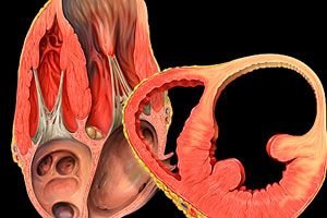
To understand what a cardiac aneurysm is, you need to delve a little deeper into anatomy and remember what the human motor is – the heart.
So, the heart is nothing more than one of the many organs in our body. It is hollow inside, and its walls are made of muscle tissue. The heart wall consists of 3 layers:
- endocardium (inner epithelial layer),
- myocardium (middle muscular layer),
- epicardium (the outer layer, which is connective tissue).
Inside the heart there is a solid partition that divides it into two parts: left and right. Each part is in turn divided into an atrium and a ventricle. The atrium and ventricle of each part of the heart are connected to each other by a special opening with a valve open to the ventricles. The bicuspid valve on the left side is called mitral, and the tricuspid valve on the right side is called tricuspid.
Blood from the left ventricle enters the aorta, and from the right ventricle - the pulmonary artery. The backflow of blood is prevented by the semilunar valves.
The work of the heart consists of constant rhythmic contraction (systole) and relaxation (diastole) of the myocardium, i.e. there is an alternating contraction of the atria and ventricles, pushing blood into the coronary arteries.
All of the above is typical for a healthy organ. But if, under the influence of certain causes, a section of the muscular part of the heart becomes thinner, it becomes unable to resist the pressure of the blood inside the organ. Having lost the ability to resist (usually due to insufficient oxygen supply, decreased muscle tone or damage to the integrity of the myocardium), such a section begins to stand out against the background of the entire organ, protruding outward and, in some cases, sagging in the form of a sac with a diameter of 1 to 20 cm. This condition is called a cardiac aneurysm.
The pressure of blood on the walls of the heart remains uniform and constant. But the healthy part of the muscular wall can restrain it, while the weakened (deformed) one cannot. If the functionality and resistance of the septum separating the ventricles or atria of the two halves of the heart is impaired, it can also bulge to the right (since it is physiologically determined that the left ventricle works more than the right), but inside the organ.
The ischemic muscle wall loses the ability to contract normally, remaining predominantly in a relaxed state, which cannot but affect the blood flow and nutrition of the entire body, and this in turn leads to the appearance of other symptoms that are dangerous to health and life.
So, we have figured out what the heart is and how such a dangerous cardiac pathology as an aneurysm of certain areas of the heart occurs. And we even found out that the most “popular” cause of the development of this disease is another life-threatening cardiac pathology – myocardial infarction, as a result of which necrotic areas and scars form on the main heart muscle, disrupting the supply of oxygen and nutrients to the muscle and reducing its resistance.
Symptoms cardiac aneurysms
The fact that cardiac aneurysm can have different sizes, localization and causes of pathology development causes significant differences in the manifestation of the disease in different people. However, in order to catch the disease at the very beginning, without waiting for the aneurysm to grow to critical sizes (clinically significant is a decrease in muscle resistance even in a small area of 1 cm), you need to know and pay attention to at least those symptoms that are characteristic of any type of cardiac aneurysm.
The first signs by which a cardiac aneurysm of any localization is determined include:
- Pain in the heart area or a feeling of heaviness (pressure) behind the sternum on the left. The pain is paroxysmal. When a person rests and is calm, the pain subsides.
- Malaise and weakness resulting from insufficient oxygen supply to the neuromuscular system. This occurs due to a decrease in the volume of blood pumped due to insufficient contractile function of the myocardium in the area of the aneurysm.
- Heart rhythm disturbances, called arrhythmia, and the sensation of a strong heartbeat, which a person does not feel in a normal state (according to patients' complaints, the heart is pounding hard). The cause of this condition is insufficient conductivity of nerve impulses in the area of the aneurysm and a large load on the diseased organ. Arrhythmias increase under the influence of stress or heavy physical exertion.
- Disturbances in the breathing rhythm, difficulty breathing or simply shortness of breath, which in the acute course of the disease can be accompanied by attacks of cardiac asthma and pulmonary edema. High pressure inside the heart is gradually transmitted to the vessels supplying blood to the lungs. As a result, oxygen exchange is disrupted and it becomes more difficult for a person to breathe. Hence the disrupted breathing rhythm.
- Pale skin tone. The cause is again a disruption in the supply of oxygen to the body's tissues. First of all, resources are directed to vital organs (brain, heart, kidneys), and the skin remains less saturated with blood.
- Cold extremities and rapid freezing associated with circulatory problems.
- Decreased skin sensitivity, appearance of "goosebumps".
- A dry, paroxysmal cough not associated with a cold or infection. It is also called cardiac. It can be a consequence of congestion in the pulmonary vessels, or it can appear as a result of compression of the lung by a large aneurysm.
- Increased sweating.
- Vertigo, or, in common parlance, dizziness, which can occur with varying frequency.
- Swelling that can be seen on the face, arms or legs.
- Fever for a long time (in acute aneurysm).
- The veins in the neck area become very engorged, making them more visible.
- Hoarse voice.
- Accumulation of fluid in the abdominal or pleural cavity, enlarged liver, dry pericarditis, which is an inflammatory process in the pericardium, accompanied by fibrous changes, obstruction of various blood vessels (can be detected during diagnostic measures for chronic aneurysm).
The symptoms of cardiac aneurysm may be superimposed on various manifestations of other existing pathologies of the cardiovascular and respiratory systems, which significantly complicates the diagnosis of the disease. And the symptoms themselves, depending on the size of the aneurysm, may be expressed to varying degrees. With a small or congenital cardiac aneurysm, the disease may proceed for a long time without any suspicious symptoms and remind of itself much later.
Where in the heart are aneurysms most often diagnosed?
As already mentioned, the most common form of myocardial pathology is considered to be an aneurysm of the left ventricle of the heart. It is this area that is loaded with work more than others. Experiencing the greatest load, the left ventricle is more prone to damage due to myocardial infarction. And therefore, an aneurysm is detected on it most often. This can also be facilitated by heart injuries or infectious pathologies.
During diagnostic procedures, the doctor may observe a protrusion of the left ventricle wall. Most often, the location of the left ventricular aneurysm is its anterior wall. But there are frequent cases of the disease, where the location of the aneurysm (protrusion) is the apex of the heart on the left side.
This pathology is not typical for children due to the absence of causes in this category of patients that can lead to the development of this disease.
Less common in patients is aneurysm of the heart vessels. This can be either an aneurysm of the ascending aorta of the heart or a protrusion of the wall of the aortic sinuses.
In the first case, the disease is caused mainly by inflammatory processes that arise as a consequence of infectious diseases. Patients' complaints are reduced to aching pains in the chest, shortness of breath and edemas of various localizations due to compression of the vena cava by the protruding wall of the aorta.
Aneurysm of the aortic sinuses is associated with a decrease in the lumen of the coronary arteries, as a result of which, under the pressure of blood, the weakened wall for some reason begins to sag, putting pressure on the right side of the heart. Fortunately, pathologies of the heart vessels associated with weakening of the walls are rare.
Ventricular septal aneurysm is not very common, as it is considered a congenital heart disease. However, it is not always detected during pregnancy or immediately after the birth of the child. Sometimes, congenital underdevelopment of the septum between the ventricles of the heart causes the aneurysm to bulge after some time.
Most often, this pathology is detected by chance, in particular during echocardiography, since it is characterized by an asymptomatic course.
An aneurysm can also choose other areas of the heart as its location (the right ventricle or atrium, the posterior wall of the left ventricle), but this happens quite rarely.
Heart aneurysm in children
As strange as it may sound, heart diseases are not only common among elderly and mature people. Young people, teenagers and even very small children can also suffer from these pathologies.
Pathological protrusion of a section of the heart muscle in children is associated with developmental defects of one or more heart valves, the interventricular or interatrial septum, resulting in the formation of an aneurysm at this site.
Such a rare pathology as an aneurysm of the interatrial septum, which can remind of itself even in adulthood, occurs in the prenatal period due to underdevelopment or changes in the structure of the septum of the heart, separating the left and right atrium. By analogy, an aneurysm of the interventricular septum is formed.
In childhood, these types of heart diseases are quite rare (no more than 1% of all patients), however, they pose a great danger to the child's life. It is good if the pathology is detected during an ultrasound of a pregnant woman. Then the child is immediately registered with a cardiologist after birth, and after the baby turns one year old, they begin to prepare him for an operation to remove the aneurysm.
The probability of developing a cardiac aneurysm is higher in children born with low birth weight and premature babies. This is due to the fact that heart defects in these categories of children are much more common, and they are more likely associated with underdevelopment of the muscular or vascular system of the heart.
While the child is small, a congenital cardiac aneurysm may not manifest itself in any way, but as the child grows older and his motor activity increases, and therefore the load on the heart, the following symptoms may appear:
- diffuse pain in the chest area,
- shortness of breath and difficulty breathing after physical exertion,
- the appearance of periodic pain in the heart area,
- cough without any cause and without sputum production,
- rapid fatigue, weakness and drowsiness,
- regurgitation during feeding (in infants), nausea (in older children),
- headaches with active movement, dizziness,
- severe sweating regardless of air temperature.
During diagnosis, doctors also determine such manifestations of the disease as:
- abnormal pulsation in the area of the 3rd rib on the left, when listening it resembles the sound of swaying waves,
- thrombi that adhere to the walls of large arteries of the heart, arising due to circulatory disorders,
- arrhythmia as a result of sports and stress.
A ruptured cardiac aneurysm is especially dangerous for both adults and children due to severe thinning of the muscle walls. That is why doctors forbid children with such a diagnosis from playing sports, since this is associated with a significant increase in the load on the heart muscle. In the future, patients are advised to lead a healthy lifestyle, avoid stressful situations and stick to a balanced diet.
 [ 25 ], [ 26 ], [ 27 ], [ 28 ], [ 29 ], [ 30 ], [ 31 ], [ 32 ], [ 33 ]
Stages
The stage of aneurysm can be determined by the degree of damage to the heart wall. If there is complete atrophy of the contractile ability of the heart muscle (akinesia), this is a severe stage of the disease with serious circulatory disorders.
If there is either a depression or a bulge of the aneurysm wall depending on the stage of the cardiac cycle (systole or diastole), such a condition is considered borderline. Although circulatory disorder is observed in this case, the symptoms of the disease and its prognosis will be different.
Forms
Cardiac aneurysms can be classified according to different parameters:
- time of formation,
- form,
- mechanisms of formation,
- sizes,
- "material" of the aneurysm wall.
Classification of cardiac aneurysms by time of formation is made only in relation to pathologies caused by myocardial infarction. The following types of post-infarction aneurysms are distinguished:
- Acute and most common form of the disease. In this case, aneurysm formation occurs during the first 2 weeks after a heart attack that caused damage to the myocardial walls. Patients experience a temperature increase above 38 degrees for a long time, breathing problems in the form of shortness of breath, heartbeat becomes rapid and its rhythm is disrupted. Blood and urine tests indicate the development of an inflammatory process.
Acute cardiac aneurysm is dangerous due to the increased risk of rupture of the pathological protrusion of the heart wall or blood vessels, which threatens the patient's life.
- Subacute cardiac aneurysm. It can appear in the period from 2-3 weeks to 2 months after myocardial infarction. The wall of this aneurysm is denser and less susceptible to rupture due to fluctuations in blood pressure inside the ventricle than the acute type of aneurysm. However, the pathological protrusion can press on other organs, causing disruptions in their work. And the decrease in the contractile function of one of the walls of the heart can have a negative effect on blood circulation.
- Chronic cardiac aneurysm. This is a kind of unpleasant surprise that the patient receives 2 or more weeks after a heart attack. Sometimes the chronic form of aneurysm is a consequence of an untreated acute one.
Once formed, such an aneurysm is not prone to rapid growth or rupture under load. But its formation is fraught with the appearance of blood clots, chronic symptoms of heart failure, arrhythmia. This is the form with the most pronounced symptoms of malaise.
An echocardiogram allows classifying cardiac aneurysms by shape. According to its data, an aneurysm can be:
- Diffuse
- Mushroom-shaped
- Saccular
- Stratifying
- "An aneurysm within an aneurysm."
Diffuse (flat) aneurysm is characterized by small dimensions, and its bottom is at the same level as the healthy myocardium. However, the protrusion can increase and change shape over time. And yet, flat chronic cardiac aneurysm is considered a pathology with the most favorable prognosis.
Mushroom-shaped resembles a jug standing on its neck. Saccular - a protrusion with a wide base and a small mouth. Resembles a diffuse aneurysm, but larger in size. Both mushroom-shaped and saccular forms are considered dangerous, since there is a high risk of blood clots forming inside the aneurysm or rupture of its wall.
Dissecting aneurysm of the aorta of the heart is a longitudinal dissection of the aortic walls, accompanied by an increase in the diameter of the main cardiac artery. Most often it is formed as a result of frequently elevated blood pressure. Its symptoms and prognosis depend on the location of the dissection.
“Aneurysm within an aneurysm” is the rarest type of pathology, when an additional protrusion is formed on the wall of an existing diffuse or saccular aneurysm, characterized by a particularly thin wall and a tendency to rupture under the slightest load.
According to the size of aneurysm, they can be:
- Clinically insignificant – up to 1 cm.
- Small – 1-2 cm.
- Large 3-5 cm.
According to the mechanism of formation, aneurysms are divided into:
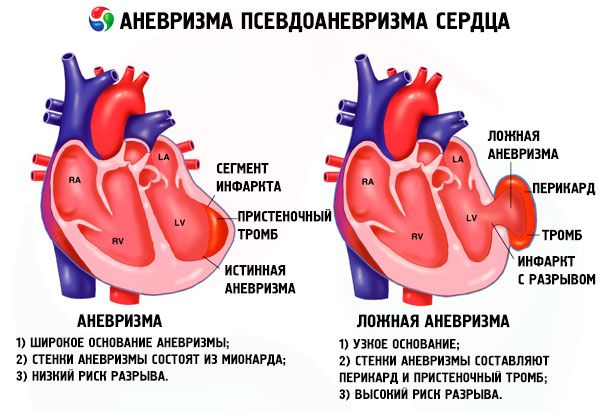
- True
- False
- Functional.
A true cardiac aneurysm is formed directly from weakened tissue of the heart itself. All of the above applies specifically to this type of aneurysm.
A false aneurysm of the heart is a pathological bulging formation consisting mainly of adhesive tissue and a leaflet of the pericardium (pericardial sac). The presence of blood in such an aneurysm is explained by a defect in the heart wall.
A functional aneurysm develops against the background of reduced contractile function of a section of the myocardium, which bends only during systole.
The aneurysm wall may consist of the following materials:
- muscle tissue,
- connective tissue (fibrin),
- a combination of two types of tissue (connective tissue formed in place of the necrotic myocardium).
In this regard, aneurysms are divided into muscular, fibrous and fibromuscular.
Complications and consequences
Aneurysm of the heart is not just an ailment, but a real threat to the patient's life. The most dangerous complication of an aneurysm is its rupture. Usually, every minute and second counts. If immediate measures are not taken to save the patient, death is inevitable, especially if the aneurysm was large.
Tissue rupture is typical mainly for acute aneurysms that develop after a myocardial infarction. The tissues of the heart muscle damaged by the infarction are considered to be the weakest during the first to second week. It is during this period that a rupture of the cardiac aneurysm can be expected.
Another terrible consequence of an aneurysm is the development of diseases caused by the blockage of blood vessels by thrombi that formed in the aneurysm cavity and at some point began to move through the circulatory system. What diseases a broken thrombus can cause depends on its size and direction of movement.
Getting into the pulmonary artery and getting stuck in it, the thrombus thereby provokes the development of a dangerous disease called thromboembolism, which threatens the patient with death if measures are not taken in time to restore normal blood circulation.
Once in the peripheral vessels, the thrombus clogs them, leading to complications such as gangrene of the extremities (more often of the legs than of the arms).
A blood clot entering the intestinal or renal artery can provoke the development of no less dangerous pathologies, such as mesenteric thrombosis (mortality rate of about 70%) and renal infarction (a serious pathology, which, however, can be successfully treated).
A stroke can also be the result of a thrombus breaking off and getting into the brachiocephalic trunk. Among other things, the same thrombus sometimes becomes the culprit of recurrent myocardial infarction.
As a complication of cardiac aneurysm, patients usually experience heart rhythm disturbances. And any arrhythmia is a threat of hypoxia of various important organs in the human body, leading to disruption of their functioning.
One of the most common consequences of an aneurysm is also considered to be heart failure (most often of the left ventricle of the heart), which manifests itself in the form of weakness, chilliness, pale skin, dizziness, shortness of breath, dry cardiac cough, edema syndrome localized in the arms and legs. If, as the disease progresses, pulmonary edema occurs, this threatens the patient not only with the fear of death, but also with the fatal outcome itself.
What is the danger of a heart vessel aneurysm? A small aneurysm may only slightly affect blood circulation, but if its size increases significantly over time under the pressure of the blood flow, this may lead to atrophy of the ribs and sternum, and also contribute to compression of the atrium and ventricle located on the right side of the heart. The latter threatens overflow of the jugular veins, the development of edema syndrome, and an increase in the size of the liver.
Large aneurysms of the aortic sinuses can compress the pulmonary trunk. This situation is life-threatening for patients. In most cases, doctors simply do not have time to do anything, death occurs so quickly.
The most dangerous is still considered to be the acute form of aortic aneurysm, which in most cases is the result of a left ventricular infarction or interatrial septum. Very often, patients do not even have time to get to the operating room. Chronic and subacute forms of the pathology are characterized by a lower mortality rate, although they still pose a danger to the life and health of the patient if you do not seek help from a medical institution in time.
As we can see, cardiac aneurysm is a pathology that is not worth joking with. And the sooner a diagnosis is made and appropriate treatment is undertaken, the greater the chances of a person to avoid the life-threatening and health-threatening consequences of a dangerous pathology affecting the heart and adjacent vessels.
[ 39 ], [ 40 ], [ 41 ], [ 42 ], [ 43 ], [ 44 ], [ 45 ], [ 46 ]
Diagnostics cardiac aneurysms
The formation of an aneurysm is most often diagnosed on the walls of the left ventricle after a myocardial infarction in people over 40 years old. And its main danger is that the weakened tissue can rupture and blood will spill outside the heart, which, if delayed, often leads to the death of the patient.
Who to contact?
Treatment cardiac aneurysms
The choice of treatment method depends on the size and type of aneurysm, as well as the patient's age and condition. It is not possible to correct the situation with medication and physiotherapy, since drugs capable of returning damaged muscles to their original shape and elasticity have not yet been found.
Prevention
Although surgical treatment of cardiac aneurysms is the preferred method of combating the disease, as we have seen, it is not always possible. Drug treatment is also preferred for small, relatively harmless aneurysms.
But the thing is that conservative treatment is not enough. In order for the aneurysm not to increase in size and not to rupture, the patient will have to reconsider his entire lifestyle and limit himself in some things. Living with a heart aneurysm means constant monitoring of the heart and fulfilling the conditions necessary to prevent complications of the aneurysm.
First of all, prevention of complications of cardiac aneurysm involves giving up bad habits, and in particular smoking and drinking alcohol, which increase the load on the heart. Nicotine causes spasm of the coronary vessels, heart rhythm disturbances, narrowing of the vessels due to cholesterol deposition on them. Alcohol, on the contrary, dilates the vessels, increasing the blood flow through the damaged walls of the myocardium, provoking a heart attack.
Particular attention should be paid not only to adequate rest, which is necessary for any disease, but also to nutrition and physical activity. Nutrition for cardiac aneurysm is dietary (therapeutic diet No. 10), which involves refusing salty and spicy foods, fried foods, fresh bread, fatty meat or fish, products containing coarse fiber, strong tea and caffeine-containing products. A diet based on vegetarian and light meat dishes with a sufficient amount of vegetables, fruits and dairy products is designed to normalize blood circulation and ease the work of a diseased heart.
Physical activity in case of cardiac aneurysm should be minimized, because what is useful for a healthy person can be dangerous for a patient with cardiac pathologies. We are talking not only about heavy physical activity associated with sports or work, but also about active movement (running, climbing stairs and even fast walking). Such activity causes increased breathing and heart rate, which is dangerous for weakened aneurysm tissue prone to rupture.
However, you should not give preference to a hypodynamic lifestyle, so as not to earn additional health problems. Daily quiet walks in the fresh air and simple physical exercises will not harm a weak heart, but will satisfy its need for oxygen.
Monitoring heart function also involves regularly measuring blood pressure and taking steps to normalize it.
The need to ease the work of a sick heart requires both weight loss (if it is above normal) and timely consultation with a doctor if alarming symptoms occur (even if they are not related to cardiac activity).
Forecast
The prognosis for cardiac aneurysm, especially after myocardial infarction, can hardly be called favorable. Without appropriate treatment, such patients die within 2-3 years after the aneurysm has formed.
The best prognosis, of course, is for flat aneurysms, but saccular and mushroom aneurysms, which in most cases have complications in the form of thrombus formation and heart failure, are a very common cause of death for patients. The prognosis is worsened by concomitant diseases such as diabetes or renal failure, as well as the patient's advanced age.
It is impossible to give a definitive answer to the question of how long patients with a cardiac aneurysm live. Everything depends on the type and size of the aneurysm, the methods of its treatment, and the patient's age when the cardiac aneurysm formed. For example, if the aneurysm formed in the interatrial septum in childhood and was not removed, the patient will most likely live about 40-45 years. Those who cross this threshold become disabled due to progressive heart failure.
If the patient is on medication, everything depends on the accuracy of the doctor's instructions, not only regarding medication, but also lifestyle in general. After heart surgery, most patients live more than 5 (about 75%) and even more than 10 (from 30 to 60%) years. But again, throughout their lives, they will have to limit themselves in physical activity and in some far from healthy pleasures.
As for disability, such a scenario is considered quite possible both in the case of an incurable surgical aneurysm of the heart and in the case of some complications after surgery. A disability group is given mainly for chronic aneurysms, especially if they are complicated by severe heart failure or there are concomitant pathologies that worsen the patient's condition.
The decision of the Medical and Social Expertise Commission regarding the group may be influenced by various factors. Patients of pre-retirement age and those for whom surgery is impossible for good reasons are most likely to receive disability. If a patient with limited ability to work simply refuses surgery, the Medical and Social Expertise Commission will insist on its implementation before it can make a final verdict.
Patients with an aneurysm can receive both working and non-working 3rd group. Everything depends on their condition and ability to work. In some cases, patients are simply sent for retraining or provided with another workplace where the cardiac aneurysm will not interfere with the fulfillment of work obligations.