
All iLive content is medically reviewed or fact checked to ensure as much factual accuracy as possible.
We have strict sourcing guidelines and only link to reputable media sites, academic research institutions and, whenever possible, medically peer reviewed studies. Note that the numbers in parentheses ([1], [2], etc.) are clickable links to these studies.
If you feel that any of our content is inaccurate, out-of-date, or otherwise questionable, please select it and press Ctrl + Enter.
Treacher Collins syndrome
Medical expert of the article

Last reviewed: 04.07.2025
Intrauterine disturbances in bone development processes cause serious craniofacial deformities, and one of the varieties of such pathology is Treacher Collins syndrome (TCS) or mandibulofascial, that is, maxillofacial dysostosis.
Disease code according to ICD 10: class XVII (congenital anomalies, deformations and chromosomal disorders), Q75.4 - mandibulofacial dysostosis.
Causes Treacher Collins syndrome
This syndrome was named after the outstanding British ophthalmologist Edward Treacher Collins, who described the main features of the pathology more than a hundred years ago. However, European doctors more often call this type of facial and jaw bone anomaly Franceschetti's disease or syndrome - based on the extensive research of the Swiss ophthalmologist Adolf Franceschetti, who introduced the term "mandibulofascial dysostosis" in the middle of the last century. In medical circles, the name Franceschetti-Collins syndrome is also used.
Treacher Collins syndrome is caused by mutations in the TCOF1 gene (at the 5q31.3-33.3 chromosome locus), which codes for a nucleolar phosphoprotein responsible for the formation of the craniofacial part of the human embryo. As a result of a premature decrease in the amount of this protein, the biogenesis and functions of rRNA are disrupted. According to geneticists from the Human Genome research program, these processes lead to a reduction in the proliferation of embryonic cells of the neural crest - a ridge along the neural groove, which closes into a neural tube during embryonic development.
Formation of facial tissues occurs due to transformation and differentiation of cells of the upper (head) part of the neural crest, which migrate along the neural tube to the area of the first and second branchial arches of the embryo. And the deficiency of these cells causes craniofacial deformations. The critical period for the occurrence of anomalies is from 18 to 28 days after fertilization. Upon completion of the migration of neural crest cells (in the fourth week of gestation), almost all loose mesenchymal tissues in the facial area are formed, which later (from 5 to 8 weeks) differentiate into skeletal and connective tissues of all parts of the face, neck, larynx, ear (including the inner ear) and future teeth.
Pathogenesis
The pathogenesis of Treacher Collins syndrome is often familial, and the anomaly is inherited in an autosomal dominant manner, although there are cases of autosomal recessive transmission of the defect (with mutations in other genes, in particular, POLR1C and POLR1D). The most unpredictable thing about maxillofacial dysostosis is that the mutation is inherited by children only in 40-48% of cases. That is, in 52-60% of patients, the causes of Treacher Collins syndrome are not associated with the presence of an anomaly in the family, and it is believed that the pathology occurs as a result of sporadic gene mutations de novo. Most likely, new mutations are the consequences of teratogenic effects on the fetus during pregnancy.
Among the teratogenic causes of this syndrome, experts name large doses of ethanol (ethyl alcohol), radiation, cigarette smoke, cytomegavirus and toxoplasma, as well as glyphosate-based herbicides (Roundal, Glyfor, Tornado, etc.). And the list of iatrogenic factors includes acne and seborrhea drugs with 13-cis-retinoic acid (Isotretinoin, Accutane); the anticonvulsant drug Phenytoin (Dilantin, Epanutin); psychotropic drugs Diazepam, Valium, Relanium, Seduxen.
Symptoms Treacher Collins syndrome
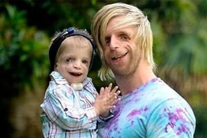
For the most part, the clinical signs of mandibulofascial dysostosis and the degree of their expression depend on the characteristics of the manifestation of gene mutations. And the first signs of this anomaly in most cases are visible in a child immediately after birth: the face with Treacher Collins syndrome has a characteristic appearance. Moreover, morphological anomalies are usually bilateral and symmetrical.
The most obvious symptoms of Treacher Collins syndrome are:
- underdevelopment (hypoplasia) of the facial bones of the skull: zygomatic, zygomatic processes of the frontal bone, lateral pterygoid plates, paranasal sinuses, lower jaw and protrusions of the bone epiphyses (condyles);
- underdevelopment of the bones of the lower jaw (micrognathia) and a mandibular angle that is more obtuse than usual;
- the nose is of normal size, but appears large due to hypoplasia of the superciliary arches and underdevelopment or absence of the zygomatic arches in the temporal region;
- the eye slits are downward, that is, the shape of the eyes is abnormal, with the outer corners drooping downwards;
- defects of the lower eyelids (coloboma) and partial absence of eyelashes on them;
- irregularly shaped auricles with a wide range of deviations, including their location in the corner of the lower jaw, absence of lobes, blind fistulas between the tragus of the ear and the corner of the mouth, etc.;
- narrowing or closure (atresia) of the external auditory canal and anomalies of the middle ear ossicles;
- absence or hypoplasia of the parotid salivary glands;
- pharyngeal hypoplasia (narrowing of the pharynx and airways);
- non-fusion of the hard palate (cleft palate), as well as absence, shortening or immobility of the soft palate.
Such anatomical anomalies in all cases have complications. These are functional hearing disorders in the form of conductive hearing loss or complete deafness; visual impairment due to improper formation of the eyeballs; defects of the palate cause difficulties with feeding and swallowing. There are dental occlusion disorders (malocclusion) associated with jaw defects, which, in turn, causes problems with chewing and articulation. Pathologies of the soft palate explain the nasal voice.
Complications and consequences
The consequences of maxillofacial anomalies in Treacher Collins syndrome are that at birth the child's intellectual abilities are normal, but due to hearing defects and other disorders, secondary mental retardation is observed.
In addition, children with such defects acutely feel their inferiority and suffer, which negatively affects their nervous system and psyche.
Diagnostics Treacher Collins syndrome
Postnatal diagnosis of Treacher Collins syndrome is essentially based on clinical signs. Craniofacial dysostosis is easily identified when the syndrome is fully expressive, but when minimally expressed symptoms of pathology are present, problems with establishing the correct diagnosis may arise.
In this case, special attention should be paid to the assessment of all functions associated with anomalies, especially those affecting breathing (due to the risk of sleep apnea). The effectiveness of feeding and hemoglobin oxygen saturation should also be assessed and monitored.
Later, on the 5th-6th day after birth, the extent of hearing damage will need to be determined using audiological testing, which should be carried out in the maternity hospital.
An examination is prescribed, during which instrumental diagnostics are performed by fluoroscopy of craniofacial dysmorphology; pantomography (panoramic X-ray of the bone structures of the facial skull); full cranial computed tomography in various projections; CT or MRI of the brain to determine the state of the internal auditory canal.
The earliest – prenatal – diagnosis of maxillofacial anomalies in the presence of Treacher Collins syndrome in the family history is possible by chorionic villus biopsy at 10-11 weeks of pregnancy (the procedure threatens miscarriage and infection of the uterus).
Blood tests are also taken from family members; at 16-17 weeks of pregnancy, amniotic fluid is analyzed (transabdominal amniocentesis); at 18-20 weeks of pregnancy, fetoscopy is performed and blood is taken from the fetal vessels of the placenta.
But most often, ultrasound is used in prenatal diagnosis of this syndrome in the fetus (at 20-24 weeks of pregnancy).
What tests are needed?
Differential diagnosis
These same methods are used by specialists when differential diagnostics are needed to recognize the mild Treacher Collins syndrome and distinguish it from other congenital anomalies of the craniofacial bones, in particular: Apert, Crouzon, Nager, Peters-Hewels, Hellermann-Steph syndromes, as well as hemifacial microsomia (Goldenhar syndrome), hypertelorism, premature fusion of the cranial sutures (craniosynostosis) or impaired fusion of the facial bones (craniosynostosis).
Who to contact?
Treatment Treacher Collins syndrome
As in all cases of genetically determined congenital defects, the treatment of severe forms of Treacher Collins syndrome is exclusively palliative, since there are simply no therapeutic methods for such pathologies. The spectrum and degree of deformations in this syndrome are extensive and, therefore, the nature and intensity of medical intervention also has many options.
Hearing aids are used to correct and improve hearing, and speech therapy sessions are used to improve speech.
Surgical interventions are required at an early age in severe cases of narrowing of the airways (a tracheostomy is performed) and larynx (a gastrostomy is performed for feeding). Surgical correction of the palate may also be required.
Mandibular lengthening surgeries are performed at the age of 2-3 years or later. Soft tissue reconstruction includes lower eyelid coloboma correction and auricular plastic surgery.
Prevention
Forecast
What is the prognosis for this pathology? It depends on the degree of deformation and the intensity of symptoms. Treacher Collins syndrome is a lifelong diagnosis.
 [ 25 ]